báo cáo khoa học: " Thin anterior uterine wall with incomplete uterine rupture in a primigravida detected by palpation and ultrasound: a case report" pptx
Bạn đang xem bản rút gọn của tài liệu. Xem và tải ngay bản đầy đủ của tài liệu tại đây (636.89 KB, 4 trang )
CAS E REP O R T Open Access
Thin anterior uterine wall with incomplete uterine
rupture in a primigravida detected by palpation
and ultrasound: a case report
Shigeki Matsubara
*
, Kazuhiko Shimada, Tomoyuki Kuwata, Rie Usui, Mitsuaki Suzuki
Abstract
Introduction: Uterine rupture is an obstetric complication associated with significant maternal and fetal morbidity
and mortality. This disorder usually occurs with a scarred uterus, especially in a uterus with prior Cesarean section.
Uterine sacculation or diverticulum may also lead to a thin uterine wall during pregnancy.
Case presentation: A 27-year-old Japanese primigravid woman was admitted to our hospital due to weak, irregular
uterine contractions in her 38
th
week of gestation. She had no past history of uterine surgery or known diseases. A
hard mass was palpable in her abdomen. An ultrasound revealed that the anterior uterine wall was thin and bulging,
with a fetal minor part beneath it which corresponded to the palpated mass. A Cesarean section was performed
which revealed a thin anterior uterine wall with incomplete uterine rupture. The woman and baby were healthy.
Conclusions: Although extremely rare, an unscarred primigravid uterus can undergo incomplete rupture even
without discernable risk factors or labor pains. Abdominal palpation and ultrasound may be useful in detecting this
condition.
Introduction
Uterine rupture is an obstetric complication associated
with significant maternal and fetal morbidity and mor-
tality. This disorder usually occurs in a scarred uterus,
especially secondary to prior Cesarean section, and is
therefore considered a disease of multigravida [1,2]. A
few reports have indicated that a uterine rupture can
occur in primigravida, although this is extremely rare
[2,3], with etiological or risk factors including a h istory
of uterine surgery, labor augmentation or underlying
connective tissue disease [2-4]. A thin uterine wall, as a
result of uterine sacculation [ 5,6] or uterine diverticu-
lum [7], may also induce uterine rupture.
We report the case of a primigravid woman with a
thin anterior uterine wall; a feature compatible with
incomplete uterine rupture. Underlying etiological fac-
tors were indiscernible. Her condition was detected by
abdominal palpation and then ultrasound. This case
report suggests that an unscarred primigravid uterus can
show the features of incomplete rupture even in the
absence of discernable risk f actors and that abdominal
palpation and ultrasound may be useful in diagnosis.
Case presentation
A 27-year-old Japanese primigravid woman was admitted
to our hospital because of slight uterine contractions at 38
+6
weeks of gestation. Her past history was unremarkable.
She had received no uterine sur gery or procedure. There
were no symptoms or history suggesting the presence of
Ehlers-Danlos syndrome in either herself or her family
memb ers. We did not perform drug screening, inc luding
cocaine (a possible cause of uterine rupture). She had
received regular pregnancy checks on up to 14 occasions
and an ultrasound examination had been performed at
every visit, according to our institute protocol. An ultra-
sound had revealed no abnormal uterine structure,
although special attention had not been paid to the uterine
wall. On admission, a speculum examination revealed that
her cervix was normally positioned and not ventrally
located. A digital examination revealed a cervical ostium
opening of 2.0 cm with 1.5 cm effacement. A cardiotoco-
gram showed a normal fetal heart rate (FHR) pattern with
* Correspondence:
Department of Obstetrics and Gynecology and Jichi Perinatal Education
Center, Jichi Medical University, 3311-1 Yakushiji, Shimotsuke, Tochigi
329-0498, Japan
Matsubara et al. Journal of Medical Case Reports 2011, 5:14
/>JOURNAL OF MEDICAL
CASE REPORTS
© 2011 Matsubara et al; licensee BioMed Central Ltd. This is an Open Access articl e distributed under the terms of the Creative
Commons Attr ibution License ( which permits unrestricted use, distribution, and
reprodu ction in any medium, provided the original work is properly cited.
weak uterine contractions of 20 second duration once per
hour. There was no tenderness or palpable mass. An
attending doctor p erformed an ultrasound exa mination,
which revealed n ormal placentation w ithout myoma and
an amniotic fluid index of 15 cm (normal range: 5-25 cm).
However, he did not comment on the uterine wall thick-
ness. Although she preferred to remain in hospital, the
irregular contractions occurring only once per hour led to
her decision to discharge at 39
+2
weeks of gestation. Dur-
ing her t ime in our hospital, a cardiotocogram was per-
formed six times and mild variable decele rations with
normal variability were observed on two occasions. Other-
wise, FHR patterns were normal. Prior to her discharge,
the cervical ostium opening was 3.0 cm with 1.0 cm effa-
cement, with -2 station of the presenting part, the head.
This roughly indicated that active labor had not yet begun.
The attending doctor palpated her abdomen and inciden-
tally noted a hard thumbhead-sized mass protruding at
the midline, 10 cm below her umbilicus. An abdominal
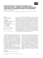
ultrasound revealed a bulging thin uterine wall of <1 mm
with a fetal minor part beneath it (Figure 1), which corre-
sponded to the hard palpable mass. Slight tenderness was
observed at the thin uterine wall. Although primigravidity,
an unscarred uterus and the absence of regular contrac-
tions reduced the possibility of an impending uterine rup-
ture, ultrasound findings led us to suspect it and an
emergent Cesarean section was performed.
Her vesicouterine fold was located in the normal posi-
tion and her bladder did not overlay the lower uterine
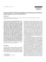
segment. Cephalad to the vesicouterine fold, an approxi-
mately 8 × 8 cm area of the uterine anterior wall was thin
and a fetal hand and head were visible through it (Figure
2A and 2B). A transverse incision was made cephalad to
this thin wall area, and a 2604 g (appropriate-for-date
according to the Japanese standard) female baby was
delivered with Apgar scores of 8 and 9 at one to five min-
utes, respectively. The placenta was delivered sponta-
neously. We excised the thin part of t he uterine wall and
reconstructed the site with a total blood loss of 370 ml.
Theuterinebodywaslocatednormally, without distor-
tion or posterior incarceration. A histological examina-
tion of the excised uterine wall was not performed. An
abdominal and vaginal ultrasound seven days post-par-
tum revealed a well-involuted uterus and the absence of
discernable uterine anomalies. Her post-partum course
was unremarkable. Considering that a short inter-delivery
period of <18 months would increase the risk of rupture
of the scarred uterus [8], we advised her to avoid further
pregnancy for at least one year. The patient and her baby
were healthy six months after the birth.
Discussion
This case suggested two clinical issues. Firstly, an
unscarred pre-labor primigravid uterus can show a very
thin uterine wall, compatible with incomplete uterine
rupture, without apparent etiological or risk factors. Sec-
ondly, abdomi nal palpation and ultraso und may be use-
ful to detect this condition even in the absence of
symptoms or signs.
Uterine ruptures are divided into complete and
incomplete. In the former, the uterine serosa together
with the uterine muscular layer is perforated and thus
the amniotic cavity directly communicates with the
abdomi nal cavity. In the latter, the uterine muscular
layer is lost but the uterine serosa is preserved. Incom-
plete rupture is also referred to as uterine dehiscence.
Data obtained from cases of complete rupture of a pri-
migravid unscarred uterus may be applicable to, or may
at least provide information on, incomplete rupture.
Therefore, our discussion is based on data on complete
rupture of a primigravid unscarred uterus, since reports
focusing on incomplete rupture of a primigravid
unscarred uterus are lacking. Two disorders, uterine sac-
culation and uterine diverticulum, may also induce a
thin uterine wall, thus causing u terine rupture. While
uterine rupture, complete or i ncomplete, denotes the
final uterine feature, uterine sacculation and diverticu-
lum denote the disease entities. To avoid confusion,
‘rupture’ has been used to denote the final uterine con-
dition, if not specified. Sacculation and diverticulum will
be touched on separately.
The first important clinical issue is that an unscarred
pre-labor primigravid uterus can show a very thin uter-
ine wall, compatible with incomplete uterine rupture,
without apparent etiological or risk factors. Walsh and
Baxi [2], reviewing the literature over six decades (1946-
2006), found 36 primigravid uterine ruptures, and we
have found a further 21 cases [3,9-12]. Of these 21
Figure 1 An abdominal ultrasound image of the uterine wall
and the fetal minor part. The small arrow indicates a thin uterine
wall, which is slightly bulging. Beneath the thin uterine wall a fetal
minor part (large arrow) is visible, which was palpated as a hard
mass through the abdomen.
Matsubara et al. Journal of Medical Case Reports 2011, 5:14
/>Page 2 of 4
cases, 15 were repo rted in a case series from Nepal [11],
with all ruptures occurring after labor of >48 hours, and
12 having received no antenatal care. Of 57 (36+21)
cases previously reported, 55 w omen had some discern-
able etiological or risk factors for rupture, including a
past history of uterine surgery, congenital uterine anom-
aly, adherent placenta, labor, or oxytocin and/or prosta-
glandin use [2-4,9-12]. The etiological factors were
described as indiscernible in the remaining two cases;
one of which was described in a case series and lacked
detailed data [1]. In the remaining case - that of a 21-
year-old Indian woman - she had stated that she was
primigravida, but she had received no antenatal care
until rupture and no further evaluation to identify any
underlying condition was performed [13]. Therefore, the
data was insufficient to claim ‘unknown etiology’.There
have been no reported cases of primigravid unscarred
uterine rupture of unknown etiology, employing its
strict definition. Etiological or risk factors were not dis-
cernable in our case report. She had no past uterine sur-
gery, no detectable uterine anomalies and no abnormal
placentation. Her family and past history rejected the
possibility of Ehlers-Danlos syndrome, which is reported
to cause primigravid uterine rupture [4]. She experi-
enced weak uterine contractions, false pains for approxi-
mately 86 hours; however, they were very weak and may
not have been the cause of the rupture.
Another possible cause of a thin uterine wall is uterine
muscular weakness. Uterine sacculation or uterine diver-
ticulum may lead to uterine wall weakness. Uterine
sacculation is defi ned as a transitory pouch or sac-like
structure developing from a portion of the gravid uterus
[6]. The typical form of sacculation results from an
incarc erated retroverte d uterus [5,6]. A ventrally-located
cerv ical ostium and vagina may cause physicians to sus-
pect this diagnosis. Magnetic resonance imaging (MRI)
may provide a pre-operative diagnosis [5]. In this condi-
tion, the anterior uterine wall becomes stretched and
thinned. The bladder and uterine cervix are also
stretched and a Cesarean section without diagnosis may
present difficu lties in identifying the bladder and cervix,
and therefore in opening the lower uterine segment [5].
In our case report, the diagnosis of this type of uterine
sacculation, accompanied by incarcerated retroverted
uterus, can be precluded. Uterine incarceration was
absent and her lower uterine segment was located in the
normal position. Other conditions, such as previous
surgery, a primary myometrial defect, uterine malforma-
tion or placental abnormalities are listed as possible
causes of uterine sacculation [6,14]. However, in our
case report, these underlying conditions were not identi-
fied. We therefore could not determine whether ute rine
wall weakness of unknown etiology was present in our
case report.
Uterine diverticulum is frequentl y misunderstood and
reported as uterine sacculation [7]. Uterine diverticulum
has a narrow connection with the uterine cavity and a
thicker wall than in sacculation [7]. While uterine saccu-
lation is usually observed during pregnancy [6,14], diver-
ticulum is usually detected in non-pregnant women.
Uterine diverticula as complications during pregnancy
are rare. Rajiah et al. [7] reported a primigravid woman
in whom an MRI revealed uterine diverticulum in the
22
nd
week of gestation. A Cesarean section was per-
formed in the 31
st
week. The diverticulum originated
from the posterolateral wall of the uterine body and did
not contai n the fetus. The diverticulum was not excised
due to surgical risks. In the post -partum period, the
diverticulum was observed via ultrasound. The authors
considered that the underlying etiology for the diverticu-
lum may have been congenital because this patient was
primigravida with no prior cervical or uterine procedure.
᧦
᧦
A
B
Figure 2 Operative findings during a Cesarean section. A: The asterisk indicates the thin anterior uterine wall. Arrows indicate the
peritoneum. The left side of the photograph is the caudal side of the patient. B: After delivering the fetus and removing the placenta, the thin
wall was gently pushed from inside the uterus with a finger. The finger tip (large arrow) is clearly visible through the thin wall. The asterisk
indicates the surgeon’s right hand. Small arrows indicate the uterine incision site.
Matsubara et al. Journal of Medical Case Reports 2011, 5:14
/>Page 3 of 4
Sun et al. [15] reported a case in which the gestational sac
implanted in a diverticulum: the pregnancy was termi-
nated at an estimated seven to eight weeks. The authors
considered that abnormal development of the parameso-
nephric duct may cause a congenital uterine deformity,
leading to a formation of diverticulum. In our case report,
the thin uterine wall did not show the morphological fea-
tures of typical diverticulum: the narrow neck characteris-
tic of typical diverticulum was not observed. Thus, this
diagnosis may be precluded. Although the morphological
features of the thin uterine wall in our case report differed
from those of uterine diverticulum, the possible underlying
etiol ogy of diverticulum suggested by Sun et al. [15] may
also hold true here. However, we do not have any evi-
dence. If we had not ex cis ed the thin uteri ne wall, and if
this portion had later bulged and eventually made a diver-
ticulum, then the thin uterine wall observed may have
proved to be a feature of the future occurrence of uterine
diverticulum. The clinical course of uterine diverticulum,
especially that of unknown etiology, is n ot yet clear and
further cases are required.
The second important clinical issue is that abdominal
palpation and ultrasound may be useful in detecting this
condition. Although abdominal pain [2,3] and non-reas-
suring FHR patterns, especially fetal bradycardia, have
been reported to be signs of impending or actual uterine
rupture in some cases, our case report showed no
abdominal pain and no abnormal FHR pattern, except for
mild variable deceleration. It was fortunate that an
attending doctor happened to palpate the abdomen even
in the absence of symptoms or signs and noticed a hard
thumbhead-sized mass. To the best of our knowledge,
this is the first reported case in which palpation inciden-
tally revealed a protruding fe tal minor part, with ultra-
sound identifying th is condit ion as impending complete
uterine rupture, leading to an emergent Cesarean section.
Congenita l weakness of the uterine wall may result in
a t hin uterine wall and this has been discussed in rela-
tion to uterine sacculation and diverticulum. However,
it remains undetermined as to why and when the uter-
ine wall b ecomes thin. Putting aside the etiology of the
thin uterine wall observed here, physicians must be
aware that an unscarred primigravid uterus can undergo
incomplete rupture.
Conclusions
An unscarred primigravid uterus can undergo incom-
plete rupture even without etiological or risk factors, or
the prese nce of any symptoms or signs. Abdominal pal-
pation and ultrasound may be useful for diagnosis.
Consent
Written informed consent was obtained from the patient
for publication of this case report and any accompanying
images. A copy of the w ritten consent is available for
review by the Editor-in-Chief of this journal.
Abbreviations
FHR: fetal heart rate; MRI: Magnetic resonance imaging
Authors’ contributions
SM, KS and RU diagnosed, investigated, followed up on and managed the
patient’s care. SM and TK wrote the manuscript. MS provided important
suggestions regarding medical content. All authors read and approved the
final manuscript.
Competing interests
The authors declare that they have no competing interests.
Received: 30 December 2009 Accepted: 17 January 2011
Published: 17 January 2011
References
1. Kieser KE, Baskett TF: A 10-year population-based study of uterine
rupture. Obstet Gynecol 2002, 100:749-753.
2. Walsh CA, Baxi LV: Rupture of the primigravid uterus: a review of the
literature. Obstet Gynecol Surv 2007, 62:327-334.
3. Dow M, Wax JR, Pinette MG, Blackstone J, Cartin A: Third-trimester uterine
rupture without previous Cesarean: a case series and review of the
literature. Am J Perinatol 2009, 26:739-744.
4. Walsh CA, Reardon W, Foley ME: Unexplained prelabor uterine rupture in
a term primigravida. Obstet Gynecol 2007, 109:455.
5. Gottschalk EM, Siedentopf JP, Schoenborn I, Gartenschlaeger S,
Dudenhausen JW, Henrich W: Prenatal sonographic and MRI findings in a
pregnancy complicated by uterine sacculation: case report and review
of the literature. Ultrasound Obstet Gynecol 2008, 32:582-586.
6. Frei KA, Duwe DG, Bonel HM, Dürig P, Schneider H: Posterior sacculation
of the uterus in a patient presenting with flank pain at 29 weeks of
gestation. Obstet Gynecol 2005, 105:639-641.
7. Rajiah P, Eastwood KL, Gunn ML, Dighe M: Uterine diverticulum. Obstet
Gynecol 2009, 113:525-527.
8. Bujold E, Gauthier RJ: Risk of uterine rupture associated with an
interdelivery interval between 18 and 24 months. Obstet Gynecol 2010,
115:1003-1006.
9. Wada S, Kudo M, Minakami H: Spontaneous uterine rupture of a twin
pregnancy after laparoscopic adenomyomectomy: a case report. J Minim
Invasive Gynecol 2006, 13:166-168.
10. Villa G, Mabrouk M, Guerrini M, Mignemi G, Colleoni GG, Venturoli S,
Seracchioli R: Uterine rupture in a primigravida with adenomyosis
recently subjected to laparoscopic resection of rectovaginal
endometriosis: case report. J Minim Invasive Gynecol 2008, 15:360-361.
11. Padhye SM: Rupture uterus in primigravida: morbidity and mortality.
Kathmandu Univ Med J (KUMJ) 2007, 5:492-496.
12. Banas T, Klimek M, Fugiel A, Skotniczny K: Spontaneous uterine rupture at
35 weeks’ gestation, 3 years after laparoscopic myomectomy, without
signs of fetal distress. J Obstet Gynaecol Res 2005, 31:527-530.
13. Abbi M, Misra R: Rupture of uterus in a primigravida prior to onset of
labor. Int J Fertil Womens Med 1997, 42:418-420.
14. Friedman A, DeFazio J, DeCherney A: Severe obstetric complications after
aggressive treatment of Asherman syndrome. Obstet Gynecol
1986,
67:864-867.
15. Sun X, Xue M, Xiao S, Wan Y, Wang B: Uterine diverticulum complicating
pregnancy diagnosed by ultrasound and uteroscopy. Int J Gynaecol
Obstet 2010, 109:247-248.
doi:10.1186/1752-1947-5-14
Cite this article as: Matsubara et al.: Thin anterior uterine wall with
incomplete uterine rupture in a primigravida detected by palpation and
ultrasound: a case report. Journal of Medical Case Reports 2011 5:14.
Matsubara et al. Journal of Medical Case Reports 2011, 5:14
/>Page 4 of 4