Báo cáo y học: "Salivary gland derived peptides as a new class of anti-inflammatory agents: review of preclinical pharmacology of C-terminal peptides of SMR1 protein" pptx
Bạn đang xem bản rút gọn của tài liệu. Xem và tải ngay bản đầy đủ của tài liệu tại đây (2.06 MB, 11 trang )
REVIEW Open Access
Salivary gland derived peptides as a new class of
anti-inflammatory agents: review of preclinical
pharmacology of C-terminal peptides of SMR1
protein
Ronald D Mathison
1*
, Joseph S Davison
1
, A Dean Befus
2
, Daniel A Gingerich
3
Abstract
The limitations of steroidal and non steroidal anti-inflammatory drugs have prompted investigation into other
biologically based therapeutics, and identification of immune selective anti-inflammatory agents of salivary origin.
The traditional view of salivary glands as acces sory digestive structures is changing as their importance as sources
of systemically active immunoregulatory and anti-inflammatory factors is recognized. Salivary gland involvement in
maintenance of whole body homeostasis is regulated by the nervous system and thus constitutes a “neuroendo-
crine axis”. The potent anti-inflammatory activities, both in vivo and in vitro, of the tripeptide Phe-Glu-Gly (FEG) are
reviewed. FEG is a carboxyl terminal peptide of the prohormone SMR1 identified in the rat submandibular salivary
gland, The D-isomeric form (feG) mimics the activity of its L-isomer FEG. Macropharmacologically, feG attenuates
the cardiov ascular and inflammatory effects of endotoxemia and anaphylaxis, by inhibition of hypotension,
leukocyte migration, vascular leak, and disruption of pulmonary function and intestinal motility. Mechanistically, feG
affects activated inflammatory cells, especially neutrophils, by regulating integrins and inhibiting intracellular
production of reactive oxyge n species. Pharmacodynamically, feG is active at low doses (100 μg/kg) and has a long
(9-12 hour) biological half life. As a therapeutic agent, feG shows promise in diseases characterized by over exuber-
ant inflammatory responses such as systemic inflammatory response syndrome and other acute inflammatory
diseases. Arthritis, sepsis, acute pancreatitis, asthma , acute respiratory inflammation, inflammatory bowel disease,
and equine laminitis are potential targets for this promising therapeutic peptide. The term “Immune Selective
Anti-Inflammatory Derivatives” (ImSAIDs) is proposed for salivary-derived pept ides to distinguish this class of agents
from corticosteroids and nonsteroidal anti-inflammatory drugs.
Introduction
Saliva, best known for its digestive and protective proper-
ties in the maintenance of t he health and integrity of the
oral and gastric mucosa [1], is becoming increasingly
recognized for its important role in regulating whole
body homeostasis [2]. Although over the past half cen-
tury many bioactive proteins a nd peptides have been
identified in saliva [3,4], salivary glands are still viewed
primarily as accessory digestive structures that provide
lubrication and digestive enzymes. Ho wever, it is now
becoming clear that salivary endocrine factors play an
important role in the modulation of systemic immune
and inflammatory reactions. Classically, the salivary
glands are generally considered as exocrine glands that
dispense their protein and fluid externally into a lumen
or a duct. However, investigations dating from 60 years
ago suggested an unorthodox view that salivary and other
exocrine glands, such as the pancreas, are capable of
endocrine secretion, dispensing their secretions intern-
ally,i.e.directlyintothebloodstream.Ithasbeensug-
gested that these glands be called “duacrine” glands [5].
Salivary glands produce various immunoregulatory
[6,7] and anti-inflammatory [8] agents. The importance
of the salivary gland in maintaining h omeostasis has
* Correspondence:
1
Faculty of Medicine, University of Calgary, 3330 Hospital Drive NW, Calgary,
Alberta, T2N 4N1, Canada
Full list of author information is available at the end of the article
Mathison et al. Journal of Inflammation 2010, 7:49
/>© 2010 Mathison et al; licensee BioMed Central Ltd. This is an Open Access article distributed under the terms of the Creative
Commons Attribution License ( which permits unrestricted use, distribution, and
reproduction in any medium, provided the original work is pro perly cited.
been clarified in recent decades by demonstration of
neuroendocrine interactions between the nervous, endo-
crine, and immune systems [9]. The salivary glands, as
well as the thymus and cervical lymph nodes, are inner-
vated by noradrenergic fibers from the sympathetic
trunk [10,11], which were shown to modulate lympho-
cyte function within lymph nodes and thymus [12,13].
This paper reviews the published pharmacologic and
immunopharmacologic evidence that salivary gland
derived peptides, with particular emphasis on the D-iso-
meric tripeptide feG, deserve consideration as poten-
tially therapeutically useful anti-inflammatory agents.
The Neuroendocrine Axis
The existence of salivary-derived, systemically acting,
anti-inflammatory factors and the regulation of salivary
gland function by the sympathetic nervous system were
demonstrated in anaphylaxis and endotoxemia models in
rats. Superior cervical ganglionectomy significantly
reduced mortality and greatly attenuated the influx of
histamine, neutrophils, and serum-derived proteins, into
bronchoalveolar fluid in anaphylaxis-induced pulmonary
inflammation in rats [14]. However, the protective effect
of superior cervical ganglionectomy was completely abol-
ished in rats with concurrent bilateral sialadenectomy of
the submandibular salivary glands [15]. These findings
reveal that submandibular salivary glands produce sys-
temically important immunomodulatory factor s and that
the cervical sympathetic nerves tonic ally inhibit the
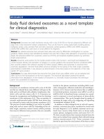
release of some of these factors. In an endotoxin-induced
acute hypotension model, either bilateral superior cervi-
cal ganglionectomy or submandibular sialadenectomy
resulted in significantly larger drops in blood pressure
compared to intact controls [16] (Figure 1). These results
indicate that the submandibular gland elaborates factors
that protect against acute hypotension induced by endo-
toxin and that these factors are under the control of the
cervical sympathetic nervous system.
Bioactivity of Salivary Gland Extracts: SGP-T
On the basis of the findings that salivary glands parti ci-
pate in modulat ing systemic inflammatory responses,
bioactive factors were sought in saliva. Extracts of sub-
mandibular glands were subjected to molecular weight
cut-off filtration and tested for bioactivity. A novel
seven amino acid peptide with sequence Thr-Asp-Ile-
Phe-Glu-Gly (TDIFEGG) was isolated, named subman-
dibular peptide-T (SGP-T), and shown to express
ant i-allergic and anti-e ndotoxin activities[16,17]. SGP-T
was identified as the car boxyl terminal of SMR1, a 146-
amino acid, multipotent prohormone product of the
VCSa1 (variable coding sequence A1) gene [ 18], which
is also identified as RATSMR1A, Smr1, SMR1 protein
and VCS-alpha 1. Recent studies have shown that SMR1
is secreted into saliva in response to intraperitoneal
administ ration of b-adrenergic and choli nergic agonists,
and removal of the cervical sympathetic ganglia that
innervate the salivary glands resulted in increased levels
of SMR1 protein in the submandibular glands [19].
These observations are in keeping with a cervical sym-
pathetic trunk - submandibular gland axis propounded
previously [15].
In ovalbumin (OA) sensitized rats SGP-T at dosages
of 35 and 100 μg/kg injected 10 minutes p rior to OA
challenge protected against anaphylactic hypotension
[20]. Interestingly, neither lower nor higher doses (10 or
350 μg/kg) of SGP-T were protective. In OA sensitized
rats challenged intra-intestinally with OA, pretreatment
with SGP-T d ose-dependently reduced the incidence
and duration of disrupted intestinal motility and pre-
vented the development of diarrhea [20]. SGP-T treat-
ment also significantly suppressed endotoxin-induced
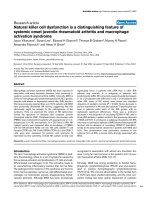
fever in rats [21]. Neutrophil migration into carrageenan
soaked sponges was inhibited by SGP-T injected intrave-
nously at 100 μg/kg at -1, 0, or 4 hours a fter implanta-
tion [22]. Interestingly, dose-response assays showed a
bell-shaped dose response curve; neither lower
(10 μg/kg) or higher (350 μg/kg) inhibited neutrophil
migration (Figure 2). SGP-T treatment also pro moted a
bell-shaped dose-dependent recovery in the ability of
neutrophils obtained from carrageenan-soaked sponges
to generate superoxide anion. In another st udy endo-
toxin-induced l eukocyte rolling and adhesion, quantified
in vivo by intravital microscopy of mesenteric venules in
Figure 1 Neuroendocrine axis and modulation of responses to
lipopolysaccharide: Intravenous administration of
lipopolysaccharide (LPS) induces rapid reduction in blood pressure
in rats. Either bilateral removal of the submandibular salivary glands
(sialadenectomized) or the superior cervical ganglia
(ganglionectomized) exacerbate the LPS-induced hypotension.
(mean ± sem, n = 6 to 8). Adapted from [16].
Mathison et al. Journal of Inflammation 2010, 7:49
/>Page 2 of 11
anesthetized rats, was prevented by pre-treatment with
SGP-T [23].
Before considering the pharmacology of SGP-T and its
analogues a brief summary of the VCSa1 gene family
and its products is presented as this subject was recently
reviewed [24].
The VCSa1 Gene Family
The Vcsa1 gene that encodes the rat SMR1 protein is a
member of the variable coding sequence multigene
family, which share a common gene structure but exhi-
bit extensive sequence variation in the coding region of
the g enes [25]. The VCS genes, which are divided into
two subgroups VCSA and VCSB, are found exclusively
in mammals [26]. The VCSA family, containing the
Vcsa1 gene, has emerged recently, and exclusively in
rodents, whereas the proline-rich VCSB family is found
in all placental mammals [27]. Human members o f the
VCSB family include PROL1, SMR3B (PROL3),and
SMR3A (PROL5) [24], and encode salivary and lacrimal
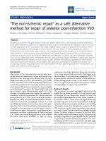
secreted proline-ric h proteins [28-30]. The SMR1 pro-
tein product of the rat Vcsa1 gene is cleaved into at
least two biologically active peptides, sialorphin
(QHNPR) a nd SGP-T (TDIFEGG) (Figure 3). Whereas
the N-terminal QHNPR sequence is conserved in all
products of the rat VCSA family members, the C-term-
inal TDIFEGG sequence is a bsent due to mutation or
truncation of the C-terminus [27]. With the absence of
the VCSA subgroup of genes in non-rodent mammals,
sialorphin and SGP-T may not be present, although
homologues of these peptides are encoded by VCSB
genes. The human VCSB gene PROL1 encodes a protein
that contains a QRFSR motif (opiorphin) that is func-
tionally equivalent to rat sialorphin [31], although a
homologue of TDIFEGG (SGP-T) has not been identi-
fied yet. Sialorphin participates in diverse physiological
processes, such as pain perception, antidepressant
effects, sexual behavior, a nd erectile function [4,32-34],
and these actions appear to be related the inhibition of
neutral endopeptidase (NEP)[4]. Human opiorphin has
similar activity [35]. Vcsa1 e xpression is hormonally
regulated by androgens [33,36], and the expression of
opiorphin family genes may be similarly regulated [37].
Pharmacology of the Tripeptide D-PHE-D-GLU-
GLY (feG)
During SGP-T isolation and testing procedures, the trun-
cated sequence Phe-Glu-Gly (FEG) was identified, which
itself showed bioactivity, as did its D-isomeric form (feG)
[17]. This tripeptide sequence was synthesi zed and char-
acterized pharmacologically in various models.
Animal Models
Several rat models of systemic inflammatory disease, and
in vitro or ex vivo immunop harmacologic assays were
utilized to test the bioactivity of feG as follows.
• Endotoxemia models. Injectio n of lipopolysaccharide
(LPS) in rats results in rapid transient decreases in
blood pressure, increases in circulating leukocytes,
migration of leukocytes into peritoneal fluid, accum ula-
tion of neutrophils in cardiac tissue, disrupted intrinsic
rhythmicity of migrating myoelectric complexes (MMC)
in intestines, etc.
• Anaphylaxis. Rats sensitized to ovalbumin (OA) or
larvae of Nippostrongylis braziliensis (Nb) and
Figure 2 SGP-T and neutrophil chemotaxis:Neutrophil
chemotaxis into carrageenan-soaked sponges over a 24 hour period
in rats is inhibited by SGP-T injected intravenously, in a bell-shaped
dose-dependent manner, at dosages indicated. (mean ± sem, n = 3
to 12). Adapted from [22].
Figure 3 eptide Products from Submandibular Rat-1 (SMR1)
Prohormone: The SMR1 precursor protein contains sialorphin near
the N-terminal, and SGP-T (submandibular gland peptide T) near the
C-terminal. FEG and FEG(NH
2
) are biologically active derivatives of
SGP-T.
Mathison et al. Journal of Inflammation 2010, 7:49
/>Page 3 of 11
subsequently challenged with these same antigens by
injection, orally, or intra-nasally depending on t he pur-
poses of the experiment, develop rapid drops in blood
pressure; accumulation of leukoc ytes in cardiac tissue ;
increases in vascular permeability; increased circulating
leukocytes; diarrhea and disrupted MMCs; and IgE-
mediated migration of eosinophils, neutrophi ls, and
monocytes into airways.
• Pulmonary bronchoconstriction (measured by speci-
fic lung resistance) and airway hyper-responsiveness to
methacholine or carbachol in sheep naturally allergic to
Ascaris suum or in rats sensitized with either OA or
with larvae of Nb and chal lenged by aerosol administra-
tion of the sensitizing antigens was measured aft er aero-
sol challenge with the antigen.
• Spinal cord injury in rats induced by 60 second clip
compression of the spinal cord was measured by lesion
site histology a nd histochemistry as well as recovery of
locomotor function.
• Pancreatitis induced in mice by intravenous injection
of caerulein was measured histologically, by determina-
tion of plasma amylase and lipase activity, and by
immunoassays.
• In vitro and ex vivo studies on leukocyte migration,
adhesion, cell surf ace marker expression, and reactive
oxygen species production.
Hypotension
An early observation was that treatment w ith feG, like
its predecessor SGP-T, inhibited the decrease in blood
pressure associated with anaphyl actic shock [38]. Chal-
lenge of sensitized rats with OA administered orally
evoked a rapid drop in ventricular peak systolic pressure
(VPSP) of 50 to 70 mm Hg. In normal rats or in unchal-
lenged OA sensitized rats intravenous administration of
SGP-T, FEG, or feG had no effect on resting VPSP at
any dosage. However, in OA challenged rats, intrave-
nous administration of each of the peptides 10 minutes
prior to challenge significantly protected against the
drop in VPSP compared to saline treated controls.
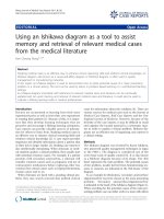
Importantly, oral administration of feG 20 minutes
before OA challenge also produced a dose-dependent
inhibition of cardiovascular shock (Figure 4).
Leukocyte migration
Neutrophil migration into carrageenan-soaked sponges
24 hours after subcutaneous implantation in rats was
inhibited by intraperitoneal injection of feG at
100 μg/kg [39] (Figure 5). Neutrophil inflitration was
significantly reduced by feG treatment in an acute pan-
creatitis model in mice [40] and also in a spinal cord
injury model in rats [41].
Oral challenge in OA sensitized rats induces systemic
effects including increased circulating leukocytes, leuko-
cyte infiltration into the heart , increased vascular perme-
ability, and pulmonary inflammation [42]. Changes in
vascular permeability occurred within 30 minutes, periph-
eral bl ood neutrophilia appeared by 3 hours, a nd signifi-
cant accumulation of neutrophils in the heart, detected by
a 75% increase in myeloperoxidase (MPO) content, was
seen at 24 hours after oral OA challeng e. Treatment with
feG intraperitoneally 20 minutes before antigen challenge
significantly inhibited the increase in vascular permeabil-
ity, circulating leukocytes and neutrophils, and neutrophil
Figure 4 feG and cardiovascular anaphylaxis:Anaphylaxis
induced by ovalbumin (OA) challenge in previously sensitized rats
causes rapid reduction in blood pressure (control). feG treatment
orally at the time of OA challenge dose-dependently inhibited
anaphylaxis-induced hypotension. (mean ± sem, n = 5 to 6).
Adapted from [38].
Figure 5 feG and neutrophil migration: Neutrophils migrate into
carrageenan-soaked surgical sponges implanted subcutaneously in
rats. feG, at a dosage of 100 μg/kg injected intraperitoneally at the
time of sponge implantation, significantly inhibited neutrophil
migration measured 24 hours after implantation. (mean ± sem,
n = 6 to 10). Adapted from [39].
Mathison et al. Journal of Inflammation 2010, 7:49
/>Page 4 of 11
infiltration into the heart. Intraperitoneal injection of feG
at 100 μg/kg at the time of oral OA challenge of sensitized
rats almost completely inhibited the increase in circulating
neutrophils detected 18 hours after challenge [43]. Pul-
monary airway inflammatio n in OA sensitized rats was
also inhibited by feG. Oral treatment with feG 30 minutes
to 6 hours
aft er oral OA challenge significantly inhibited
neutrophil and eosinophil numbers in airways 24 hours
after challenge [44] (Figure 6). In another study, oral treat-
ment with feG at dosages of 250 and 1,000 μg/kg 30 min-
utes before OA challenge inhibited influx of neutrophils,
monocytes, and eosinophils into bronchoalveolar lavage
fluid (BAL) but had no effect on lymphocytes [45].
Infusion of LPS in rats also causes accumulation of
neutrophils in heart tissue in addition to acute hypoten-
sion [46]. Intravenous treatment with a carboxamide
derivative, feG(NH
2
), at the time of LPS infusion, dose-
dependently inhibited accumulation of neutrophils in
atrial slices 24 hours after intravenous LPS (Figure 7).
Orally administered feG (100 μg/kg) also significantly
reduced the number of macrophages and neutrophils
recovered in peritoneal lavage fluid 24 hours after LPS
challenge [47].
Intestinal effects
Oral challenge with OA in sensitized rats also results in
disrupted intrinsic rhythmicity MMCs in the small
intestine, and in diarrhea in 85% of challenged animals
[38,48]. Oral dosage of feG at 350 μg/kgatthetimeof
OA challenge totally abolished the intestinal anaphylac-
tic reaction and diarrhea in all rats tested. In a similar
study feG given orally 30 minutes before OA challenge
dose dependently inhibited anaphylaxis-induced intest-
inal motility, with maximal inhibition achieved at the
highest dosage-100 μg/kg [49]. Interesting ly, feG dosage
(100 μg/kg) up t o 8 hours before challenge afforded sig-
nificant protection against intestinal anaphylaxis, sug-
gesting a long biological half life (Figure 8) [49].
Infusion of LPS in rats also has acute effects on the
intestine by disrupting the standard MMCs and pro-
duces a pattern of intense, irregular myoelectricity [50]
Figure 6 Allergen i nduced by aerosol challenge with
ovalbumin (OA) in previously sensitized rats causes pulmonary
airway inflammation: feG treatment orally 30 minutes, 3 hours, or
6 hours after OA challenge inhibited the influx of eosinophils and
neutrophils into airways. Adapted from [44].
Figure 7 Neutrophil accumulation in heart tissue:Intravenous
administration of lipopolysaccharide (LPS) in rats causes
accumulation of neutrophils in heart tissue as detected by
myeloperoxidase (MPO) activity in atrial slices 24 hours after LPS
infusion. Intravenous treatment with a carboxamide derivative, feG
(NH
2
), at the time of LPS infusion, dose-dependently inhibited MPO
in atrial slices. (mean ± sem, n = 4 to 8). Adapted from [46].
Figure 8 feG and intestinal allergic responses:Oralchallenge
with ovalbumin (OA) in sensitized rats results in disrupted intrinsic
rhythmicity of migrating myoelectric complexes (MMC) in the small
intestine. feG injected intravenously at 100 μg/kg up to 8 hours
before challenge significantly reduced disruption in MMCs,
suggesting a long biological half life (mean ± sem, n = 4 to 8).
Adapted from [49].
Mathison et al. Journal of Inflammation 2010, 7:49
/>Page 5 of 11
Intravenous injection of feG 20 minutes before LPS
dose-dependently reduced the length of time of disrup-
tion of jeju nal MMCs. Inte restingly, the carboxamide
derivative, feG(NH
2
), was found to be mor e potent than
feG in this endotoxemia model. feG given orally 20 min-
utes before LPS challenge inhibited disruption of MMCs
in a bell shaped, dose- dependent manner, with 65 μg/kg
providing maximal inhibition.
Effects on pulmonary inflammation and function
Effects of feG treatment were further studied in pul-
monary inflammation models in rats sensit ized with
either OA or with larvae of Nippostrongylis braziliensis
(Nb) and challenged by aerosol a dministration of the
sensitizing antigens [45], Oral t reatment with feG at 1
mg/kg 30 minut es prior to OA challenge si gnificantly
reduced airway hyper-responsiveness to methacholine
measure d 24 hours after challenge. In Nb sensitized rats
feG significantly reduced tracheal smooth muscle con-
traction in response to aerosol Nb challenge.
In asthma tic sheep naturally sensitized to Ascaris
suum, bronchoconstriction, determined by measuring
specific lung resistance (SRL), and airway hyper-
responsiveness to carbachol were measured in instru-
mented sheep after aerosol challenge with the antigen
[51]. Bronchoconstriction (SRL) increased rapidly up to
500% immediately after aerosol challenge, decreased to
baseline values over 3 hours, but was followed by a sec-
ondary increase in SRL 5 hours after challenge. Treat-
ment with feG intravenously (1 mg/kg) or orally (2 mg/
kg) had no effect on the early, acute phase increase in
SRL, but inhibited the late phase increase by 72% and
78% respectively relative to challenged untreated con-
trols (Figure 9). Inhaled feG, at a dose of 30 mg/sheep,
reduced early (by 83%) as well late (by 88%) broncho-
constriction. Airway hyper-responsiveness to carbachol,
measured 24 hours after antigen challenge, was signifi-
cantly inhibited by pre-challenge treatment with feG
intravenously, orally, or by aerosol delivery.
In cats sensitized to Bermuda grass allergen, adminis-
tration of feG orally at 1 mg/kg immediately prior to
allergen challenge resulted in a significant reduc tion in
accumulation of eosinophils in bronchoalveolar lavage
fluid [52]. However, daily treatment for 2 weeks in
experimentally asthmatic cats had no measurable effect
on airway inflammation [53]. This latter result suggests
that further studies will be necessary to evaluate dosing
regimens and formulation for feG (see Pharmacody-
namic/pharmacokinetic considerations below).
Vascular Permeability
The effects of feG on vascular permeability induced by
antigen challenge and histamine have been studied in
both rats and dogs. With both species intradermal injec-
tion of feG (10
-6
Mto10
-9
M) significantly reduced the
increase in vascular leak of a dy e (Evans blue) provoke d
by both active cutaneous anaphylaxis and histamine by
up to 40% at high doses to ~20% at lower doses (unpub-
lished observations).
Other disease models: acute pancreatitis, spinal cord
injury
In acute pancreatitis, induced in mice by 12 hourly
injections of caerulein, a single dose of feG (100 μg/kg)
was administered intraperitoneally at induction (prophy-
lactic) or 3 hours post induction (therapeutic) [40].
Plasma lipase activity was reduced in feG gro ups treated
both prophylactically and therapeutically; amylase was
reduced in feG groups t reated prophylactically (Figure
10). H istologically, f eG treatment reduced pancreatitis-
induced edema and acinar cell necrosis.
In a clip compression model of spinal cord injury in
rats, leukocyte infiltration, free radical formation, and
oxidative damage at the lesion site were quantified [41].
Neutrophil infiltration, detected by MPO activity, and
activated phagocytic macrophages, identified by ED-1
expression, were present within 24 hours of injury.
Intravenous feG treat ment 2, 6, or 12 hours after injury
reduced MPO activity, ED-1 expression, oxidative
enzymes, free radical production, lipid peroxidation, and
cell death (caspase-3 e xpression) in injured cord lesion
sites. These anti-inflammatory and anti-oxidative actions
of feG treatment correlated with improved neurological
outcomes after spinal cord injury. In a similar spinal
Figure 9 feG and asthma in sheep: In asthmatic sheep naturally
sensitized to Ascaris suum, bronchoconstriction determined by
measuring specific lung resistance (SR
L
) increased rapidly
immediately after aerosol challenge, decreased to baseline values
over 4 hours, but was followed by a secondary increase in SR
L
5
after hours post challenge. Inhaled feG at a dose of 30 mg/sheep
reduced early as well as late increases in SR
L
, whereas treatment
with feG intravenously (1 mg/kg) or orally (2 mg/kg) inhibited only
late phase bronchoconstriction. (mean ± sem, n = 4 to 8). Adapted
from [51].
Mathison et al. Journal of Inflammation 2010, 7:49
/>Page 6 of 11
cord injury model feG given intravenously at 200 μg/kg
twice daily for 5 days improved locomotor and al lodynia
scores relative to controls over 7 weeks following cord
injury [54] (Figure 11).
Pharmacodynamic/pharmacokinetic considerations
From a pharmacodynamic perspective, it appears that
feG has a long biological half life. Single intravenous
dosages of feG inhibit endotoxin-provoked accumulation
of neutrophils in cardiac tissue for at least 24 hours [46]
(see Figure 7). Single oral dos age of feG in OA sensi-
tized challenged rats a lso inhibits neutrophil and
eosinophil migration into airways for at least 24 hours
[44] (see Figure 6). L ikewi se in asthmatic sheep intrave-
nous, oral, or aerosol administration of feG blocks air-
way responsiveness for at least 24 hours after antigen
challenge [51].
Bell shaped dose-response relationships were ob served
in variou s assays, so frequently as to not be dismissible
as coincidental. First observed with SGP-T inhibition of
anaphylaxis-induced hypotension in rats [55] and inhibi-
tion of neutrophil migration into carrageenan soaked
sponges [22] (see Figure 5), feG treatment also resulted
in a biphasic dose-response curve in an intes tinal endo-
toxemia model [38]. In vitro incubation of human neu-
trophils with feG within a window of molar
concentrations between 10
-11
to 10
-9
M down regulated
platelet activating factor- (PAF) induced expression of
CD 11b (AlphaM integrin chain) and PAF-induced neu-
trophil migration [39] (Figure 12). Within these same
molar concentrations feG inhibited fibrinogen and fibro-
nectin binding of peritoneal leukocytes from rats that
had been infused with LPS 18 hours earlier. Binding of
leukocytes from LPS treated rats to atrial slices was
inhibited by feG in vitro at concentrations of 10
-9
Mbut
not 10
-7
M [46]. These findings suggest that dosage of
feG may b e critical to achie ve the desired thera peutic
effect.
Pharmacokinetic studies, to our knowledge, have not
been performed on feG i n any species. Howev er, results
of preliminary pharmacokinetic and toxicokinetic studies
have been performed on a closely-related salivary tripep-
tide (D-cyclohexylalanine-D-glutamate-glycine; (cha)eG)
in rats, dogs, and monkeys (proprie tary, in-house data,
2010). In rats and dogs oral dosages of 2,500 μg/kg of
Figure 10 feG and acute pancreatitis. In acute pancreatitis,
induced in mice by 12 hourly injections of caerulein, a single
intraperitoneal dose of feG (100 μg/kg) administered at start of
caerulein induction or 3 hours after start of induction, inhibited
plasma lipase and amylase activity. Adapted from [40].
Figure 11 feG and spinal cord injury: In a spinal cord injury
model induced by 60 second clip compression of the spinal cord,
rats given feG intravenously at 200 μg/kg twice daily for 5 days had
higher BBB locomotor scores compared to controls (p = 0.043) over
7 weeks following cord injury. Adapted from [54].
Figure 12 feG and human neutrophils: Incubation of human
neutrophils with feG within a window of molar concentrations
between 10
-11
to 10
-9
M downregulated platelet activating factor-
(PAF) induced neutrophil migration in vitro. (mean ± sem, n = 3
to 7). Adapted from [39].
Mathison et al. Journal of Inflammation 2010, 7:49
/>Page 7 of 11
(cha)eG were required to achieve detectable plasma con-
centrations (>5 ng/mL). Oral bioavailability was esti-
mated to be less than 1% in the rat. In monkeys
detectable plasma levels of (cha)eG persisted for 24
hoursfollowingasingleintravenousdosageof10mg/
kg, with an apparent terminal half life of approximately
9 h ours, consistent with pharmacodynamic findings in
rats (see Figure 8). However, noting that in vitro feG is
active within a window of concentrations of about
0.0035 to 0.35 ng/mL, and that in model studies in rats
feG dosage of 100 μg/kg was consistently found to be
effective regardless of route of administration, it must
be concluded that the systemic bioactivi ty of fe G occurs
at concentrations well below minimum detectable
plasma concentration s of current assays. In other words,
thedosageriddleisunlikelytobesolvedby
pharmacokinetics.
Mechanism studies: Effect of feG on neutrophil
chemotaxis, adhesion, and function
Results of in vivo studies point to the neutrophil as the
primary t arget cell for the immunopharmacologic
actions of feG and other bioactive factors p roduced by
the salivary gland. Early results showed that SGP-T
treatment inhibited neutrophil chemotaxis [22] as well
as rolling [23].
Effect on adhesion
In periton eal neutrophils collected from OA sensitized
rats 24 hours after challenge, pre-treatment with feG
had no effect on expression of the alpha integrin CD
11b but down regulated expression of the beta 1 integ-
rin CD49 d (Alpha-4 integrin chain) [42]. In vitro incu-
bation of human neutrophils with feG inhibited PAF
induced neutrophil migration (see Figure 12) as well as
expression of CD 11b [39]. In normal (unstimulated)
neutrophils feG had no effect on neutrophil adhesion to
gelatin, whereas in PAF-activated cells feG at 10
-11
and
10
-10
M significantly inhibited adhesion of human neu-
trophils. However, within molar concentrations of 10
-11
to 10
-9
M, feG had no effect on PAF-stimulated super-
oxide release or on phagocytotic activity, suggesting that
feG modulates primarily neutrophil adhesion and migra-
tory responses. Peritoneal neutrophils f rom OA sensi-
tized rats 24 hours after challenge were also tested for
expression of CD11b and CD16b (Fc-gamma RIIIb: Low
affinity immunoglobulin gamma Fc reg ion receptor
IIIB). feG treatment (100 μg/kg orally) inhibite d CD 11b
antibody binding to peritoneal neutrophils in unchal-
lenged but not in OA challenged rats. CD 16b binding,
however, was inhibited by feG treatment in both chal-
lenged and unchallenged rats. In vitro (microtiter plates)
feG inhibits adhesion of rat peritoneal leukocytes, but
only if the cells were stimulated with PAF[43], indicating
that feG’s a ctions require cell activation. f eG treatment
also completely blocked the expression of the beta
1-integrin CD49 d on circulating neutrophils which was
up regulated by OA challenge, but had no effect on
CD11b expression. These and other findings led to the
conclusion, that when administered in vivo feG prevents
inflammation-induced reduction in cell adhesion as well
as restoring its inhibitory effect in vitro.
Effect on oxidative activity
Neutrophils, which play a key role in the development
and perpetuation SIRS, inactivate and destroy virulent
pathogens through the release of superoxide and
enzymes and by phagocytosis [56]. In OA sensitized rats
the extracellular release of superoxide anion by circulat-
ing neutrophils 18 hours after OA challenge was not
modified by either OA challenge o r feG treatment [57],
confirming similar findings in previous studies [39].
However, incubation of the cells with phorbol myristate
acetate (PMA), a protein kinase C (PKC) activator,
increased intracellul ar release of reactive oxygen species
as determined by flow cytometry for a marker of oxygen
free radicals, 123-dihydrorhodamine. feG treatment at
the time of challenge inhibited intracellular superoxide
production by PMA-stimulated blood neutrophils 18
hours afte r challenge (Figure 13). These findings led to
the speculation that feG reduces the capaci ty of neutro-
phils to generate r eactive oxygen species by preventing
the deregulation of PKC consequent to an allergi c
reaction.
Saliva, in addition to its role as a digestive aid, contri-
butes significantly to lubrication, protection, defence and
Figure 13 feG and the oxidative burst: - Dose-response for
phorbol myristate acetate- (PMA) stimulated intracellular oxidative
activity of circulating neutrophils 18 hours after ovalbumin (OA)
challenge in OA sensitized rats. feG was injected intraperitoneally at
100 μg/kg at the time of challenge. Oxidative activity was measured
using flow cytometry for a marker of oxygen free radicals, 123-
dihydrorhodamine. (mean ± sem, n = 6 to 7). Adapted from [57].
Mathison et al. Journal of Inflammation 2010, 7:49
/>Page 8 of 11
wound healing in the mouth. The importance of salivary
glands and their secretions are poorly appreciated, and
they are only taken seriously when saliv ary gland dys-
function results in decreased saliva flow. In humans this
dysfunction contributes to difficulties in tasting, eating,
swallowing, and speaking, and resul ts in sores of the soft
tissues of the mouth and periodontal disease. These
pathologies also manifest in human patients with a vari-
etyofsystemicdiseasesincluding-Sjögren’ s syndrome,
rheumatoid arthritis, juvenile idiopathic (rheumatoid)
arthritis, systemic lupus erythematosus (an inflammatory
connective tissue disease), systemic sclerosis (scelo-
derma), primary bilary cirrhosis (an autoimmune disease
of the liver), sarcidosis (a multisystem granulomatous dis-
order), infections with human immunodeficiency virus,
herpes virus, hepatitis C, ectodermal dysplasia, chronic
pancreatitis and depression [58].
Nonetheless, it should be recognized that the relation-
ship between salivary glands and systemic health is
bidirectional. “Oral infection may represent a significant
risk-factor for systemic diseases, and hence the control
of oral disease is essential in the prevention and man-
agement of these systemic conditions” [59]. Chronic
inflammatory p eriodontal diseases are among the most
prevalent chronic infections in humans, and many inves-
tigators have established a significant, albeit modest,
positive association between periodontal disease and
cardiovascular disease, which includes a therosclerosis,
myocardial infarct ion and stroke. In addition, epidemio-
logical associations have been made between periodontal
diseases and chronic diseases suc h as diabet es, respira-
tory diseases and osteoporosis [60].
Likewise in veterinary medicine epidemiologic studies
reveal that oral disease is the most common disease in
all age groups of dogs and cats [61]. Moreover, there is
evidence that oral infection also has systemic effects
including renal, hepatic, pulmonary, and cardiac dis-
eases; osteoporosis, adverse pregnancy effects, and dia-
betes mellitus [62], and can lead to systemic
inflammation [63]. The severity of periodontal disease
was found to be positively correlated with histological
changes in kidneys, myocardium, and liver [64].
In this review we focused on SGP-T and its derivatives
namely FEG and its D-i someri c derivative feG, which in
themselves demonstrate the significant physiological and
immunological modulation exerted by salivary gland
peptides. These peptides have si gnificant anti-inflamma-
tory actions, as shown in animal models of endotoxic
shock (Figures 1 &7), allergic and anaphylactic reactions
(Figures 4, 6, 8 &9), pancrea tic (Figure 10) and spinal
cord injury (Figure 11).
feG, and its analogues, exhibit a distinctly differ-
ent mechanism of anti-inflammatory action from
corticosteroids and nonsteroidal anti-inflammatory
drugs (NSAIDs). NSAIDs and corticosteroids have
become the mainstay of anti-inflammatory agents in
human and veterinary medicine . NSAIDs are popular
owing to their immune sparing effect, especially since
the discovery that they act by inhibiting cyclooxygen-
ase (COX), an enzyme that catalyses the arachidonic
acid cascade resulting in production of pro-inflamma-
tory eicosanoids [65]. In contrast to enzymatic block-
ade, the tripeptide feG has multimodal activity and
acts directly on activated leukocytes, specifically down
regulating expression of integrins, thereby inhibiting
chemotaxis (Figures 2 &12) and cell migration (Figure
5). Furthermore, feG inhibits the function of neutro-
phils by specifically inhibiting intracellular superoxide
production by activated neutrophils (Figure 13), prob-
ably as a consequence of interruption of the signaling
cascade that induces superoxide generation [66].
Hence feG and its analogues appear to represent a
new class of anti-inflammatory agents which act on
immune cells, th e central regulators of all inflammation.
The term “Immune Selective Anti-Inflammatory Deriva-
tives” (ImSAIDs) is propose d for salivary-derived pep-
tides to distinguish this class of agents from
corticosteroids and NSAIDs. A closely-related salivary
tripeptide ((cha)eG) is currently under investigation as
an anti-asthmatic therapeutic in humans.
Conclusions
Based on its mechanism of action and demonstrable in
vivo pharmacolo gic activity, feG deserves evaluation in a
number of situations characterized by over-exube rant or
chronic inflammatory responses of human and veterin-
ary significance associated with several m ajor o rgan sys-
tems:
• Whole body and circulat ory: sepsis, e ndotoxem ia,
SIRS [67];
• Gastrointestinal: pancreatitis, hepatitis, gastroenter-
itis, enteritis;
• Oral cavity: stomatitis
• Respiratory: asthma, acute pulmonary inflamma-
tion of diverse etiologies;
• M uscu lo-Skeletal: fibromyalgia, rheumatoid arthri-
tis, equine laminitis (now characterized as a neutro-
phil-mediated inflammatory disease [68]);
• Nervous: spinal cord injury, peripheral nerve
injury;
• Urinary tract: cystitis
Aside from these therapeutic potentials, feG may
eventually prove to be useful as a vetraceutical or a
nutraceutical [the term coined by Stephen DeFelice
Mathison et al. Journal of Inflammation 2010, 7:49
/>Page 9 of 11
[69]] to reduce the incidence and severity of systemic
and localized inflammations caused by intense exercise,
poor oral health and other causes.
List of Abbreviations
BAL: bronchoalveolar lavage fluid; CD11b: AlphaM integrin chain; CD16b: Fc-
gamma RIIIb - Low affinity immunoglobulin gamma Fc region receptor IIIB;
CD49 d: Alpha-4 integrin chain; (cha)eG: D-cyclohexylalanine-D-glutamate-
glycine COX: cyclooxygenase; FEG: Phenylalanine-Glutamate-Glycine; feG: D-
phenylalanine-D-glutamate-Glycine; IgE: immunoglobulin E; ImSAIDs:
Immune Selective Anti-Inflammatory Derivatives; LPS: lipopoylsaccharide;
MMC: migrating myoelectric complexes; MPO: myeloperoxidase;Nb:
Nippostrongylus brasiliensis; NSAID: non steroidal anti-inflammatory drugs; OA:
ovalbumin; PAF: platelet activating factor; PKC: protein kinase C; PMA:
phorbol myristate acetate; SGP-T: submandibular peptide-T; SIRS: systemic
inflammatory response syndrome; SRL: specific lung resistance; SMR1:
submandibular rat-1; VCS-1: variable coding sequence-1; VPSP: ventricular
peak systolic pressure
Competing interests
DAG is a research veterinarian and a minority shareholder in a company
which has commercial rights to salivary-derived peptides for veterinary use.
RM and JSD have shares in a privately held company that is developing
peptides and their analogues for therapeutic use.
Authors’ contributions
DAG conducted the literature search, wrote the first draft of the manuscript,
and composed and edited the figures. RM contributed literature searches
and the rewriting and editing. JSD and ADB provided important discussion
and editorial comments. All authors read and approved the final manuscript.
Acknowledgements
The financial assistance of Allergen NCE Inc. is gratefully acknowledged.
Author details
1
Faculty of Medicine, University of Calgary, 3330 Hospital Drive NW, Calgary,
Alberta, T2N 4N1, Canada.
2
550A Heritage Medical Research Centre, Faculty
of Medicine and Dentistry, University of Alberta, Edmonton, Alberta, T6G 2S2,
Canada.
3
Turtle Creek Biostatistical Consulting, 2219 Wilmington Road,
Lebanon, OH 45036, USA.
Received: 19 August 2010 Accepted: 28 September 2010
Published: 28 September 2010
References
1. Pedersen AM, Bardow A, Jensen SB, Nauntofte B: Saliva and
gastrointestinal functions of taste, mastication, swallowing and
digestion. Oral Dis 2002, 8:117-129.
2. Tenovuo J: Antimicrobial agents in saliva - protection for the whole
body. Journal of Dental Research 2002, 81:807-809.
3. Barka T: Biologically active polypeptides in submandibular glands. J
Histochem Cytochem 1980, 28:836-859.
4. Rougeot C, Messaoudi M, Hermitte V, Rigault AG, Blisnick T, Dugave C,
Desor D, Rougeon F: Sialorphin, a natural inhibitor of rat membrane-
bound neutral endopeptidase that displays analgesic activity. Proc Natl
Acad Sci USA 2003, 100:8549-8554.
5. Isenman L, Liebow C, Rothman S: The endocrine secretion of mammalian
digestive enzymes by exocrine glands. Am J Physiol 1999, 276:E223-232.
6. Kongshavn PA, Lapp WS: Immunosuppressive effect of male mouse
submandibular gland extracts on plaque-forming cells in mice: abolition
by orchiectomy. Immunology 1972, 22:227-230.
7. Kemp A, Mellow L, Sabbadini E: Suppression and enhancement of in vitro
lymphocyte reactivity by factors in rat submandibular gland extracts.
Immunology 1985, 56:261-267.
8. Amico-Roxas M, Caruso A, Leone MG, Scifo R, Vanella A, Scapagnini U:
Nerve growth factor inhibits some acute experimental inflammations.
Arch Int Pharmacodyn Ther 1989, 299:269-285.
9. Mathison R, Davison JS, Befus AD: Neuroendocrine regulation of
inflammation and tissue repair by submandibular gland factors. Immunol
Today 1994, 15:527-532.
10. Felten DL, Felten SY, Bellinger DL, Carlson SL, Ackerman KD, Madden KS,
Olschowki JA, Livnat S: Noradrenergic sympathetic neural interactions
with the immune system: structure and function. Immunol Rev 1987,
100:225-260.
11. Nance DM, Hopkins DA, Bieger D: Re-investigation of the innervation of
the thymus gland in mice and rats. Brain Behav Immun 1987, 1:134-147.
12. Madden KS, Felten SY, Felten DL, Sundaresan PR, Livnat S: Sympathetic
neural modulation of the immune system. I. Depression of T cell
immunity in vivo and vitro following chemical sympathectomy. Brain
Behav Immun 1989, 3:72-89.
13. Alito AE, Romeo HE, Baler R, Chuluyan HE, Braun M, Cardinali DP:
Autonomic nervous system regulation of murine immune responses as
assessed by local surgical sympathetic and parasympathetic
denervation. Acta Physiol Pharmacol Latinoam 1987, 37:305-319.
14. Ramaswamy K, Mathison R, Carter L, Kirk D, Green F, Davison JS, Befus D:
Marked antiinflammatory effects of decentralization of the superior
cervical ganglia. J Exp Med 1990, 172:1819-1830.
15. Mathison R, Hogan A, Helmer D, Bauce L, Woolner J, Davison JS, Schultz G,
Befus D: Role for the submandibular gland in modulating pulmonary
inflammation following induction of systemic anaphylaxis. Brain Behav
Immun 1992, 6:117-129.
16. Mathison R, Befus D, Davison JS: Removal of the submandibular glands
increases the acute hypotensive response to endotoxin. Circ Shock 1993,
39:52-58.
17. Mathison RD, Befus AD, Davison JS: A novel submandibular gland peptide
protects against endotoxic and anaphylactic shock. Am J Physiol 1997,
273:R1017-1023.
18. Rosinski-Chupin I, Tronik D, Rougeon F: High level of accumulation of a
mRNA coding for a precursor-like protein in the submaxillary gland of
male rats. Proc Natl Acad Sci USA 1988, 85:8553-8557.
19. Morris KE, St Laurent CD, Hoeve RS, Forsythe P, Suresh MR, Mathison RD,
Befus AD: Autonomic nervous system regulates secretion of anti-
inflammatory prohormone SMR1 from rat salivary glands. Am J Physiol
Cell Physiol 2009, 296:C514-524.
20. Mathison R, Tan D, Oliver M, Befus D, Scott B, Davison JS: Submandibular
gland peptide-T (SGP-T) inhibits intestinal anaphylaxis. Dig Dis Sci 1997,
42:2378-2383.
21. Mathison RD, Malkinson T, Cooper KE, Davison JS: Submandibular glands:
novel structures participating in thermoregulatory responses. Can J
Physiol Pharmacol 1997, 75:407-413.
22. Nkemdirim M, Kubera M, Mathison R: Modulation of neutrophil activity by
submandibular gland peptide-T (SGP-T). Pol J Pharmacol 1998, 50:417-424.
23. Mathison RD, Sank C, Davison JS: Inhibition of leukocyte rolling by
submandibular gland peptide-T (SGP-T). Proc West Pharmacol Soc 1999,
42:39-40.
24. Morris K, Kuo B, Wilkinson MD, Davison JS, Befus AD, Mathison RD: Vcsa1
gene peptides for the treatment of inflammatory and allergic reactions.
Recent Pat Inflamm Allergy Drug Discov 2007, 1:124-132.
25. Rosinski-Chupin I, Kuramoto T, Courty Y, Rougeon F, Serikawa T:
Assignment of the rat variable coding sequence (VCS) gene family to
chromosome 14. Mamm Genome 1995, 6:153-154.
26. Rosinski-Chupin I, Rougeon F: The gene encoding SMR1, a precursor-like
polypeptide of the male rat submaxillary gland, has the same
organization as the preprothyrotropin-releasing hormone gene. DNA Cell
Biol 1990, 9:553-559.
27. Rougeot C, Rosinski-Chupin I, Rougeon F: Novel genes and hormones in
salivary glands: From the gene for the submandibular rat1 protein
(SMR1) precursor to receptor sites for SMR1 mature peptides. Biomedical
Reviews 1998, 9:17-32.
28. Dickinson DP, Thiesse M: cDNA cloning of an abundant human lacrimal
gland mRNA encoding a novel tear protein.
Curr Eye Res 1996, 15:377-386.
29. Isemura S: Nucleotide sequence of gene PBII encoding salivary proline-
rich protein P-B. J Biochem (Tokyo) 2000, 127:393-398.
30. Isemura S, Saitoh E: Nucleotide sequence of gene PBI encoding a protein
homologous to salivary proline-rich protein P-B. J Biochem (Tokyo) 1997,
121:1025-1030.
Mathison et al. Journal of Inflammation 2010, 7:49
/>Page 10 of 11
31. Wisner A, Dufour E, Messaoudi M, Nejdi A, Marcel A, Ungeheuer MN,
Rougeot C: Human Opiorphin, a natural antinociceptive modulator of
opioid-dependent pathways. Proc Natl Acad Sci USA 2006,
103:17979-17984.
32. Davies KP, Tar M, Rougeot C, Melman A: Sialorphin (the mature peptide
product of Vcsa1) relaxes corporal smooth muscle tissue and increases
erectile function in the ageing rat. BJU Int 2007, 99:431-435.
33. Messaoudi M, Desor D, Nejdi A, Rougeot C: The endogenous androgen-
regulated sialorphin modulates male rat sexual behavior. Horm Behav
2004, 46:684-691.
34. Tong Y, Tar M, Davelman F, Christ G, Melman A, Davies KP: Variable coding
sequence protein A1 as a marker for erectile dysfunction. BJU Int 2006,
98:396-401.
35. Davies KP: The role of opiorphins (endogenous neutral endopeptidase
inhibitors) in urogenital smooth muscle biology. J Sex Med 2009, 6(Suppl
3):286-291.
36. Rosinski-Chupin I, Huaulme JF, Rougeot C, Rougeon F: The transcriptional
response to androgens of the rat VCSA1 gene is amplified by both
binary and graded mechanisms. Endocrinology 2001, 142:4550-4559.
37. Tong Y, Tar M, Melman A, Davies K: The opiorphin gene (ProL1) and its
homologues function in erectile physiology. BJU Int 2008, 102:736-740.
38. Mathison R, Lo P, Moore G, Scott B, Davison JS: Attenuation of intestinal
and cardiovascular anaphylaxis by the salivary gland tripeptide FEG and
its D-isomeric analog feG. Peptides 1998, 19:1037-1042.
39. Mathison RD, Befus AD, Davison JS, Woodman RC: Modulation of
neutrophil function by the tripeptide feG. BMC Immunol 2003, 4:3.
40. Rifai Y, Elder AS, Carati CJ, Hussey DJ, Li X, Woods CM, Schloithe AC,
Thomas AC, Mathison RD, Davison JS, Toouli J, Saccone GT: The tripeptide
analog feG ameliorates severity of acute pancreatitis in a caerulein
mouse model. Am J Physiol Gastrointest Liver Physiol 2008, 294:G1094-1099.
41. Bao F, John SM, Chen Y, Mathison RD, Weaver LC: The tripeptide
phenylalanine-(d) glutamate-(d) glycine modulates leukocyte infiltration
and oxidative damage in rat injured spinal cord. Neuroscience 2006,
140:1011-1022.
42. Turesin F, Sadr A, Davison JS, Mathison R: The tripeptide FEG ameliorates
systemic inflammatory responses to rat intestinal anaphylaxis. BMC
Physiol 2002, 2:13.
43. Mathison RD, Christie E, Davison JS: The tripeptide feG inhibits leukocyte
adhesion. J Inflamm (Lond) 2008, 5:6.
44. Dery RE, Mathison R, Davison J, Befus AD: Inhibition of allergic
inflammation by C-terminal peptides of the prohormone submandibular
rat 1 (SMR-1). Int Arch Allergy Immunol
2001, 124:201-204.
45. Dery RE, Ulanova M, Puttagunta L, Stenton GR, James D, Merani S,
Mathison R, Davison J, Befus AD: Frontline: Inhibition of allergen-induced
pulmonary inflammation by the tripeptide feG: a mimetic of a neuro-
endocrine pathway. Eur J Immunol 2004, 34:3315-3325.
46. Mathison R, Woodman R, Davison JS: Regulation of leukocyte adhesion to
heart by the tripeptides feG and feG(NH2). Can J Physiol Pharmacol 2001,
79:785-792.
47. Mathison R, Lo P, Tan D, Scott B, Davison JS: The tripeptide feG reduces
endotoxin-provoked perturbation of intestinal motility and
inflammation. Neurogastroenterol Motil 2001, 13:599-603.
48. Mathison R: Submandibular gland peptides and the modulation of
anaphylactic and endotoxic reactions. Biomedical Reviews 1998, 9:101-106.
49. Mathison RD, Davison JS, Befus AD: The tripeptide feG reduces
perturbation of intestinal motility provoked by anaphylaxis. Proc West
Pharmacol Soc 2001, 44:157-158.
50. Tan D, Rougeot C, Davison JS, Mathison R: The carboxamide feG(NH2)
inhibits endotoxin perturbation of intestinal motility. Eur J Pharmacol
2000, 409:203-205.
51. Mathison R, Davison JS, Befus AD, Abraham WM: The tripeptide feG
inhibits asthmatic reactions in sheep. In Immunology 2004. Edited by:
Monduzzi E. Montreal Medimond International Proceedings; 2004:515-519.
52. DeClue AE, Schooley E, Nafe LA, Reinero CR: feG-COOH blunts eosinophilic
airway inflammation in a feline model of allergic asthma. Inflamm Res
2009, 58:457-462.
53. Eberhardt JM, AE D, CR R: Chronic use of the immunomodulating
tripeptide feG-COOH in experimental feline asthma. Vet Immunol
Immunopathol 2009, 132:175-180.
54. John SM, Bao F, Chen Y, Mathison RD, Weaver LC: Effects of a novel
tripeptide on neurological outcomes after spinal cord injury. Neuroreport
2006, 17:1793-1796.
55. Mathison RD, Befus AD, Davison JS: Reduction in cardiovascular
anaphylaxis by submandibular gland peptide-T. Proc West Pharmacol Soc
1997, 40:73-74.
56. Fujishima S, Aikawa N: Neutrophil-mediated tissue injury and its
modulation. Intensive Care Med 1995, 21:277-285.
57. Mathison RD, Davison JS: The tripeptide feG regulates the production of
intracellular reactive oxygen species by neutrophils. J Inflamm (Lond)
2006, 3:9.
58. von Bültzingslöwen I, Sollecito TP, Fox PC, Daniels T, Jonsson R,
Lockhart PB, Wray D, Brennan MT, Carrozzo M, Gandera B, Fujibayashi T,
Navazesh M, Rhodus NL, Schiødt M: Salivary dysfunction associated with
systemic diseases: systematic review and clinical management
recommendations. Oral Surg Oral Med Oral Pathol Oral Radiol Endod 2007,
103:e1-15.
59. Williams RC, Barnett AH, Claffey N, Davis M, Gadsby R, Kellett M, Lip GY,
Thackray S: The potential impact of periodontal disease on general
health: a consensus view. Curr Med Res Opin 2008, 24:1635-1643.
60. Cullinan MP, Ford PJ, Seymour GJ: Periodontal disease and systemic
health: current status. Aust Dent J 2009, 54(Suppl 1):S62-69.
61. Lund EM, Armstrong PJ, Kirk CA, Kolar LM, Klausner JS: Health status and
population characteristics of dogs and cats examined at private
veterinary practices in the United States. J Am Vet Med Assoc 1999,
214:1336-1341.
62. Niemiec BA: Periodontal disease. Top Companion Anim Med 2008, 23:72-80.
63. Reiter AM, Brady CA, Harvey CE: Local and systemic complications in a cat
after poorly performed dental extractions. J Vet Dent 2004, 21:215-221.
64. DeBowes LJ, Mosier D, Logan E, Harvey CE, Lowry S, Richardson DC:
Association of periodontal disease and histologic lesions in multiple
organs from 45 dogs. J Vet Dent 1996, 13:57-60.
65. Vane JR: Inhibition of prostaglandin synthesis as a mechanism of action
for aspirin-like drugs. Nat New Biol 1971, 231:232-235.
66. Zarbock A, Ley K: Mechanisms and consequences of neutrophil
interaction with the endothelium. Am J Pathol 2008, 172:1-7.
67. Brady CA, Otto CM: Systemic inflammatory response syndrome, sepsis,
and multiple organ dysfunction. Vet Clin North Am Small Anim Pract 2001,
31:1147-1162, v-vi.
68. Belknap JK: The pharmacologic basis for the treatment of developmental
and acute laminitis. Vet Clin North Am Equine Pract 2010, 26:115-124.
69. FIM Rationale and Proposed Guidelines for the Nutraceutical Research &
Education Act - NREA. [ />html].
doi:10.1186/1476-9255-7-49
Cite this article as: Mathison et al.: Salivary gland derived peptides as a
new class of anti-inflammatory agents: review of preclinical
pharmacology of C-terminal peptides of SMR1 protein. Journal of
Inflammation 2010 7:49.
Submit your next manuscript to BioMed Central
and take full advantage of:
• Convenient online submission
• Thorough peer review
• No space constraints or color figure charges
• Immediate publication on acceptance
• Inclusion in PubMed, CAS, Scopus and Google Scholar
• Research which is freely available for redistribution
Submit your manuscript at
www.biomedcentral.com/submit
Mathison et al. Journal of Inflammation 2010, 7:49
/>Page 11 of 11