Báo cáo y học: "The role of toll-like receptors in acute and chronic lung inflammation" pdf
Bạn đang xem bản rút gọn của tài liệu. Xem và tải ngay bản đầy đủ của tài liệu tại đây (486.8 KB, 14 trang )
REVIEW Open Access
The role of toll-like receptors in acute and
chronic lung inflammation
Erin I Lafferty
1
, Salman T Qureshi
1,2*
, Markus Schnare
3*
Abstract
By virtue of its direct contact with the environment, the lung is constantly cha llenged by infectious and non-infec-
tious stimuli that necessitate a robust yet highly controlled host response coordinated by the innate and adaptive
arms of the immune system. Mammalian Toll-like receptors (TLRs) function as crucial sentinels of microbial and
non-infectious antigens throughout the respiratory tract and mediate host innate immunity. Selective induction of
inflammatory responses to harmful environmental exposures and tolerance to innocuous antigens are required to
maintain tissue homeostasis and integrity. Conversely, dysregulated innate immune responses manifest as sustained
and self-perpetuating tissue damage rather than controlled tissue repair. In this article we review aspects of Toll-like
receptor function that are relevant to the development of acute lung injury and chronic obstructive lung diseases
as well as resistance to frequently associated microbial infections.
Introduction
As an essential interface between the environment and
the internal milieu, the lungs are continuously exposed to
dust, pollen, chemicals, and microbial pathogens. Pneu-
monia and related patterns of lower respiratory tract
infection are a frequent consequence of this interaction
and account for a significant proportion of h uman mor-
bidity and mortality throughout the world [1,2]. To con-
tain potential environmental threats, the lungs are
equipped with complex and multifaceted host defences.
During tidal ventilation, the complex geometry of the
nasal passages and branching pattern of the central air-
ways impede the penetration of relatively large or heavy
infectious particles while tight i ntercellular junctions
ensure the structural integrity of th e lung epithelium.
This barrier is e nhanced by airway goblet cells that
secrete mucus and ciliated epithelial cells that constantly
transport this viscous layer towards the bronchi and away
from the alveoli to facilitate expulsion of trapped parti-
cles [3]. A variety of soluble host defence mediators such
as secretory IgA, antimicrobial peptides, surfactant pro-
teins, lactoferrin, and lysozyme also bolster the mucosal
defences of the lower respiratory tract. Finally, resident
alveolar macrophages (AMs) and airway mucosal dendri-
tic cells (DCs) provide constant surveillance for poten-
tially pathogenic factors while inhibiting T cell responses
to myriad non-pathogenic antigens [4]. These normal
host defences ensure that most encounters between the
respiratory tract and pathogens are inconsequential;
nevertheless, in response to prolonged, intense, or highly
virulent microbial exposure, an inflammatory response or
productive infection is likely to occur. To rapidly initiate
an acute inflammatory response in these circumstances,
the lung epithelium, myeloid cells, and associated lym-
phoid tissue are all equipped with a series of highly con-
served pattern recognition receptor (PRRs) including
Toll-like receptors (TLRs), NOD-like receptors (NLRs),
and RIG-I like receptors (RLRs). PRR activation leads to
the release of cytokines and chemokines that attract leu-
kocytes to the site of infection and trigger the maturation
and trafficking of antigen presenting cells for induction
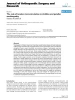
of adaptive immunity (figure 1). The purpose of this arti-
cle is to review the role of TLRs in the pathogenesis or
consequences of acute lung injury (ALI) and chronic
inflammatory lung diseases including asthma, chronic
obstructive pulmonary disease (COPD), and cystic fibro-
sis (CF).
* Correspondence: ;
marburg.de
1
Division of Experimental Medicine, McGill University, Montréal, Québec H3A
1A3, Canada
3
Institute of Immunology, Philipps-University of Marburg, Germany
Full list of author information is available at the end of the article
Lafferty et al. Journal of Inflammation 2010, 7:57
/>© 2010 Lafferty e t al; licensee BioMed Central Ltd. This i s an Open Access article distributed under the terms of the Creative Commons
Attribution License ( which permits unrestricted use, distribution, and reproduct ion in
any medium, provided the origin al work is properly cited.
Ligands of TLRs
Microbial ligands
Constant interactions between the respiratory tract and
theenvironmentposeamajorchallengetohostimmu-
nity and necessitate robust surveillance mechanisms to
distinguish innocuous from pathogenic exposures. One
strategy that is used by TLRs for selective induction of a
host response is recognition of unique microbial struc-
tures termed pathogen-associated molecular patterns
(PAMPs) [5-8]. Eleven functional TLR genes that play
diverse roles in host defense, inflammation, autoimmu-
nity, and neoplasia have been discovered in mouse and
man (mouse TLR10 is a pseudogene and human TLR11
encodes a truncated protein) [5]. Prototypic examples of
PAMPs include lipopolysaccharide (LPS), a outer mem-
brane component of Gram-negative bacteri a that stimu-
lates TLR4 [8,9], bacterial lipoproteins that stimulate
TLR2 in conjunction with either TLR1 or TLR6 [10],
and flagellin, the protein monomer of bacterial flagella
that activates TLR5 [11]. Nucleic acids are recognized
by endosomal TLRs; double-stranded DNA with
unmethylated CpG motifs activates TLR9 in a host spe-
cies-specific manner while TLR3 and TLR7/8 are acti-
vated by dsRNA including synthetic poly (I:C) [12] and
ssRNA, respectively [13,14].
Host-derived ligands
Following the discovery that TLRs discriminate self from
non-self through their intracellular localization or recog-
nition of distinct ligand signatures, evidence was gath-
ered in support of the hypothesis that endogenous host
molecules termed danger associated molecular patterns
(DAMPs) also stimulate TLRs. The first suggestion of
this process came from studies of heat shock protein
Host Environment Stimulus
Age Genetics Lifestyle Exposure Microbial Non-Microbial
Chronic
inflammation
Acute
inflammation
Innate Immunity
Toll-like receptors (TLRs)
NOD-like receptors (NLRs)
RIG-like receptors (RLRs)
Adaptive Immunity
CD4 and CD8 T cells
Antigen-specific B cells
Host recovery
and protection
from
reinfection
EXCESSIVE innate
immune signaling:
Death due to
inflammation
DEFICIENT innate
immune signaling:
Death due to infection
Immune
status
Figure 1 Innate and adaptive immunity in acute and chronic lung inflammation. A variety of host and environmental factors contribute to
the development of acute and chronic lung inflammation. Recognition of pathogen associated molecular patterns (PAMPs) or endogenous
damage associated molecular patterns (DAMPs) by host pattern recognition receptors (PRRs), including Toll-like receptors (TLRs), elicits innate
immune responses that subsequently instruct adaptive immunity. Recovery from the inciting stimulus depends on robust yet tightly regulated
innate and adaptive immune responses. Deficient innate immune signaling leads to excess pathogen burden while an exaggerated response
can cause severe tissue injury and death of the host.
Lafferty et al. Journal of Inflammation 2010, 7:57
/>Page 2 of 14
(hsp) [15]; subsequently, a number of other endogenous
ligands including the extra domain A of fibron ectin and
hyaluronic acid were also shown to activate TLRs
[16,17]. Recognition of endogenous ligands by TLRs
may also contribute to the onset and initiation of auto-
immune responses. For example, the high mobility
group box protein 1 (HMGB1) protein that nor mally
resides in the cell nucleus can activate TLR2 and induce
hallmarks of lupus-like disease when released from
apoptotic cells as a complex with nucleosomes [18].
TLR signaling
The activation of TLRs results in acute inflammation and
controls the adaptive immune response at various levels.
Partially overlapping intracellular signaling pathways
downstream of each TLR activate specific transcription
factors that regulate the expression of genes responsible
for inflammatory and immune responses. Four adaptors
that harbour a Toll-Interleukin-1 Receptor (TIR) domain,
including MyD88, TIRAP (MAL), TRIF (TICAM1), and
TRAM, connect the TLRs to the cytoplasmic signaling
machinery [5]. MyD88 was initially identified as part of
the interleukin (IL) -1R and IL-18R signalling pathways
and was subsequently implicated in signal ling by almost
all TLRs to trigger NF-B, Interferon Regulatory Factor
(IRF) 5, and Mitogen Activated Protein (MAP) kinase
activation. A notable exception is TLR3 that mediates the
activation of IRFs exclusively through t he adaptor mole-
cule TRIF [19]. The function of TIRAP is to recruit
MyD88 to TLR2 and TLR4 at the plasma membrane,
while TRAM recruits TRIF to TLR4 for activation of
IRF3. A fifth adaptor protein, SARM, negatively regulates
TRIF-dependent signaling [20,21]. Activat ion of different
intracellular signaling mechanisms t hrough TLRs results
in the induction of distinct gene programs and cytokine
expression patterns that control the recruitment of
downstream molecules and regulate the identity,
strength, and kinetics of gene and protein expression.
More detailed reviews of the TLR signalling pathways
have been published elsewhere [22-24].
The potent stimulatory responses mediated by TLR
signaling must be tightly regulated at numerous levels in
order to prevent the deleterious consequ ences of e xces -
sive innate immune activation [25]. For example, soluble
forms of TLR4 and TLR2 may function as decoy recep-
tors to terminate ligand interactions with membrane
bound TLRs [26]. Furthermore, IRAK-M has 30-40%
homology to the other IRAK-family members and stabi-
lizes the T LR-MyD88-IRAK4 complex, leading to a
unique negative regulatory influence on T LR signaling
[27,28]. TLR signaling is also inhibited by transmem-
brane receptors like ST2, SIGIRR, and TRAILR while
proteins such as Tollip [29], SARM [21], an inducible
splice variant of MyD88 (MyD88s) [30], and the
suppressor of cytokine signaling1(SOCS1)[31]are
responsible for modulation of intracellular T LR
signaling.
In addition to TLRs, a variety of other PRRs including
the cytoplasmic NLRs and RLRs play important roles in
the induction of lung inflammation. For example, the cyto-
plasmic NALP3 protein, a member of the NLR family that
triggers assembly of the caspase-1 inflammasome and pro-
duction of mature IL-1b, was recently implicated in the
development of asbestos or silica-induced pulmonary
fibrosis [32]. RLRs on immune and non-immune cells
recognize viral RNA species and induce host responses
through the adaptor IPS-1. Several putative cytosolic
detectors of double-stranded DNA including DAI (ZBP1-
DLM1) and AIM2 have also been identified; however their
roles in lung diseases have not been established. A detailed
discussion of these important non-TLR innate immune
receptors is beyond the scope of this article; however,
interested readers may consult other sources [33].
Expression and function of TLRs in lung cells or
tissue
TLRs are widely expressed on both resident lung cells as
well as i nfiltrating cells of myeloid and lymphoid origin.
Primary bronchial epithelial cells express mRNA for
TLR1-10 and secrete the chemokine CXCL8 (IL-8) in
response to various TLR ligands [34]. Human AMs have
been shown to express low levels of TLR3, TLR5, and
TLR9 and higher levels of TLR1, TLR2, TLR4, TLR7, and
TLR8 [35,36]. Lung endothelial cells express TLR4 that is
crucial for neutrophil recruitment and capillary seques-
tration following systemic LPS administra tion [37]. Neu-
trophils that localize to the lung vasculature in response
to LPS express TLR1, TLR2, TLR4, TLR5, and TLR9
[38]. Several DC subsets have been identified in the
mouse and human lung and can be distingui shed accord-
ing to their surface marker expression and anatomical
location [39,40]. Lung DCs act as sentinels that are acti-
vated by TLR ligation in order to bridge innate and adap-
tive immunity. Lung plasmacytoid DCs (pDCs) express
uniquely high levels of TLR7 and TLR9 that suppress the
allergic response and regulatory lung DCs give rise to
regulatory T cells [41]. Notably, in some cases the level
of TLR transcription in cells does not correlate with
functional responses [35,36]. For example, following sti-
mulation with LPS or mycobacterial DNA, human AMs
produced higher levels of the i nflammatory cytokine
TNF-a while interstitial macrophages produced higher
levels of the immunoregulatory cytokines IL-6 and IL-10
despite similar levels of TLR mRNA [35]. Finally, lung
tissue cells may also be activated through cooperative
interactions with TLR responsive lymphoid cells, as
exemplified by airway smooth muscle cell activation via
IL-1b release from LPS-stimulated monocytes [42]. Thus,
Lafferty et al. Journal of Inflammation 2010, 7:57
/>Page 3 of 14
responsiveness to TLR ligands in the lung is shaped by
cell intrinsic mechanisms as well as cooperative actions
of both resident and recruited cell populations.
Acute Lung Injury (ALI)/Acute Respiratory Distress
Syndrome (ARDS)
ALI or ARDS is a life-t hreatening condition that is char-
acterized by increased inflammatory cytokine expression
and cell infiltration into the lungs, non-cardiogenic pul-
monary edema, and diffuse alveolar damage that cul-
minates in respiratory failure [43,44]. ALI can be a
consequence of bacterial or viral infection or may be trig-
gered by non-infectious insults including environmental
toxin exposure ( ozone, heavy metals), trauma, or hyper-
oxia. TLRs mediate ALI through recognition of microbial
PAMPs or through detection of endogenous DAMPs
(hsp, hyaluronan, fibrinogen, HMGB1 [16,45-50], both of
which trigger inflammation [51-57]. Depending on the
specific nature and intensity of the inciting stimulus , this
response can be beneficial (maintenance of tissue integ-
rity and repair) or detrimental (increased fibrosis and
fluid in the lungs) for host recovery (figure 1) [43,57,58].
In this review we will focus on the contribution of TLR
signaling to a subset of clinically relevant causes of ALI.
Non-infectious causes of ALI/ARDS
Hemorrhagic shock (HS) is common in trauma patients
and can prime the host immune response to elicit
excessive inflammation, neutrophil influx and tissue
injury in response to a secondary stimulus, causing ALI
through the so-called ‘two-hit hypothesis’ [59-61]. Well
characterized mouse models of HS-induced ALI using
LPS as the secondary stimulus have determined that
cross talk between TLR2 and TLR4 elicits heightened
inflammatory mediator expression, such as CXCL1,
leading to increased neutrophil influx and pulmonary
edema [55,60,6 2-64]. Early inflammati on in HS-induced
ALI is dependent on upregulation of TLR4 by LPS,
while later inflammation is mediated by heightened
TLR2 expression on AMs and endothelial cells [64].
Deletion of either TLR2 or TLR4 in mice conferred pro-
tection from ALI and confirmed the presence of cross
talk between these two receptors [63,65].
Hyperoxia (high concentrations of inspired oxygen) is
a common therapy in critically ill patients; however, this
treatment can also cause severe ALI by upregulating the
production of reactive oxygen species [44,66-69]. TLR4
protects the host from hypero xia-induced ALI by main-
taining lung integrity and inducing the expression of
protecti ve anti-apoptotic factors such as Bcl2 and Phos-
pho-Akt [70,71]. TLR4 or TLR2/ TLR4 double knockout
mice exposed to hyperoxia have significantly greater
lung inflammation and permeability and are more sus-
ceptible to lethal ALI compared to wild type mice
[71,72]. Conversely, TLR3-def icient mice are protected
from ALI due to decreased neutro phil recruitment,
induction o f pro-apoptotic factors, and increased surfac-
tant pro tein expression that clears injury-induced cellu-
lar debris [73-75].
Bleomycin is a potent anticancer agent that ultimately
leads to cell death through generation of oxygen radicals
and DNA breaks [76]. Bleomycin toxicity is usually asso-
ciated with diffuse pulmonary fibrosis but may also
cause ALI by triggering the degradation of high molecu-
lar weight hyaluronan (HA) in the extracellular matrix
[77-79]. In contrast to intac t HA that mediates homeos-
tasis, accumulation of low molecular weight HA frag-
ments is detrimental because it induces relentless
pulmonary inflammation in AMs [72,78]. Loss of TLR2
and TLR4 or the adaptor molecule MyD 88 leads to
increased tissue injury, epithelial cell apoptosis and
decreased surviv al following bleomycin exposur e as well
as decreased chemokine expression and defective neu-
trophil recruitment to the lungs [72]. Further mechanis-
tic studies showed that TLR2 and TLR4 not only trigger
basal NF-B activation at the lung epithelium through
interactions with intact HA in order to maintain cell
integrity and decrease lung injury, but also mediate
macrophage inflammatory responses to HA fragments
following chemically induced tissue injury [72,80].
Infectious causes of ALI/ARDS
Pneumonia is the most common cause of ALI or ARDS
[81]. During the past decade, novel and highly virulent
respiratory viruses, such as the Severe Acute Respiratory
Syndrome Coronavirus (SARS CoV), hav e emerged as
important causes of excessive lung damage in infected
humans [82]. The 2003 global SARS epidemic had a
50% mortality rate with 16% of all infected individuals
developing ALI [83 ,84]. The lung patholog y in these
patients mirrored ALI caused by other factors, consist-
ing primarily of diffuse alveolar damage caused by virus-
alveolar cell interaction [85]. The contribution of TLRs
to SARS pathogen esis is not well under stood [86]; how-
ever, using different mouse models of related CoV infec-
tion, a protective role for TLR4 [87] and MyD88 [88]
has been suggested while TLR7 may be important for
viral clearance through production of type I IFN [89].
Highly pathogenic strains of influenza virus are another
important cause of ALI/ARDS in humans. Compared to
seasonal influenza strains that bind cells of the upper
respiratory tract, highly pathogenic H5N1 influenza virus
infects alveolar type II cells, macrophages, and non-
ciliated cuboidal epithelium of the terminal bronchi lead-
ing to a lower respiratory tract infection and ALI/ARDS
[90,91]. Modeling of H5N1 infection in mice repro duced
the pattern of damage seen in humans including
increased neutrophilia, alveolar and interstitial edema,
Lafferty et al. Journal of Inflammation 2010, 7:57
/>Page 4 of 14
lung hemorrhage, and elevated TNF-a and IL-6 expres-
sion in the airway lining fluid [92,93]. Mice that survived
beyond the acute phase of infection had large regions of
lung interstitial and intra-alveolar fibrosis and ALI [93].
The role of TLRs has been intensively studied in
influenza infection. TLR7 expression on pDCs plays a
cell-specific role against influenza through MyD88-
dependent IFN-a induct ion [13,94]. Des pite the im por-
tance of TLR7/MyD88 signaling, MyD88-deficient mice
canstillproducetypeIIFN,control viral replication,
and recover from the infection [95]. An increased lung
viral load was seen only when MyD88 and IPS-1 (the
adaptor molecule for the cytosolic RIG-I pathway) were
both absent, suggesting that these pathways can compen-
sate for one another during influenza infection [95].
Though not essential for survival, MyD88 does play a dis-
tinct role in the adaptive immune response to influenza
through regulation of B-cell isotype switching [95,96].
TheroleofTLR3intheimmuneresponsetoinflu-
enza has been debated in the literature. Although several
studies have shown that dsRNA is not produced during
influenza replication [97,98], very low and potentially
undetectable levels of this viral intermediate could still
elicit a substantial immune response through TLR3
[99,100]. The finding that TLR3 is upregulated in
human bronchial and alveolar epithelial cells during
influenza infection suggests that it may play an impor-
tant role in immune signaling [101]. Deletion of TLR3
leads to downregulation of inflammatory cyt okine and
chemokine production and an elevated viral load during
the late phase of influenza infection. Surprisingly, TLR3
mutant mice have an increased survival rate compared
to wild type mice suggesting that TLR3 signaling is det-
rimental to the host, despite its role in reducing viral
replication [102,103]. In addition to the TLRs, RIG-I,
NLRP3, and NOD2 have also been implicated in the
immune response to influenza [104-108]; however, the
relative contribution of these PRRs to influenza-specific
host defense requires additional study.
TLRs in chronic pulmonary diseases
Cystic Fibrosis (CF)
CF is an autosomal recessive disorder caused by muta-
tions in the cystic fibrosis transmembrane conductance
regulator (CFTR) gene [109]. The airways of CF patients
are characterized by chronic bacterial colonization and
associated neutrophilic inflammation. P. aeruginosa
infection is the major cause of morbidity and mortality
among CF-affected individuals, producing acute pneumo-
nia or chronic lung disease with periodic acute exacerba-
tions [3,110,111]. A predisposition to chronic and
progressive P. aeruginosa infection occurs despite the
finding that both CF and non-CF lung epithelial cells
express functional TLRs that can mediate inflammatory
responses to microbes. For example, in one study com-
paring human CFTE29o (trachea; homozygous for the
delta F508 CFTR mutatio n) and 16HBE14o (bronchial
non-CF) cells, comparable mRNA and surface protein
expression of TLR1-5 and TLR9 was observed [112].
TLR6 mRNA, but not protein, expression was observed
in both cell lines; however, for unclear reasons only the
CF line respon ded to TLR2/TLR6 agonist MALP-2 [112].
Despite this similar TLR expression pattern, a more
recent study showed increased inflammatory responses
following stimulation with clinical Pseudomonas isolates
in a C F airway epithelial cell line (IB3-1) compared to a
“ CF-corrected” line stably expressing wild type CFTR
[113]. A detailed analysis showed that these responses
were dependent on bacterial flagellin and TLR5 expres-
sion. Peripheral blood mononuclear cells from CF
patients also responded more vigorously to stimulation
with P. aeruginosa and TLR ligands compared to healthy
controls and expressed higher levels of TLR5 mRNA,
suggesting that CFTR mutations modulate the host
inflammatory response through undetermined mechan-
isms [113]. In another study, a selective increase in TLR5
expression was found on airway, but not circulating, neu-
trophils from CF patients compared to pat ients with
bronchiectasi s and healthy co ntrol subjects [38]. The
functional relevance of neutrophil TLR5 expression was
reflected by its correlation with lung function values in P.
aeruginosa-infected CF patients. Neutrophils also had
increased flagellin dependent IL-8 secretion, phagocyto-
sis, and respiratory burst activity that were attributed to
chronic infection rather than as a primary consequence
of mutant CFTR [38]. TLR5-deficient mice showed
impaired bacterial clearance, reduced airway neutrophil
recruitment and MCP-1 production after low dose chal-
lenge with flagellated P. aeruginosa that was not observed
after challenge with an isotypic non-flagellated strain,
confirming a specific contribution of TLR5-dependent
pathways to the host inflammatory response [114].
In addition to TLR5-dependent recognition of flagellin,
P. aeruginosa LPS is detected by TLR4 and the P. aerugi-
nosa ExoS toxin i s recognized by both TLR2 and TLR4
[11,115-117]. Loss of a single TLR does not confer sus-
ceptibility to P. aeruginosa infection while deletion of the
adaptor molecule MyD88 does confer hypersusceptibility,
increased lung bacterial load, and deficient neutrophil
recruitment [114,117-123]. Interestingly, MyD88 may
play an essential role only during the early phase of infec-
tion (4-8 hours) as inflamm ation and control of bacterial
load 48 hours after low dose infection occurred through
an undetermined MyD88-independent mechanism [119].
Both TLR2 and TL R4 signal through MyD88-dependen t
and -independent pathways while TLR5 signals exclusively
through MyD88. Studies to determine the relative contri-
bution of TLR2, TLR4, and TLR5 have had conflicting
Lafferty et al. Journal of Inflammation 2010, 7:57
/>Page 5 of 14
results, possibly due to the complex pathogenesis of pseu-
domonal infection [123-125].
Staphylococcus aureus and Burkholderia cenocepacia
have been associated with early and advanced CF lung
disease, respectively [3]. B. cenocepacia provokes lung
epithelial damage and T NF-a secretion that l eads to
severe pneumonia and sepsis in CF patients [126,127].
Excess inflammation and mortality in B. cenocepacia
infection occurred through flagellin-dependent activation
of TLR5 and MyD88 [128,129]. Another study showed
that, despite higher bacterial load, MyD88-deficient mice
had less inflammation and decreased mortality compared
to wild type mice infected with B. cenocepacia [130].
Chronic Obstructive Pulmonary Disease (COPD)
COPD includes disorders of the respiratory system that
are characterized by abnormal infla mmation as well as
expiratory airflow limitation that is not fully reversible. In
humans, the main risk factor for COPD is smoking and
the disease prevalence rises with age [131]. Although the
pathogenesis of COPD is not well understood, various
aspects of lung innate immunity are impaired including
mucociliary clearance, AM function, a nd expression of
airway antimicrobial polypeptides [132]. As a re sult,
microbial pathogens frequently establish residence in the
lower respiratory tract and induce a vicious circle of
inflammation and infection that may contribute to pro-
gressive loss of lung function [133] (figure 1).
There is accumulating evidence that impaired innate
immunity is likely to contribute to the pathogenesis of
COPD [134]. An essential role for TLRs in the mainte-
nance of lung structural homeostasis under ambient
conditions was recently described [135]. In this study,
TLR4- and MyD88-deficient mice developed sponta-
neous age-related emphysema that was associated with
increased oxidant stress, cell death, and elastolytic activ-
ity. A detailed mechanistic analysis showed that TLR4
maintains a critical oxidant/antioxidant balance in the
lung by modulating the expression and activit y of
NADPH oxidase 3 in structural cells. In light of this
finding, the free radicals and oxidant properties of
tobacco smoke have been hypothesized to subvert innate
immunity and cause lung cell necrosis and tissue
damage [136,137]. Indee d, mice with short-term cigar-
ette smoke exposure develop neutrophilic airway inflam-
mation that is dependent on TLR4, MyD88, and IL-1R1
signaling [138]. Consistent with these findings, C3H/HeJ
mice that have naturally defectiv e TLR4 signaling
develop less chronic inflammation after 5 weeks of
cigarette smoke exposure [139]. Finally, long-term cigar-
ette smoke exposure induced strain-dependent emphy-
sema in mice in one study, although no specific
association to TLRs was described [140].
Several studies have evaluated TLR expression and
function in AMs from COPD patients, smokers, and
non-smokers. Using flow cytometry, one group showed
reduced TLR2 expression on AMs of COPD patients and
smokers compared to non-smokers following ex vivo
ligand stimulation. Upregulation of TLR2 mRNA and
protein expression was observed only in AMs from non-
smokers while no significa nt differences in TLR4 expres-
sion were demonstrated among these three groups [141].
Another report showed comparable AM expression of
TLR2, TLR4 or the co-receptors MD-2 or CD14 between
smokers and non-smokers [142], yet AM stimulation
with TLR2 or TLR4 ligands elicited lower mRNA and
protein expression of inflammatory cytokines (TNF-a,
IL-1b, IL-6) or chemokines (IL-8, RANTES) in smokers
that was associated with suppressed IRAK-1 and p38
phosphorylation and impaired NF-B activation [142].
From this data, the authors concluded that chronic LPS
exposure via cigarette smoking selectively reprograms
AMs and alters the inflammatory response to TLR2 and
TLR4 ligands [142]. Finally, another study showed
reduced TLR4 mRNA expression in nasal and tracheal
epithelial cells of smokers compared to h ealthy non-
smoking control subjects with no differences in TLR2
expression [143]. Relative t o non-smokers, patients with
mild or moderate COPD showed increased expression of
TLR4 and HBD-2, a TLR4 inducible antimicrobial pep-
tide, while those with advanced COPD had a reduction in
TLR4 and HBD-2 expression [143]. Modulation of TLR4
expression by cigarette smoke extract was studied
in vitro and revea led a dose-dependent reduction in
TLR4 mRNA and protein expression as w ell as reduced
IL-8 secretion in the A549 alveolar epithelial cells [14 3].
Taken together, these findings point to dynamic regula-
tion of airway epithelial and AM TLRs in response to
diverse environmental stimuli. The differences in TLR
expression across studies could be related to variable LPS
content in tobacco smoke, bacterial colonization, or a
persistent inflammatory state. Increased TLR4 expression
in mild or mod erate COPD may reflect a robust host
response, while the decreased TLR4 expression level in
association with severe COPD may reflect a loss of innate
immunity or an adaptive regulatory response.
The interaction of cigarette smoke and PRR activation
has been studied using mouse models. In one study, AMs
from mice that had been exposed to cigarette smoke for
eight weeks showed decreased cytokine (TNF-a,IL-6)
and chemokine (R ANTES) production following in vitro
stimulation with double-stranded RNA, LPS, or NLR
agonists [144]. No alteration of TLR3 or TLR4 expression
was observed; however, there was decrease d nuclear
translocation of the transcription factor NF-B. The
functional impairment of cytokine release was specific to
Lafferty et al. Journal of Inflammation 2010, 7:57
/>Page 6 of 14
AMs and reversible after cessation of smoke exposure
[144]. A subsequent report found a synergistic interac-
tion of cigarette smoke and dsRNA or influenza virus
that leads to emphysema in mice through epithelial and
endothelial cell apoptosis as well as proteolysis [145].
This pr ocess was mediated by IL-12, IL-18, and IFN-g as
well as activation of antiviral response pathways includ-
ing the intracellular signaling adaptor protein IPS-1 and
the kinase PKR.
Defective innate immunity may predispose to acute
exacerbations of COPD that are characterized by acutely
worsening dyspnea, cough, sputum production, and
accelerated airflow obstruction [146]. Bacterial coloniza-
tion (Streptococcus pneumoniae, Haemophilus influen-
zae) or viral infection (Inf luenza A and B, Respiratory
Syncytial Virus) of the lower respiratory tract are pri-
mary causes of acute COPD exacerbations [146-152].
Virulent pneumococci express the toxin pneumolysin
that is able to physically interact with TLR4 [153-159].
Consistent with this finding, nasopharyngeal infection of
TLR4-deficient mice with S. pneumoniae causes
enhanced bacterial load, dissemination, and death com-
pared to wild type mice [158]. Interestingly, the role of
TLR4 seems to be specific to the nasopharynx as TLR4-
deficient mice exhibit only a modest impairment of host
defense following direct pneumococcal infection of the
lower respiratory tract [160]. TLR2 is also upregulated
following pneumococcal infection and enhances host
inflammatory responses [161,162]. Despite a modest
reduction of inflammatory mediator production, TLR2-
deficient mice can still clear high and low infectious
doses of S. pneumoniae, suggesting that another PRR
compensatesforthelossofTLR2inthismodel
[160,163]. TLR9-deficient mice are slightly more suscep-
tible to pneumococcal infection compared to wild type
animals [164]. Conversely, abrogation of MyD88 signal-
ing leads to uncontrolled airway pneumococcal growth,
sys temic bacterial dissem ination and decreased immune
mediator (TNF-a and IL-6) expression [158,165,166].
The severe susceptibility phenotype of MyD88-deficient
mice compared to mice with a single deletion of TLR9
or combined deletion of TLR2 and TLR4 highlights the
crucial role of t his downstream adaptor in host defense
against S. pneumoniae [158,160,163,164,167].
Non-typeable H. influenzae (NTHi) is another bacter-
ium that commonly colonizes the respiratory epithelium
and causes COPD exacerbations [168-171]. While NTHi
produces both TLR4 and TLR2 ligands, TLR4/MyD88 is
the dominant immune signaling pathway in vit ro and
mediates bacterial clearance in vivo [172]. TLR4 signal-
ing in response to NTHi is entirely MyD88 dependent
as TRIF KO mice had an identical bacterial load com-
pared to wild type mice [172]. TLR3 may also play a
role in inflammatory mediator production in the
immune response to NTHi although its relative contri-
bution to bacterial clearance is not clear [173].
Asthma
Asthma is a potentially life-threatening chronic inflam-
matory airway disease that is characterized by episodic
bronchoconstriction, mucus hypersecretion, goblet cell
hyperplasia and tissue remodelling that may begin in
childhood. The underlying immune response in asthma
is targeted against environmental antigens including pol-
len or dust particles and is characterized by the presence
of antigen-specific Th2 cells in the lung that facilitate
production of antigen specific IgE [174,175]. Viral and
bacterial infections have been associated with induction
or protection against asthma, suggesting that innate
immunity plays an important role in disease pathogen-
esis [176]. On the basis of several epidemiologic, human,
and animal studies, the timing and extent of LPS expo-
sure, and presumably TLR4 activation, appears to deter-
mine whether a protective Th1 response or a permissive
Th2 response develops in the lung [177]. For example, it
was demonstrated that low dose administration of intra-
nasal LPS induces a Th2 biased immune response in the
lung whereas elsewhere in the body LPS is a strong
inducer of a Th1 immune response [178]. Nevertheless,
experimental treatment of mice w ith microbes [179] or
TLR agonists [180,181] inhibits allergic sensitization,
eosinophilic inflammation, and airways hyperresponsive-
ness. Recently, experimental intranasal infection of preg-
nant mice with Acinetobacter lwoffii F78 was shown to
confer protection against ovalbumin-induced asthma in
the progeny. Using knockout mice, the pro tective effect
was shown to be dependent on maternal TLR expres-
sion and suggests that microbial recognition during
pregnancy somehow primes the fetal lung environment
for a Th1 response later in life [182].
Lung resident cells that express TLR4 also play an
important role in the induction of allergen specific Th2
cells via recognition of house dust mite (a ubiquitous
indoor allergen) that leads t o the production of thymic
stromal lymphopoietin, granulocyte-macrophage colony-
stimulating factor, IL-25 and IL-33. This cytokine milieu
can bias lung DCs towards a Th2 activating phenotype
that drives the polarization of naïve lymphocytes [183].
In addition, eosinophil derived neurotoxin can induce
TLR2-dependent DC maturat ion, leading to Th2 polar i-
zation by secretion of high amounts of IL-6 and IL-10
[184] while basophils may also instruct T cells to
become Th2 cells [185].
TLRs have been shown via genetic association studies
as well as single and multiple gene knockout studies to
play a role in the development of allergic asthma. For
example TLR7 and TLR8 are associated with human
asthma [186] while ligands of TLR7 and TLR8 can
Lafferty et al. Journal of Inflammation 2010, 7:57
/>Page 7 of 14
prevent airway remodel ing caused by experimentally
induced asthma [187,188]. TLR10 single nucleotide
polymorphisms have also been associated with asthma
in two independent samples [189] although the ligand
for TLR10 has not been defined. Finally, in a multi-
centre asthma study, TLR4 and TLR9 were both asso-
ciated with wheezing and TLR4 was also associated with
allergen specific IgE secretion [190]. Based on this
observation, TLR9 ligands are currently in clinical trials
for the treatment or prevention of asthma [191].
Asthma can be further exacerbated by bacterial
respiratory tract infection including Mycoplasma pneu-
moniae or Chlamydophila pneumoniae [192]. In one
study, 50% of patients suffering from their first asth-
matic episode were infected with M. pneumoniae while
10% were serologically positive for acute C. pn eumoniae
infection [193,194]. MyD88-deficient mice inf ected with
C. pneumoniae failed to upregulate cytokine and chemo-
kine expression, had delayed CD8
+
and CD4
+
T cell
recruitment, and could not clear the bacterium from the
lungs leading to severe chronic infection and signifi-
cantly increased mortality [195]. At a later stage of
infection, IL-1b,IFN-g and other inflammatory media-
torsmaybeupregulatedviaaMyD88-independent
pathway but are not suffic ient to preven t mortality from
C. pneumoniae [195]. TLR2 and TLR4-deficient mice
can recover from C. pneumoniae infection with no
impairment of bacterial clearance suggesting that other
PRRs are also involved in host defense or that TLR2/
TLR4 act in concert during C. pneumoniae infection
[195,196].
TLR2 is also upregulated in response to M. pneumo-
niae infec tion, leading to increased expression of airway
mucin [197,198]. Allergic inflammation along with the
induction of Th2 cytokines (IL-4, IL-13) leads to TLR2
inhibition during M. pneumoniae infection, thereby
decreasing the production of IL-6 and other Th1 proin-
flammatory mediators that are required for bacterial
clearance [199]. Antibiotic treatment of asthmatic
patients infected with M. pneumoniae improves their
pulmonary function and highlights the increasingly
important role that bacterial colonization and interac-
tions with the host innate immune response play in
asthma exacerbations and mortality [200,201].
Viral infection of the lower respiratory tract can also
contribute to asthma development and exacerbations.
Respiratory Syncytial Virus (RSV) is a particularly impor-
tant cause of acute bronchiolitis and wheezing in children
that may lead to the subsequent development of asthma
[202-206]. Wheezing after the acquisition of severe RSV
infection early in life has been associated with elevated
Th2 responses, eosinophilia, and IL-10 production
[207-211]. During RSV infection, the viral G protein
mediates attachment to lung epithelial cells and the F
protein leads to the fusion of the viral envelope with the
host cell plasma membrane [212]. In response to RSV
infection, TLRs are broadly upregulated in the human
tracheal epithelial cell line 9HTEo [213]. In mice, TLR4
has been shown to re cognize the F protein and activate
NF-B during RSV infection [203,214]. Accordingly,
TLR4-deficient animals exhibit impaired NK cell function
and increased viral load [205,215]. Defective TLR4 signal-
ling has also been linked to increased pathology in a study
of pre-term inf ants [216]. An e ssential role for IL-12,
rather than TLR4, in susceptibility to RSV has also been
proposed [214]; however, significant differences in experi-
mental design limit the comparison of these apparently
discordant studies [217].
In human lung fibroblasts and epithelial cells, the for-
mation of dsRNA during RSV replication can activate
TLR3-mediated immune signaling, leading to the upre-
gulation of the chemokines RANTES and IP-10 [218].
TLR3-deficient mice have a predominantly Th2
response to RSV characterized by increased airway eosi-
nophilia, mucus hypersecretion and expression of IL-5
and IL-13 [219]. RIG-I-induced IFN-b expression during
RSV infection was recently shown to trigger TLR3 acti-
vation, suggesting that TLR3 mediates a secondary
immune signaling pathway [220]. Interestingly, while
TLR3 is involved in chemokine expression it has no role
in RSV viral clearance, which is primarily mediated by
the TLR2/TLR6 heterodimer [218,219].
In summary, the emerging picture of allergic asthma
suggests that the disease can be mediated or exacerbated
in genetically predisposed individuals by infection. In
some cases these infections may induce an inflammatory
state that protects against asthma, while in others the
infection may elicit an acute allergic response or bias
the host towards a subsequent Th2 response (figure 1).
Conclusion
Innate immunity is a principal mechanism for the main-
tenanceoflungtissuehomeostasis despite continuous
exposure to environmental irritants and potentially
pathogenic microorganisms. In recent years tremendous
progress has been made with regard to how the TLRs
contribute to host defence and tissue repair. The
insights that have arisen from this work allow one to
postulate a few general principles with regard to lung
innate immunity. F irst, acute pulmonary diseases such
as ALI and bronchiolitis frequently develop into chronic
inflammatory states (fibroproliferati ve ARDS) or exhibit
a relapsing and remitting pattern (asthma). Second,
infectious diseases are principal causes of sustained lung
inflammation, as exemplified by severe influenza pneu-
monia that progresses to ARDS or severe RSV infect ion
that precedes the development of asthma. Third, defec-
tive innate immunity contributes to the development of
Lafferty et al. Journal of Inflammation 2010, 7:57
/>Page 8 of 14
chronic obstructive lung diseases while directly or indir-
ectly predisposing the host to infection, as observed in
CF patients with chronic P. aeruginosa infection or
acute exacer bations of COPD caused by S. pneumoniae.
Finally, tissue repair and remodelling are crucial to the
pathogenesis of lung inflammation as well as to host
defense, and based on current data it appears that TLR-
dependent mechanisms mediate the development of
both processes.
Despite extensive research, many questions remain
unanswered, including the relative contributions of TLR
and non-TLR PRRs to lung inflammation and protective
immunity, the precise nature of gene-environment inter-
actions in asthma pathogenesis, the molecular mechan-
isms that negatively regulate the innate immune
response during ALI, the failure of innate immunity to
sterilize the lower respiratory tract in CF, and the role
of innate immunity in tissue remodelling in asthma and
COPD. A deeper understanding of the basic biology of
TLRs will prov ide additional opportunities to elucidate
the links between innate immunity and the development
of acute and chronic inflammatory or infectious lung
diseases. Ultimately, it is our hope that such knowledge
will provide new strategies to limit the burden of
human suffering and death due to respiratory disease.
Acknowledgements
This work is supported by a McGill University Faculty of Medicine
studentship (EIL), a Canada Research Chair (SQ), grants from the Canadian
Institutes of Health Research (SQ), a grant from the Fonds de la recherche
en santé du Québec to the Research Institute of the McGill University Health
Centre and a grant from the German Research Foundation (MS).
Author details
1
Division of Experimental Medicine, McGill University, Montréal, Québec H3A
1A3, Canada.
2
Department of Medicin e, McGill University, Montréal, Québec
H3A 1A1, Canada.
3
Institute of Immunology, Philipps-University of Marburg,
Germany.
Authors’ contributions
E.I.L., S.T.Q., and M.S. wrote the manuscript and approved the final text.
Competing interests
The authors declare that they have no competing interests.
Received: 7 July 2010 Accepted: 25 November 2010
Published: 25 November 2010
References
1. Mizgerd JP: Lung infection–a public health priority. PLoS Med 2006, 3(2):
e76.
2. Mizgerd JP: Acute lower respiratory tract infection. N Engl J Med 2008,
358(7):716-727.
3. Campodonico VL, Gadjeva M, Paradis-Bleau C, Uluer A, Pier GB: Airway
epithelial control of Pseudomonas aeruginosa infection in cystic fibrosis.
Trends Mol Med 2008, 14(3):120-133.
4. Holt PG, Strickland DH, Wikstrom ME, Jahnsen FL: Regulation of
immunological homeostasis in the respiratory tract. Nat Rev Immunol
2008, 8(2):142-152.
5. Takeda K, Akira S: TLR signaling pathways. Semin Immunol 2004, 16(1):3-9.
6. Lemaitre B, Nicolas E, Michaut L, Reichhart JM, Hoffmann JA: The
dorsoventral regulatory gene cassette spatzle/Toll/cactus controls the
potent antifungal response in Drosophila adults. Cell 1996, 86(6):973-983.
7. Medzhitov R, Preston-Hurlburt P, Kopp E, Stadlen A, Chen C, Ghosh S,
Janeway CA Jr: MyD88 is an adaptor protein in the hToll/IL-1 receptor
family signaling pathways. Mol Cell 1998, 2(2):253-258.
8. Poltorak A, He X, Smirnova I, Liu MY, Van Huffel C, Du X, Birdwell D,
Alejos E, Silva M, Galanos C, Freudenberg M, Ricciardi-Castagnoli P,
Layton B, Beutler B: Defective LPS signaling in C3H/HeJ and C57BL/
10ScCr mice: mutations in Tlr4 gene. Science 1998, 282(5396):2085-2088.
9. Qureshi ST, Lariviere L, Leveque G, Clermont S, Moore KJ, Gros P, Malo D:
Endotoxin-tolerant mice have mutations in Toll-like receptor 4 (Tlr4). J
Exp Med 1999, 189(4):615-625.
10. Takeuchi O, Kawai T, Muhlradt PF, Morr M, Radolf JD, Zychlinsky A,
Takeda K, Akira S: Discrimination of bacterial lipoproteins by Toll-like
receptor 6. Int Immunol 2001, 13(7):933-940.
11. Hayashi F, Smith KD, Ozinsky A, Hawn TR, Yi EC, Goodlett DR, Eng JK,
Akira S, Underhill DM, Aderem A: The innate immune response to
bacterial flagellin is mediated by Toll-like receptor 5. Nature 2001,
410(6832):1099-1103.
12. Alexopoulou L, Holt AC, Medzhitov R, Flavell RA: Recognition of double-
stranded RNA and activation of NF-kappaB by Toll-like receptor 3. Nature
2001, 413(6857):732-738.
13. Diebold SS, Kaisho T, Hemmi H, Akira S, Reis e Sousa C: Innate antiviral
responses by means of TLR7-mediated recognition of single-stranded
RNA. Science 2004, 303(5663):1529-1531.
14. Heil F, Hemmi H, Hochrein H, Ampenberger F, Kirschning C, Akira S,
Lipford G, Wagner H, Bauer S: Species-specific recognition of single-
stranded RNA via toll-like receptor 7 and 8. Science
2004,
303(5663):1526-1529.
15. Erridge C: Endogenous ligands of TLR2 and TLR4: agonists or assistants?
J Leukoc Biol 2010, 87(6):989-999.
16. Termeer C, Benedix F, Sleeman J, Fieber C, Voith U, Ahrens T, Miyake K,
Freudenberg M, Galanos C, Simon JC: Oligosaccharides of Hyaluronan
activate dendritic cells via toll-like receptor 4. J Exp Med 2002, 195(1):99-111.
17. Tsan MF, Gao B: Endogenous ligands of Toll-like receptors. J Leukoc Biol
2004, 76(3):514-519.
18. Urbonaviciute V, Furnrohr BG, Meister S, Munoz L, Heyder P, De Marchis F,
Bianchi ME, Kirschning C, Wagner H, Manfredi AA, Kalden JR, Schett G,
Rovere-Querini P, Herrmann M, Voll RE: Induction of inflammatory and
immune responses by HMGB1-nucleosome complexes: implications for
the pathogenesis of SLE. J Exp Med 2008, 205(13):3007-3018.
19. Yamamoto M, Sato S, Mori K, Hoshino K, Takeuchi O, Takeda K, Akira S:
Cutting edge: a novel Toll/IL-1 receptor domain-containing adapter that
preferentially activates the IFN-beta promoter in the Toll-like receptor
signaling. J Immunol 2002, 169(12):6668-6672.
20. Carty M, Goodbody R, Schroder M, Stack J, Moynagh PN, Bowie AG: The
human adaptor SARM negatively regulates adaptor protein TRIF-
dependent Toll-like receptor signaling. Nat Immunol 2006,
7(10):1074-1081.
21. Barton GM, Kagan JC: A cell biological view of Toll-like receptor function:
regulation through compartmentalization. Nat Rev Immunol 2009,
9(8):535-542.
22. Kawai T, Akira S: TLR signaling. Semin Immunol 2007, 19(1):24-32.
23. Lee MS, Kim YJ: Signaling pathways downstream of pattern-recognition
receptors and their cross talk. Annu Rev Biochem 2007, 76:447-480.
24. O’Neill LA, Bowie AG: The family of five: TIR-domain-containing adaptors
in Toll-like receptor signalling. Nat Rev Immunol 2007, 7(5):353-364.
25. O’Neill LA: Targeting signal transduction as a strategy to treat
inflammatory diseases. Nat Rev Drug Discov 2006, 5(7):549-563.
26. Iwami KI, Matsuguchi T, Masuda A, Kikuchi T, Musikacharoen T, Yoshikai Y:
Cutting edge: naturally occurring soluble form of mouse Toll-like
receptor 4 inhibits lipopolysaccharide signaling. J Immunol 2000,
165(12):6682-6686.
27. Deng JC, Cheng G, Newstead MW, Zeng X, Kobayashi K, Flavell RA,
Standiford TJ: Sepsis-induced suppression of lung innate immunity is
mediated by IRAK-M. J Clin Invest 2006, 116(9):2532-2542.
28. Kobayashi K, Hernandez LD, Galan JE, Janeway CA Jr, Medzhitov R,
Flavell RA: IRAK-M is a negative regulator of Toll-like receptor signaling.
Cell
2002, 110(2):191-202.
Lafferty et al. Journal of Inflammation 2010, 7:57
/>Page 9 of 14
29. Zhang G, Ghosh S: Negative regulation of toll-like receptor-mediated
signaling by Tollip. J Biol Chem 2002, 277(9):7059-7065.
30. Burns K, Janssens S, Brissoni B, Olivos N, Beyaert R, Tschopp J: Inhibition of
interleukin 1 receptor/Toll-like receptor signaling through the
alternatively spliced, short form of MyD88 is due to its failure to recruit
IRAK-4. J Exp Med 2003, 197(2):263-268.
31. Mansell A, Smith R, Doyle SL, Gray P, Fenner JE, Crack PJ, Nicholson SE,
Hilton DJ, O’Neill LA, Hertzog PJ: Suppressor of cytokine signaling 1
negatively regulates Toll-like receptor signaling by mediating Mal
degradation. Nat Immunol 2006, 7(2):148-155.
32. Dostert C, Petrilli V, Van Bruggen R, Steele C, Mossman BT, Tschopp J:
Innate immune activation through Nalp3 inflammasome sensing of
asbestos and silica. Science 2008, 320(5876):674-677.
33. Takeuchi O, Akira S: Pattern recognition receptors and inflammation. Cell
2010, 140(6):805-820.
34. Sha Q, Truong-Tran AQ, Plitt JR, Beck LA, Schleimer RP: Activation of airway
epithelial cells by toll-like receptor agonists. Am J Respir Cell Mol Biol
2004, 31(3):358-364.
35. Hoppstadter J, Diesel B, Zarbock R, Breinig T, Monz D, Koch M,
Meyerhans A, Gortner L, Lehr CM, Huwer H, Kiemer AK: Differential cell
reaction upon Toll-like receptor 4 and 9 activation in human alveolar
and lung interstitial macrophages. Respir Res 2010, 11:124.
36. Maris NA, Dessing MC, de Vos AF, Bresser P, van der Zee JS, Jansen HM,
Spek CA, van der Poll T: Toll-like receptor mRNA levels in alveolar
macrophages after inhalation of endotoxin. Eur Respir J 2006,
28(3):622-626.
37. Andonegui G, Bonder CS, Green F, Mullaly SC, Zbytnuik L, Raharjo E,
Kubes P: Endothelium-derived Toll-like receptor-4 is the key molecule in
LPS-induced neutrophil sequestration into lungs. J Clin Invest 2003,
111(7):1011-1020.
38. Koller B, Kappler M, Latzin P, Gaggar A, Schreiner M, Takyar S, Kormann M,
Kabesch M, Roos D, Griese M, Hartl D: TLR expression on neutrophils at
the pulmonary site of infection: TLR1/TLR2-mediated up-regulation of
TLR5 expression in cystic fibrosis lung disease. J Immunol 2008,
181(4):2753-2763.
39. GeurtsvanKessel CH, Lambrecht BN: Division of labor between dendritic
cell subsets of the lung. Mucosal Immunol 2008, 1(6):442-450.
40. Wikstrom ME, Stumbles PA: Mouse respiratory tract dendritic cell subsets
and the immunological fate of inhaled antigens. Immunol Cell Biol 2007,
85(3):182-188.
41. Plantinga M, Hammad H, Lambrecht BN: Origin and functional
specializations of DC subsets in the lung. Eur J Immunol 2010,
40(8):2112-2118.
42. Morris GE, Whyte MK, Martin GF, Jose PJ, Dower SK, Sabroe I: Agonists of
toll-like receptors 2 and 4 activate airway smooth muscle via
mononuclear leukocytes. Am J Respir Crit Care Med 2005, 171(8):814-822.
43. Imai Y, Kuba K, Neely GG, Yaghubian-Malhami R, Perkmann T, van Loo G,
Ermolaeva M, Veldhuizen R, Leung YH, Wang H, Liu H, Sun Y, Pasparakis M,
Kopf M, Mech C, Bavari S, Peiris JS, Slutsky AS, Akira S, Hultqvist M,
Holmdahl R, Nicholls J, Jiang C, Binder CJ, Penninger JM: Identification of
oxidative stress and Toll-like receptor 4 signaling as a key pathway of
acute lung injury. Cell 2008, 133(2):235-249.
44. Rubenfeld GD, Caldwell E, Peabody E, Weaver J, Martin DP, Neff M, Stern EJ,
Hudson LD: Incidence and outcomes of acute lung injury. N Engl J Med
2005, 353(16):1685-1693.
45. Beg AA: Endogenous ligands of Toll-like receptors: implications for
regulating inflammatory and immune responses. Trends Immunol 2002,
23(11):509-512.
46. Johnson GB, Brunn GJ, Kodaira Y, Platt JL: Receptor-mediated monitoring
of tissue well-being via detection of soluble heparan sulfate by Toll-like
receptor 4. J Immunol 2002, 168(10):5233-5239.
47. Park JS, Svetkauskaite D, He Q, Kim JY, Strassheim D, Ishizaka A, Abraham E:
Involvement of toll-like receptors 2 and 4 in cellular activation by high
mobility group box 1 protein. J Biol Chem 2004, 279(9):7370-7377.
48. Smiley ST, King JA, Hancock WW: Fibrinogen stimulates macrophage
chemokine secretion through toll-like receptor 4. J Immunol 2001,
167(5):2887-2894.
49. Tsung A, Sahai R, Tanaka H, Nakao A, Fink MP, Lotze MT, Yang H, Li J,
Tracey KJ, Geller DA, Billiar TR: The nuclear factor HMGB1 mediates
hepatic injury after murine liver ischemia-reperfusion. J Exp Med 2005,
201(7):1135-1143.
50. Vabulas RM, Ahmad-Nejad P, Ghose S, Kirschning CJ, Issels RD, Wagner H:
HSP70 as endogenous stimulus of the Toll/interleukin-1 receptor signal
pathway. J Biol Chem 2002, 277(17):15107-15112.
51. Jiang Y, Xu J, Zhou C, Wu Z, Zhong S, Liu J, Luo W, Chen T, Qin Q, Deng P:
Characterization of cytokine/chemokine profiles of severe acute
respiratory syndrome. Am J Respir Crit Care Med 2005, 171(8):850-857.
52. Kaczorowski DJ, Mollen KP, Edmonds R, Billiar TR: Early events in the
recognition of danger signals after tissue injury. J Leukoc Biol 2008,
83(3):546-552.
53. Rifkin IR, Leadbetter EA, Busconi L, Viglianti G, Marshak-Rothstein A: Toll-like
receptors, endogenous ligands, and systemic autoimmune disease.
Immunol Rev 2005, 204:27-42.
54. Taylor KR, Trowbridge JM, Rudisill JA, Termeer CC, Simon JC, Gallo RL:
Hyaluronan fragments stimulate endothelial recognition of injury
through TLR4. J Biol Chem 2004, 279(17):17079-17084.
55. Xiang M, Fan J: Pattern recognition receptor-dependent mechanisms of
acute lung injury. Mol Med 2010, 16(1-2):69-82.
56. Yu M, Wang H, Ding A, Golenbock DT, Latz E, Czura CJ, Fenton MJ,
Tracey KJ, Yang H: HMGB1 signals through toll-like receptor (TLR) 4 and
TLR2.
Shock 2006, 26(2):174-179.
57. Opitz B, van Laak V, Eitel J, Suttorp N: Innate immune recognition in
infectious and noninfectious diseases of the lung. Am J Respir Crit Care
Med 2010, 181(12):1294-1309.
58. Hollingsworth JW, Cook DN, Brass DM, Walker JK, Morgan DL, Foster WM,
Schwartz DA: The role of Toll-like receptor 4 in environmental airway
injury in mice. Am J Respir Crit Care Med 2004, 170(2):126-132.
59. Fan J, Kapus A, Li YH, Rizoli S, Marshall JC, Rotstein OD: Priming for
enhanced alveolar fibrin deposition after hemorrhagic shock: role of
tumor necrosis factor. Am J Respir Cell Mol Biol 2000, 22(4):412-421.
60. Fan J, Li Y, Vodovotz Y, Billiar TR, Wilson MA: Hemorrhagic shock-activated
neutrophils augment TLR4 signaling-induced TLR2 upregulation in
alveolar macrophages: role in hemorrhage-primed lung inflammation.
Am J Physiol Lung Cell Mol Physiol 2006, 290(4):L738-L746.
61. Fan J, Marshall JC, Jimenez M, Shek PN, Zagorski J, Rotstein OD:
Hemorrhagic shock primes for increased expression of cytokine-induced
neutrophil chemoattractant in the lung: role in pulmonary inflammation
following lipopolysaccharide. J Immunol 1998, 161(1):440-447.
62. Fan J: TLR Cross-Talk Mechanism of Hemorrhagic Shock-Primed
Pulmonary Neutrophil Infiltration. Open Crit Care Med J 2010, 2:1-8.
63. Hoth JJ, Hudson WP, Brownlee NA, Yoza BK, Hiltbold EM, Meredith JW,
McCall CE: Toll-like receptor 2 participates in the response to lung injury
in a murine model of pulmonary contusion. Shock 2007, 28(4):447-452.
64. Li Y, Xiang M, Yuan Y, Xiao G, Zhang J, Jiang Y, Vodovotz Y, Billiar TR,
Wilson MA, Fan J: Hemorrhagic shock augments lung endothelial cell
activation: role of temporal alterations of TLR4 and TLR2. Am J Physiol
Regul Integr Comp Physiol 2009, 297(6):R1670-1680.
65. Hoth JJ, Wells JD, Brownlee NA, Hiltbold EM, Meredith JW, McCall CE,
Yoza BK: Toll-like receptor 4-dependent responses to lung injury in a
murine model of pulmonary contusion. Shock 2009, 31(4):376-381.
66. Buccellato LJ, Tso M, Akinci OI, Chandel NS, Budinger GR: Reactive oxygen
species are required for hyperoxia-induced Bax activation and cell death
in alveolar epithelial cells. J Biol Chem 2004, 279(8):6753-6760.
67. Frank JA, Matthay MA: Science review: mechanisms of ventilator-induced
injury. Crit Care 2003, 7(3):233-241.
68. Haitsma JJ, Uhlig S, Lachmann U, Verbrugge SJ, Poelma DL, Lachmann B:
Exogenous surfactant reduces ventilator-induced
decompartmentalization of tumor necrosis factor alpha in absence of
positive end-expiratory pressure. Intensive Care Med 2002, 28(8):1131-1137.
69. Tremblay LN, Miatto D, Hamid Q, Govindarajan A, Slutsky AS: Injurious
ventilation induces widespread pulmonary epithelial expression of
tumor necrosis factor-alpha and interleukin-6 messenger RNA. Crit Care
Med 2002,
30(8):1693-1700.
70. Vaneker M, Joosten LA, Heunks LM, Snijdelaar DG, Halbertsma FJ, van
Egmond J, Netea MG, van der Hoeven JG, Scheffer GJ: Low-tidal-volume
mechanical ventilation induces a toll-like receptor 4-dependent
inflammatory response in healthy mice. Anesthesiology 2008,
109(3):465-472.
71. Zhang X, Shan P, Qureshi S, Homer R, Medzhitov R, Noble PW, Lee PJ:
Cutting edge: TLR4 deficiency confers susceptibility to lethal oxidant
lung injury. J Immunol 2005, 175(8):4834-4838.
Lafferty et al. Journal of Inflammation 2010, 7:57
/>Page 10 of 14
72. Jiang D, Liang J, Fan J, Yu S, Chen S, Luo Y, Prestwich GD,
Mascarenhas MM, Garg HG, Quinn DA, Homer RJ, Goldstein DR, Bucala R,
Lee PJ, Medzhitov R, Noble PW: Regulation of lung injury and repair by
Toll-like receptors and hyaluronan. Nat Med 2005, 11(11):1173-1179.
73. Frerking I, Gunther A, Seeger W, Pison U: Pulmonary surfactant: functions,
abnormalities and therapeutic options. Intensive Care Med 2001,
27(11):1699-1717.
74. Lewis JF, Veldhuizen RA: The future of surfactant therapy during ALI/
ARDS. Semin Respir Crit Care Med 2006, 27(4):377-388.
75. Murray LA, Knight DA, McAlonan L, Argentieri R, Joshi A, Shaheen F,
Cunningham M, Alexopolou L, Flavell RA, Sarisky RT, Hogaboam CM:
Deleterious role of TLR3 during hyperoxia-induced acute lung injury. Am
J Respir Crit Care Med 2008, 178(12):1227-1237.
76. Burger RM, Peisach J, Horwitz SB: Activated bleomycin. A transient
complex of drug, iron, and oxygen that degrades DNA. J Biol Chem 1981,
256(22):11636-11644.
77. Adamson IY, Bowden DH: The pathogenesis of bloemycin-induced
pulmonary fibrosis in mice. Am J Pathol 1974, 77(2):185-197.
78. McKee CM, Penno MB, Cowman M, Burdick MD, Strieter RM, Bao C,
Noble PW: Hyaluronan (HA) fragments induce chemokine gene
expression in alveolar macrophages. The role of HA size and CD44. J Clin
Invest 1996, 98(10):2403-2413.
79. Noble PW, McKee CM, Cowman M, Shin HS: Hyaluronan fragments
activate an NF-kappa B/I-kappa B alpha autoregulatory loop in murine
macrophages. J Exp Med 1996, 183(5):2373-2378.
80. Jiang D, Liang J, Li Y, Noble PW: The role of Toll-like receptors in non-
infectious lung injury. Cell Res 2006, 16(8):693-701.
81. Matthay MA, Zemans RL: The Acute Respiratory Distress Syndrome:
Pathogenesis and Treatment. Annu Rev Pathol 2010.
82. Looney MR: Newly recognized causes of acute lung injury: transfusion of
blood products, severe acute respiratory syndrome, and avian influenza.
Clin Chest Med 2006, 27(4):591-600, abstract viii.
83. Fowler RA, Lapinsky SE, Hallett D, Detsky AS, Sibbald WJ, Slutsky AS,
Stewart TE: Critically ill patients with severe acute respiratory syndrome.
JAMA 2003, 290(3):367-373.
84. Lew TW, Kwek TK, Tai D, Earnest A, Loo S, Singh K, Kwan KM, Chan Y,
Yim CF, Bek SL, Kor AC, Yap WS, Chelliah YR, Lai YC, Goh SK: Acute
respiratory distress syndrome in critically ill patients with severe acute
respiratory syndrome. JAMA 2003, 290(3):374-380.
85. Franks TJ, Chong PY, Chui P, Galvin JR, Lourens RM, Reid AH, Selbs E,
McEvoy CP, Hayden CD, Fukuoka J, Taubenberger JK, Travis WD: Lung
pathology of severe acute respiratory syndrome (SARS): a study of 8
autopsy cases from Singapore. Hum Pathol
2003, 34(8):743-748.
86. Roberts A, Lamirande EW, Vogel L, Jackson JP, Paddock CD, Guarner J,
Zaki SR, Sheahan T, Baric R, Subbarao K: Animal models and vaccines for
SARS-CoV infection. Virus Res 2008, 133(1):20-32.
87. Khanolkar A, Hartwig SM, Haag BA, Meyerholz DK, Harty JT, Varga SM: Toll-
like receptor 4 deficiency increases disease and mortality after mouse
hepatitis virus type 1 infection of susceptible C3H mice. J Virol 2009,
83(17):8946-8956.
88. Sheahan T, Morrison TE, Funkhouser W, Uematsu S, Akira S, Baric RS,
Heise MT: MyD88 is required for protection from lethal infection with a
mouse-adapted SARS-CoV. PLoS Pathog 2008, 4(12):e1000240.
89. Cervantes-Barragan L, Zust R, Weber F, Spiegel M, Lang KS, Akira S, Thiel V,
Ludewig B: Control of coronavirus infection through plasmacytoid
dendritic-cell-derived type I interferon. Blood 2007, 109(3):1131-1137.
90. Shinya K, Ebina M, Yamada S, Ono M, Kasai N, Kawaoka Y: Avian flu:
influenza virus receptors in the human airway. Nature 2006,
440(7083):435-436.
91. van Riel D, Munster VJ, de Wit E, Rimmelzwaan GF, Fouchier RA,
Osterhaus AD, Kuiken T: H5N1 Virus Attachment to Lower Respiratory
Tract. Science 2006, 312(5772):399.
92. Qiao J, Zhang M, Bi J, Wang X, Deng G, He G, Luan Z, Lv N, Xu T, Zhao L:
Pulmonary fibrosis induced by H5N1 viral infection in mice. Respir Res
2009, 10(1):107.
93. Xu T, Qiao J, Zhao L, Wang G, He G, Li K, Tian Y, Gao M, Wang J, Wang H,
Dong C: Acute respiratory distress syndrome induced by avian influenza
A (H5N1) virus in mice. Am J Respir Crit Care Med 2006, 174(9):1011-1017.
94. Lund JM, Alexopoulou L, Sato A, Karow M, Adams NC, Gale NW, Iwasaki A,
Flavell RA: Recognition of single-stranded RNA viruses by Toll-like
receptor 7. Proc Natl Acad Sci USA 2004, 101(15):5598-5603.
95. Koyama S, Ishii KJ, Kumar H, Tanimoto T, Coban C, Uematsu S, Kawai T,
Akira S: Differential role of TLR- and RLR-signaling in the immune
responses to influenza A virus infection and vaccination. J Immunol 2007,
179(7):4711-4720.
96. Heer AK, Shamshiev A, Donda A, Uematsu S, Akira S, Kopf M, Marsland BJ:
TLR signaling fine-tunes anti-influenza B cell responses without
regulating effector T cell responses. J Immunol 2007, 178(4):2182-2191.
97. Pichlmair A, Schulz O, Tan CP, Naslund TI, Liljestrom P, Weber F, Reis e
Sousa C: RIG-I-mediated antiviral responses to single-stranded RNA
bearing 5’-phosphates. Science 2006, 314(5801):997-1001.
98. Weber F, Wagner V, Rasmussen SB, Hartmann R, Paludan SR: Double-
stranded RNA is produced by positive-strand RNA viruses and DNA
viruses but not in detectable amounts by negative-strand RNA viruses. J
Virol 2006, 80(10):5059-5064.
99. Jacobs BL, Langland JO: When two strands are better than one: the
mediators and modulators of the cellular responses to double-stranded
RNA. Virology 1996, 219(2):339-349.
100. Marcus PI, Sekellick MJ: Defective interfering particles with covalently
linked [+/-]RNA induce interferon. Nature 1977, 266(5605):815-819.
101. Guillot L, Le Goffic R, Bloch S, Escriou N, Akira S, Chignard M, Si-Tahar M:
Involvement of toll-like receptor 3 in the immune response of lung
epithelial cells to double-stranded RNA and influenza A virus. J Biol
Chem 2005, 280(7):5571-5580.
102. Le Goffic R, Balloy V, Lagranderie M, Alexopoulou L, Escriou N, Flavell R,
Chignard M, Si-Tahar M: Detrimental contribution of the Toll-like receptor
(TLR)3 to influenza A virus-induced acute pneumonia. PLoS Pathog 2006,
2(6):e53.
103. Le Goffic R, Pothlichet J, Vitour D, Fujita T, Meurs E, Chignard M, Si-Tahar M:
Cutting Edge: Influenza A virus activates TLR3-dependent inflammatory
and RIG-I-dependent antiviral responses in human lung epithelial cells. J
Immunol 2007, 178(6):3368-3372.
104. Allen IC, Scull MA, Moore CB, Holl EK, McElvania-TeKippe E, Taxman DJ,
Guthrie EH, Pickles RJ, Ting JP: The NLRP3 inflammasome mediates in
vivo innate immunity to influenza A virus through recognition of viral
RNA. Immunity 2009, 30(4):556-565.
105. Ichinohe T, Lee HK, Ogura Y, Flavell R, Iwasaki A: Inflammasome
recognition of influenza virus is essential for adaptive immune
responses. J Exp Med 2009, 206(1):79-87.
106. Rehwinkel J, Tan CP, Goubau D, Schulz O, Pichlmair A, Bier K, Robb N,
Vreede F, Barclay W, Fodor E, Reis e Sousa C: RIG-I detects viral genomic
RNA during negative-strand RNA virus infection. Cell 2010, 140(3):397-408.
107. Sabbah A, Chang TH, Harnack R, Frohlich V, Tominaga K, Dube PH, Xiang Y,
Bose S: Activation of innate immune antiviral responses by Nod2. Nat
Immunol 2009, 10(10):1073-1080.
108. Thomas PG, Dash P, Aldridge JR Jr, Ellebedy AH, Reynolds C, Funk AJ,
Martin WJ, Lamkanfi M, Webby RJ, Boyd KL, Doherty PC, Kanneganti TD:
The intracellular sensor NLRP3 mediates key innate and healing
responses to influenza A virus via the regulation of caspase-1. Immunity
2009, 30(4):566-575.
109. Davis PB, Drumm M, Konstan MW: Cystic fibrosis. Am J Respir Crit Care Med
1996, 154(5):1229-1256.
110. Faure K, Sawa T, Ajayi T, Fujimoto J, Moriyama K, Shime N, Wiener-
Kronish JP: TLR4 signaling is essential for survival in acute lung injury
induced by virulent Pseudomonas aeruginosa secreting type III secretory
toxins. Respir Res 2004, 5:1.
111. Gibson RL, Burns JL, Ramsey BW: Pathophysiology and management of
pulmonary infections in cystic fibrosis. Am J Respir Crit Care Med 2003,
168(8):918-951.
112. Greene CM, Carroll TP, Smith SG, Taggart CC, Devaney J, Griffin S, O’
Neill SJ,
McElvaney NG: TLR-induced inflammation in cystic fibrosis and non-
cystic fibrosis airway epithelial cells. J Immunol 2005, 174(3):1638-1646.
113. Blohmke CJ, Victor RE, Hirschfeld AF, Elias IM, Hancock DG, Lane CR,
Davidson AG, Wilcox PG, Smith KD, Overhage J, Hancock RE, Turvey SE:
Innate immunity mediated by TLR5 as a novel antiinflammatory target
for cystic fibrosis lung disease. J Immunol 2008, 180(11):7764-7773.
114. Morris AE, Liggitt HD, Hawn TR, Skerrett SJ: Role of Toll-like receptor 5 in
the innate immune response to acute P. aeruginosa pneumonia. Am J
Physiol Lung Cell Mol Physiol 2009, 297(6) :L1112-1119.
115. Epelman S, Stack D, Bell C, Wong E, Neely GG, Krutzik S, Miyake K, Kubes P,
Zbytnuik LD, Ma LL, Xie X, Woods DE, Mody CH: Different domains of
Lafferty et al. Journal of Inflammation 2010, 7:57
/>Page 11 of 14
Pseudomonas aeruginosa exoenzyme S activate distinct TLRs. J Immunol
2004, 173(3):2031-2040.
116. Hajjar AM, Ernst RK, Tsai JH, Wilson CB, Miller SI: Human Toll-like receptor
4 recognizes host-specific LPS modifications. Nat Immunol 2002,
3(4):354-359.
117. Zhang Z, Louboutin JP, Weiner DJ, Goldberg JB, Wilson JM: Human airway
epithelial cells sense Pseudomonas aeruginosa infection via recognition
of flagellin by Toll-like receptor 5. Infect Immun 2005, 73(11):7151-7160.
118. Balloy V, Verma A, Kuravi S, Si-Tahar M, Chignard M, Ramphal R: The role of
flagellin versus motility in acute lung disease caused by Pseudomonas
aeruginosa. J Infect Dis 2007, 196(2):289-296.
119. Power MR, Marshall JS, Yamamoto M, Akira S, Lin TJ: The myeloid
differentiation factor 88 is dispensable for the development of a
delayed host response to Pseudomonas aeruginosa lung infection in
mice. Clin Exp Immunol 2006, 146(2):323-329.
120. Power MR, Peng Y, Maydanski E, Marshall JS, Lin TJ: The development of
early host response to Pseudomonas aeruginosa lung infection is
critically dependent on myeloid differentiation factor 88 in mice. J Biol
Chem 2004, 279(47):49315-49322.
121. Ramphal R, Balloy V, Huerre M, Si-Tahar M, Chignard M: TLRs 2 and 4 are
not involved in hypersusceptibility to acute Pseudomonas aeruginosa
lung infections. J Immunol 2005, 175(6):3927-3934.
122. Skerrett SJ, Liggitt HD, Hajjar AM, Wilson CB: Cutting edge: myeloid
differentiation factor 88 is essential for pulmonary host defense against
Pseudomonas aeruginosa but not Staphylococcus aureus. J Immunol
2004, 172(6):3377-3381.
123. Skerrett SJ, Wilson CB, Liggitt HD, Hajjar AM: Redundant Toll-like receptor
signaling in the pulmonary host response to Pseudomonas aeruginosa.
Am J Physiol Lung Cell Mol Physiol 2007, 292(1):L312-322.
124. Ramphal R, Balloy V, Jyot J, Verma A, Si-Tahar M, Chignard M: Control of
Pseudomonas aeruginosa in the lung requires the recognition of either
lipopolysaccharide or flagellin. J Immunol 2008, 181(1):586-592.
125. Raoust E, Balloy V, Garcia-Verdugo I, Touqui L, Ramphal R, Chignard M:
Pseudomonas aeruginosa LPS or flagellin are sufficient to activate TLR-
dependent signaling in murine alveolar macrophages and airway
epithelial cells. PLoS One 2009, 4(10):e7259.
126. Mahenthiralingam E, Urban TA, Goldberg JB: The multifarious,
multireplicon Burkholderia cepacia complex. Nat Rev Microbiol 2005,
3(2):144-156.
127. Speert DP: Advances in Burkholderia cepacia complex. Paediatr Respir Rev
2002, 3(3):230-235.
128. de C Ventura GM, Le Goffic R, Balloy V, Plotkowski MC, Chignard M, Si-
Tahar M: TLR 5, but neither TLR2 nor TLR4, is involved in lung epithelial
cell response to Burkholderia cenocepacia. FEMS Immunol Med Microbiol
2008, 54(1):37-44.
129. Urban TA, Griffith A, Torok AM, Smolkin ME, Burns JL, Goldberg JB:
Contribution of Burkholderia cenocepacia flagella to infectivity and
inflammation. Infect Immun
2004, 72(9):5126-5134.
130. Ventura GM, Balloy V, Ramphal R, Khun H, Huerre M, Ryffel B,
Plotkowski MC, Chignard M, Si-Tahar M: Lack of MyD88 protects the
immunodeficient host against fatal lung inflammation triggered by the
opportunistic bacteria Burkholderia cenocepacia. J Immunol 2009,
183(1):670-676.
131. Lopez AD, Shibuya K, Rao C, Mathers CD, Hansell AL, Held LS, Schmid V,
Buist S: Chronic obstructive pulmonary disease: current burden and
future projections. Eur Respir J 2006, 27(2):397-412.
132. Sethi S, Mallia P, Johnston SL: New paradigms in the pathogenesis of
chronic obstructive pulmonary disease II. Proc Am Thorac Soc 2009,
6(6):532-534.
133. Sethi S, Murphy TF: Infection in the pathogenesis and course of chronic
obstructive pulmonary disease. N Engl J Med 2008, 359(22):2355-2365.
134. Schleimer RP: Innate immune responses and chronic obstructive
pulmonary disease: “Terminator” or “Terminator 2"? Proc Am Thorac Soc
2005, 2(4):342-346, discussion 371-342.
135. Zhang X, Shan P, Jiang G, Cohn L, Lee PJ: Toll-like receptor 4 deficiency
causes pulmonary emphysema. J Clin Invest 2006, 116(11):3050-3059.
136. Cosio MG, Saetta M, Agusti A: Immunologic aspects of chronic
obstructive pulmonary disease. N Engl J Med 2009, 360(23):2445-2454.
137. Sopori M: Effects of cigarette smoke on the immune system. Nat Rev
Immunol 2002, 2(5):372-377.
138. Doz E, Noulin N, Boichot E, Guenon I, Fick L, Le Bert M, Lagente V, Ryffel B,
Schnyder B, Quesniaux VF, Couillin I: Cigarette smoke-induced pulmonary
inflammation is TLR4/MyD88 and IL-1R1/MyD88 signaling dependent. J
Immunol 2008, 180(2):1169-1178.
139. Maes T, Bracke KR, Vermaelen KY, Demedts IK, Joos GF, Pauwels RA,
Brusselle GG: Murine TLR4 is implicated in cigarette smoke-induced
pulmonary inflammation. Int Arch Allergy Immunol 2006, 141(4):354-368.
140. Guerassimov A, Hoshino Y, Takubo Y, Turcotte A, Yamamoto M, Ghezzo H,
Triantafillopoulos A, Whittaker K, Hoidal JR, Cosio MG: The development of
emphysema in cigarette smoke-exposed mice is strain dependent. Am J
Respir Crit Care Med 2004, 170(9):974-980.
141. Droemann D, Goldmann T, Tiedje T, Zabel P, Dalhoff K, Schaaf B: Toll-like
receptor 2 expression is decreased on alveolar macrophages in cigarette
smokers and COPD patients. Respir Res 2005, 6:68.
142. Chen H, Cowan MJ, Hasday JD, Vogel SN, Medvedev AE: Tobacco smoking
inhibits expression of proinflammatory cytokines and activation of IL-1R-
associated kinase, p38, and NF-kappaB in alveolar macrophages
stimulated with TLR2 and TLR4 agonists. JImmunol2007, 179(9):6097-6106.
143. MacRedmond RE, Greene CM, Dorscheid DR, McElvaney NG, O’Neill SJ:
Epithelial expression of TLR4 is modulated in COPD and by steroids,
salmeterol and cigarette smoke. Respir Res 2007, 8:84.
144. Kang MJ, Lee CG, Lee JY, Dela Cruz CS, Chen ZJ, Enelow R, Elias JA:
Cigarette smoke selectively enhances viral PAMP- and virus-induced
pulmonary innate immune and remodeling responses in mice. J Clin
Invest 2008, 118(8):2771-2784.
145. Robbins CS, Bauer CM, Vujicic N, Gaschler GJ, Lichty BD, Brown EG,
Stampfli MR: Cigarette smoke impacts immune inflammatory responses
to influenza in mice. Am J Respir Crit Care Med 2006, 174(12):1342-1351.
146. Taylor AE, Finney-Hayward TK, Quint JK, Thomas CM, Tudhope SJ,
Wedzicha JA, Barnes PJ, Donnelly LE: Defective macrophage phagocytosis
of bacteria in COPD. Eur Respir J 2010, 35(5):1039-1047.
147. Berenson CS, Wrona CT, Grove LJ, Maloney J, Garlipp MA, Wallace PK,
Stewart CC, Sethi S: Impaired alveolar macrophage response to
Haemophilus antigens in chronic obstructive lung disease. Am J Respir
Crit Care Med 2006, 174(1):31-40.
148. Bogaert D, van der Valk P, Ramdin R, Sluijter M, Monninkhof E, Hendrix R,
de Groot R, Hermans PW: Host-pathogen interaction during
pneumococcal infection in patients with chronic obstructive pulmonary
disease. Infect Immun 2004, 72(2):818-823.
149. Groenewegen KH, Wouters EF: Bacterial infections in patients requiring
admission for an acute exacerbation of COPD; a 1-year prospective
study. Respir Med 2003, 97(7):770-777.
150. Ko FW, Ip M, Chan PK, Chan MC, To KW, Ng SS, Chau SS, Tang JW, Hui DS:
Viral etiology of acute exacerbations of COPD in Hong Kong. Chest 2007,
132(3):900-908.
151. Nseir S, Cavestri B, Di Pompeo C, Diarra M, Brisson H, Lemyze M, Roussel-
Delvallez M, Durocher A: Factors predicting bacterial involvement in
severe acute exacerbations of chronic obstructive pulmonary disease.
Respiration 2008, 76(3):253-260.
152. Sethi S, Evans N, Grant BJ, Murphy TF: New strains of bacteria and
exacerbations of chronic obstructive pulmonary disease. N Engl J Med
2002, 347(7):465-471.
153. Berry AM, Alexander JE, Mitchell TJ, Andrew PW, Hansman D, Paton JC:
Effect of defined point mutations in the pneumolysin gene on the
virulence of Streptococcus pneumoniae. Infect Immun 1995,
63(5):1969-1974.
154. Berry AM, Lock RA, Hansman D, Paton JC: Contribution of autolysin to
virulence of Streptococcus pneumoniae. Infect Immun 1989,
57(8):2324-2330.
155. Braun JS, Novak R, Gao G, Murray PJ, Shenep JL: Pneumolysin, a protein
toxin of Streptococcus pneumoniae, induces nitric oxide production
from macrophages. Infect Immun
1999, 67(8):3750-3756.
156. Cockeran R, Durandt C, Feldman C, Mitchell TJ, Anderson R: Pneumolysin
activates the synthesis and release of interleukin-8 by human
neutrophils in vitro. J Infect Dis 2002, 186(4):562-565.
157. Houldsworth S, Andrew PW, Mitchell TJ: Pneumolysin stimulates
production of tumor necrosis factor alpha and interleukin-1 beta by
human mononuclear phagocytes. Infect Immun 1994, 62(4):1501-1503.
158. Malley R, Henneke P, Morse SC, Cieslewicz MJ, Lipsitch M, Thompson CM,
Kurt-Jones E, Paton JC, Wessels MR, Golenbock DT: Recognition of
Lafferty et al. Journal of Inflammation 2010, 7:57
/>Page 12 of 14
pneumolysin by Toll-like receptor 4 confers resistance to pneumococcal
infection. Proc Natl Acad Sci USA 2003, 100(4):1966-1971.
159. Srivastava A, Henneke P, Visintin A, Morse SC, Martin V, Watkins C, Paton JC,
Wessels MR, Golenbock DT, Malley R: The apoptotic response to
pneumolysin is Toll-like receptor 4 dependent and protects against
pneumococcal disease. Infect Immun 2005, 73(10):6479-6487.
160. Branger J, Knapp S, Weijer S, Leemans JC, Pater JM, Speelman P, Florquin S,
van der Poll T: Role of Toll-like receptor 4 in gram-positive and gram-
negative pneumonia in mice. Infect Immun 2004, 72(2) :788-794.
161. Schwandner R, Dziarski R, Wesche H, Rothe M, Kirschning CJ:
Peptidoglycan- and lipoteichoic acid-induced cell activation is mediated
by toll-like receptor 2. J Biol Chem 1999, 274(25):17406-17409.
162. Yoshimura A, Lien E, Ingalls RR, Tuomanen E, Dziarski R, Golenbock D:
Cutting edge: recognition of Gram-positive bacterial cell wall
components by the innate immune system occurs via Toll-like receptor
2. J Immunol 1999, 163(1):1-5.
163. Knapp S, Wieland CW, van’t Veer C, Takeuchi O, Akira S, Florquin S, van der
Poll T: Toll-like receptor 2 plays a role in the early inflammatory
response to murine pneumococcal pneumonia but does not contribute
to antibacterial defense. J Immunol 2004, 172(5):3132-3138.
164. Albiger B, Dahlberg S, Sandgren A, Wartha F, Beiter K, Katsuragi H, Akira S,
Normark S, Henriques-Normark B: Toll-like receptor 9 acts at an early
stage in host defence against pneumococcal infection. Cell Microbiol
2007, 9(3):633-644.
165. Albiger B, Sandgren A, Katsuragi H, Meyer-Hoffert U, Beiter K, Wartha F,
Hornef M, Normark S, Normark BH: Myeloid differentiation factor 88-
dependent signalling controls bacterial growth during colonization and
systemic pneumococcal disease in mice. Cell Microbiol 2005,
7(11):1603-1615.
166. Khan AQ, Chen Q, Wu ZQ, Paton JC, Snapper CM: Both innate immunity
and type 1 humoral immunity to Streptococcus pneumoniae are
mediated by MyD88 but differ in their relative levels of dependence on
toll-like receptor 2. Infect Immun 2005, 73(1):298-307.
167. Xu F, Droemann D, Rupp J, Shen H, Wu X, Goldmann T, Hippenstiel S,
Zabel P, Dalhoff K: Modulation of the inflammatory response to
Streptococcus pneumoniae in a model of acute lung tissue infection.
Am J Respir Cell Mol Biol 2008, 39(5):522-529.
168. Sapey E, Stockley RA: COPD exacerbations. 2: aetiology. Thorax 2006,
61(3):250-258.
169. Bandi V, Apicella MA, Mason E, Murphy TF, Siddiqi A, Atmar RL,
Greenberg SB: Nontypeable Haemophilus influenzae in the lower
respiratory tract of patients with chronic bronchitis. Am J Respir Crit Care
Med 2001, 164(11):2114-2119.
170. Rao VK, Krasan GP, Hendrixson DR, Dawid S, St Geme JW: Molecular
determinants of the pathogenesis of disease due to non-typable
Haemophilus influenzae. FEMS Microbiol Rev 1999, 23(2):99-129.
171. Turk DC: The pathogenicity of Haemophilus influenzae.
J Med Microbiol
1984, 18(1):1-16.
172. Wieland CW, Florquin S, Maris NA, Hoebe K, Beutler B, Takeda K, Akira S,
van der Poll T: The MyD88-dependent, but not the MyD88-independent,
pathway of TLR4 signaling is important in clearing nontypeable
haemophilus influenzae from the mouse lung. J Immunol 2005,
175(9):6042-6049.
173. Teng F, Slavik V, Duffy KE, San Mateo L, Goldschmidt R: Toll-like receptor 3
is involved in airway epithelial cell response to nontypeable
Haemophilus influenzae. Cell Immunol 2010, 260(2):98-104.
174. Renz H, Bradley K, Gelfand EW: Production of interleukin-4 and interferon-
gamma by TCR-V beta-expressing T-cell subsets in allergen-sensitized
mice. Am J Respir Cell Mol Biol 1996, 14(1):36-43.
175. Robinson D, Hamid Q, Bentley A, Ying S, Kay AB, Durham SR: Activation of
CD4+ T cells, increased TH2-type cytokine mRNA expression, and
eosinophil recruitment in bronchoalveolar lavage after allergen
inhalation challenge in patients with atopic asthma. J Allergy Clin
Immunol 1993, 92(2):313-324.
176. Tulic MK, Hurrelbrink RJ, Prele CM, Laing IA, Upham JW, Le Souef P, Sly PD,
Holt PG: TLR4 polymorphisms mediate impaired responses to respiratory
syncytial virus and lipopolysaccharide. J Immunol 2007, 179(1):132-140.
177. Chaudhuri N, Dower SK, Whyte MK, Sabroe I: Toll-like receptors and
chronic lung disease. Clin Sci (Lond) 2005, 109(2):125-133.
178. Eisenbarth SC, Piggott DA, Huleatt JW, Visintin I, Herrick CA, Bottomly K:
Lipopolysaccharide-enhanced, toll-like receptor 4-dependent T helper
cell type 2 responses to inhaled antigen. J Exp Med 2002,
196(12):1645-1651.
179. Erb KJ, Holloway JW, Sobeck A, Moll H, Le Gros G: Infection of mice with
Mycobacterium bovis-Bacillus Calmette-Guerin (BCG) suppresses
allergen-induced airway eosinophilia. J Exp Med 1998, 187(4):561-569.
180. Broide D, Schwarze J, Tighe H, Gifford T, Nguyen MD, Malek S, Van Uden J,
Martin-Orozco E, Gelfand EW, Raz E: Immunostimulatory DNA sequences
inhibit IL-5, eosinophilic inflammation, and airway hyperresponsiveness
in mice. J Immunol 1998, 161(12):7054-7062.
181. Kline JN, Waldschmidt TJ, Businga TR, Lemish JE, Weinstock JV, Thorne PS,
Krieg AM: Modulation of airway inflammation by CpG
oligodeoxynucleotides in a murine model of asthma. J Immunol 1998,
160(6):2555-2559.
182. Conrad ML, Ferstl R, Teich R, Brand S, Blumer N, Yildirim AO, Patrascan CC,
Hanuszkiewicz A, Akira S, Wagner H, Holst O, von Mutius E, Pfefferle PI,
Kirschning CJ, Garn H, Renz H: Maternal TLR signaling is required for
prenatal asthma protection by the nonpathogenic microbe
Acinetobacter lwoffii F78. J Exp Med 2009, 206(13):2869-2877.
183. Hammad H, Chieppa M, Perros F, Willart MA, Germain RN, Lambrecht BN:
House dust mite allergen induces asthma via Toll-like receptor 4
triggering of airway structural cells. Nat Med 2009, 15(4):410-416.
184. Yang D, Chen Q, Su SB, Zhang P, Kurosaka K, Caspi RR, Michalek SM,
Rosenberg HF, Zhang N, Oppenheim JJ: Eosinophil-derived neurotoxin
acts as an alarmin to activate the TLR2-MyD88 signal pathway in
dendritic cells and enhances Th2 immune responses. J Exp Med
2008,
205(1):79-90.
185. Sokol CL, Chu NQ, Yu S, Nish SA, Laufer TM, Medzhitov R: Basophils
function as antigen-presenting cells for an allergen-induced T helper
type 2 response. Nat Immunol 2009, 10(7):713-720.
186. Moller-Larsen S, Nyegaard M, Haagerup A, Vestbo J, Kruse TA, Borglum AD:
Association analysis identifies TLR7 and TLR8 as novel risk genes in
asthma and related disorders. Thorax 2008, 63(12):1064-1069.
187. Camateros P, Tamaoka M, Hassan M, Marino R, Moisan J, Marion D,
Guiot MC, Martin JG, Radzioch D: Chronic asthma-induced airway
remodeling is prevented by toll-like receptor-7/8 ligand S28463. Am J
Respir Crit Care Med 2007, 175(12):1241-1249.
188. Moisan J, Camateros P, Thuraisingam T, Marion D, Koohsari H, Martin P,
Boghdady ML, Ding A, Gaestel M, Guiot MC, Martin JG, Radzioch D: TLR7
ligand prevents allergen-induced airway hyperresponsiveness and
eosinophilia in allergic asthma by a MYD88-dependent and MK2-
independent pathway. Am J Physiol Lung Cell Mol Physiol 2006, 290(5):
L987-995.
189. Lazarus R, Raby BA, Lange C, Silverman EK, Kwiatkowski DJ, Vercelli D,
Klimecki WJ, Martinez FD, Weiss ST: TOLL-like receptor 10 genetic
variation is associated with asthma in two independent samples. Am J
Respir Crit Care Med 2004, 170(6):594-600.
190. Genuneit J, Cantelmo JL, Weinmayr G, Wong GW, Cooper PJ, Riikjarv MA,
Gotua M, Kabesch M, von Mutius E, Forastiere F, Crane J, Nystad W, El-
Sharif N, Batlles-Garrido J, Garcia-Marcos L, Garcia-Hernandez G, Morales-
Suarez-Varela M, Nilsson L, Braback L, Saraclar Y, Weiland SK, Cookson WO,
Strachan D, Moffatt MF: A multi-centre study of candidate genes for
wheeze and allergy: the International Study of Asthma and Allergies in
Childhood Phase 2. Clin Exp Allergy 2009, 39(12):1875-1888.
191. Kline JN, Krieg AM: Toll-like receptor 9 activation with CpG
oligodeoxynucleotides for asthma therapy. Drug News Perspect 2008,
21(8):434-439.
192. Pelaia G, Vatrella A, Gallelli L, Renda T, Cazzola M, Maselli R, Marsico SA:
Respiratory infections and asthma. Respir Med 2006, 100(5):775-784.
193. Allegra L, Blasi F, Centanni S, Cosentini R, Denti F, Raccanelli R, Tarsia P,
Valenti V: Acute exacerbations of asthma in adults: role of Chlamydia
pneumoniae infection. Eur Respir J 1994, 7(12):2165-2168.
194. Biscardi S, Lorrot M, Marc E, Moulin F, Boutonnat-Faucher B, Heilbronner C,
Iniguez JL, Chaussain M, Nicand E, Raymond J, Gendrel D: Mycoplasma
pneumoniae and asthma in children. Clin Infect Dis 2004,
38(10):1341-1346.
195. Naiki Y, Michelsen KS, Schroder NW, Alsabeh R, Slepenkin A, Zhang W,
Chen S, Wei B, Bulut Y, Wong MH, Peterson EM, Arditi M: MyD88 is pivotal
for the early inflammatory response and subsequent bacterial clearance
and survival in a mouse model of Chlamydia pneumoniae pneumonia. J
Biol Chem 2005, 280(32):29242-29249.
Lafferty et al. Journal of Inflammation 2010, 7:57
/>Page 13 of 14
196. Rodriguez N, Wantia N, Fend F, Durr S, Wagner H, Miethke T: Differential
involvement of TLR2 and TLR4 in host survival during pulmonary
infection with Chlamydia pneumoniae. Eur J Immunol 2006,
36(5):1145-1155.
197. Chu HW, Jeyaseelan S, Rino JG, Voelker DR, Wexler RB, Campbell K,
Harbeck RJ, Martin RJ: TLR2 signaling is critical for Mycoplasma
pneumoniae-induced airway mucin expression. J Immunol 2005,
174(9):5713-5719.
198. Kraft M, Adler KB, Ingram JL, Crews AL, Atkinson TP, Cairns CB, Krause DC,
Chu HW: Mycoplasma pneumoniae induces airway epithelial cell
expression of MUC5AC in asthma. Eur Respir J 2008, 31(1):43-46.
199. Wu Q, Martin RJ, Lafasto S, Efaw BJ, Rino JG, Harbeck RJ, Chu HW: Toll-like
receptor 2 down-regulation in established mouse allergic lungs
contributes to decreased mycoplasma clearance. Am J Respir Crit Care
Med 2008, 177(7):720-729.
200. Andersen P: Pathogenesis of lower respiratory tract infections due to
Chlamydia, Mycoplasma, Legionella and viruses. Thorax 1998,
53(4):302-307.
201. Kraft M, Cassell GH, Pak J, Martin RJ: Mycoplasma pneumoniae and
Chlamydia pneumoniae in asthma: effect of clarithromycin. Chest 2002,
121(6):1782-1788.
202. Piedimonte G: The association between respiratory syncytial virus
infection and reactive airway disease. Respir Med 2002, 96(Suppl B):
S25-29.
203. Tan WC: Viruses in asthma exacerbations. Curr Opin Pulm Med 2005,
11(1):21-26.
204. Hall CB: Respiratory syncytial virus and parainfluenza virus. N Engl J Med
2001, 344(25):1917-1928.
205. Haynes LM, Moore DD, Kurt-Jones EA, Finberg RW, Anderson LJ, Tripp RA:
Involvement of toll-like receptor 4 in innate immunity to respiratory
syncytial virus. J Virol 2001, 75(22):10730-10737.
206. Thompson WW, Shay DK, Weintraub E, Brammer L, Cox N, Anderson LJ,
Fukuda K: Mortality associated with influenza and respiratory syncytial
virus in the United States. JAMA 2003, 289(2):179-186.
207. Bont L, Heijnen CJ, Kavelaars A, van Aalderen WM, Brus F, Draaisma JT,
Geelen SM, Kimpen JL: Monocyte IL-10 production during respiratory
syncytial virus bronchiolitis is associated with recurrent wheezing in a
one-year follow-up study. Am J Respir Crit Care Med 2000,
161(5):1518-1523.
208. Ehlenfield DR, Cameron K, Welliver RC: Eosinophilia at the time of
respiratory syncytial virus bronchiolitis predicts childhood reactive
airway disease. Pediatrics 2000, 105(1 Pt 1):79-83.
209. Renzi PM, Turgeon JP, Yang JP, Drblik SP, Marcotte JE, Pedneault L, Spier S:
Cellular immunity is activated and a TH-2 response is associated with
early wheezing in infants after bronchiolitis. J Pediatr 1997,
130(4):584-593.
210. Roman M, Calhoun WJ, Hinton KL, Avendano LF, Simon V, Escobar AM,
Gaggero A, Diaz PV: Respiratory syncytial virus infection in infants is
associated with predominant Th-2-like response. Am J Respir Crit Care
Med 1997, 156(1):190-195.
211. Sigurs N: A cohort of children hospitalised with acute RSV bronchiolitis:
impact on later respiratory disease. Paediatr Respir Rev 2002, 3(3):177-183.
212. Meyer G, Deplanche M, Schelcher F: Human and bovine respiratory
syncytial virus vaccine research and development. Comp Immunol
Microbiol Infect Dis 2008, 31(2-3):191-225.
213. Xie XH, Law HK, Wang LJ, Li X, Yang XQ, Liu EM: Lipopolysaccharide
induces IL-6 production in respiratory syncytial virus-infected airway
epithelial cells through the toll-like receptor 4 signaling pathway. Pediatr
Res 2009, 65(2):156-162.
214. Kurt-Jones EA, Popova L, Kwinn L, Haynes LM, Jones LP, Tripp RA, Walsh EE,
Freeman MW, Golenbock DT, Anderson LJ, Finberg RW: Pattern
recognition receptors TLR4 and CD14 mediate response to respiratory
syncytial virus. Nat Immunol 2000, 1(5):398-401.
215. Haeberle HA, Takizawa R, Casola A, Brasier AR, Dieterich HJ, Van Rooijen N,
Gatalica Z, Garofalo RP: Respiratory syncytial virus-induced activation of
nuclear factor-kappaB in the lung involves alveolar macrophages and
toll-like receptor 4-dependent pathways. J Infect Dis 2002,
186(9):1199-1206.
216. Awomoyi AA, Rallabhandi P, Pollin TI, Lorenz E, Sztein MB, Boukhvalova MS,
Hemming VG, Blanco JC, Vogel SN: Association of TLR4 polymorphisms
with symptomatic respiratory syncytial virus infection in high-risk infants
and young children. J Immunol 2007, 179(5):3171-3177.
217. Murawski MR, Bowen GN, Cerny AM, Anderson LJ, Haynes LM, Tripp RA,
Kurt-Jones EA, Finberg RW: Respiratory syncytial virus activates innate
immunity through Toll-like receptor 2. J Virol 2009, 83(3):1492-1500.
218. Rudd BD, Burstein E, Duckett CS, Li X, Lukacs NW: Differential role for TLR3
in respiratory syncytial virus-induced chemokine expression. J Virol 2005,
79(6):3350-3357.
219. Rudd BD, Smit JJ, Flavell RA, Alexopoulou L, Schaller MA, Gruber A,
Berlin AA, Lukacs NW: Deletion of TLR3 alters the pulmonary immune
environment and mucus production during respiratory syncytial virus
infection. J Immunol 2006, 176(3):1937-1942.
220. Liu P, Jamaluddin M, Li K, Garofalo RP, Casola A, Brasier AR: Retinoic acid-
inducible gene I mediates early antiviral response and Toll-like receptor
3 expression in respiratory syncytial virus-infected airway epithelial cells.
J Virol 2007, 81(3):1401-1411.
doi:10.1186/1476-9255-7-57
Cite this article as: Lafferty et al.: The role of toll-like receptors in acute
and chronic lung inflammation. Journal of Inflammation 2010 7:57.
Submit your next manuscript to BioMed Central
and take full advantage of:
• Convenient online submission
• Thorough peer review
• No space constraints or color figure charges
• Immediate publication on acceptance
• Inclusion in PubMed, CAS, Scopus and Google Scholar
• Research which is freely available for redistribution
Submit your manuscript at
www.biomedcentral.com/submit
Lafferty et al. Journal of Inflammation 2010, 7:57
/>Page 14 of 14