Báo cáo y học: "The effect of substance P on asthmatic rat airway smooth muscle cell proliferation, migration, and cytoplasmic calcium concentration in vitro" pdf
Bạn đang xem bản rút gọn của tài liệu. Xem và tải ngay bản đầy đủ của tài liệu tại đây (2.6 MB, 9 trang )
RESEARCH Open Access
The effect of substance P on asthmatic rat airway
smooth muscle cell proliferation, migration, and
cytoplasmic calcium concentration in vitro
Miao Li, Yun-Xiao Shang
*
, Bing Wei and Yun-Gang Yang
Abstract
Airway remodeling and airway hyper-responsiveness are prominent features of asthma. Neurogenic inflammation
participates in the development of asthma. Neurokinin substance P acts by binding to neurokinin-1 receptor (NK-
1R). Airw ay smooth muscle cells (ASMC) are important effector cells in asthma. Increases in ASMC proliferation,
migration, and cytoplasmic Ca
2+
concentration are critical to airway remodeling and hyper-responsiveness. The
effects of substance P on ASMC were investigated in Wistar rats challenged with a previ ously described asthma tic
rat model. To exclude possible influ ences from other factors, the role of substance P was also investigated in
primary cultured rat ASMC. Substance P and WIN62577-induced changes in cytoplasmic Ca
2+
concentration were
observed by fluorescence microscopy, and expression of Ca
2+
homeostasis-regulating genes was assessed with
real-time PCR. We found that cytoplasmic Ca
2+
concentration increased in normal rat ASMC treated with substance
P, but decreased in asthmatic rat ASMC treated with WIN62577, an antagonist of NK-1R. Real-time PCR analysis
revealed increased Serca2 mRNA expression but decreased Ip3r mRNA expression after WIN62577 treatment in
asthmatic rat ASMC. Flow cytometric analysis (FCM) revealed that most asthmatic rat ASMC stayed at G
1
phase
after combined treatment with WIN62577 and IL-13 in vitro. Transwell analysis suggested that ASMC migration was
reduced after WIN62577 treatment. Therefore, we conclude that NK-1R is related to asthma mechanisms and a NK-
1R antagonist downregulates calcium concentration in asthmatic ASMC by increasing Serca2 mRNA and decreasing
Ip3r mRNA expression. The NK-1R antagonist WIN62577 inhibited ASMC IL-13-induced proliferation and ASMC
migration in vitro and therefore may be a new therapeutic option in asthma.
Introduction
Asthma is a chronic inflammatory disease of the lower air-
ways associated with various comorbidities and character-
ized by variable, often reversible, airway obstruction [1].
Airway hyper-responsiveness is a hallmark of asthma and
seems to be related to chronicairwayinflammation[2].
Thus, anti-inflammatory treatment with inhaled corticos-
teroids is the cornerstone of pharmacotherapy for persis-
tent asthma [1]. However, corticosteroids do not fully
suppress asthma-associated airway inflammation, particu-
larly for ast hma airway remodeling; therefore many new
therapeutic options to control airway inflammation are
being explored.
In asthmatic airways, ASMC proliferate and migrate,
especially during airway remodeling [3]. ASMCs are not
only important effector cells but also inflammatory cells in
asthma. The responsiveness of smooth muscle to diverse
stimuli is controlled by changing the concentration of
intracellar calium ion ([Ca
2+
]
i
). Elevation of [Ca
2+
]
i
results
from increased Ca
2+
influx across the plasma membrane
following activation of Ca
2+
-permeable ion channels and
the Na
+
-Ca
2+
-exchanger (NCX, 3Na
+
:1Ca
2+
), and from
release of stored Ca
2+
from the sarcoplasmic reticulum
(SR) triggered by inositol 1,4,5-triphosphate receptor
(IP3R) or ryanodine receptor (RyR) channels [4]. Impaired
replenishment of SR stores arising from reduced activity
of the sarco/endoplasmic reticulum Ca
2+
(SERCA) pump
result in incre ased Ca
2+
concentration, which can in turn
impact a wide range of Ca
2+
-dependent smooth muscle
functions [5]. Abnormal Ca
2+
handling by ASMC has been
proposed previously to be an important determinant of the
* Correspondence:
Department of Pediatrics, No.2 Hospital of China Medical University,
Shenyang 110004, China
Li et al. Journal of Inflammation 2011, 8:18
/>© 2011 Li et al; licensee BioMed Central Ltd. This is an Open Access article distributed under the terms of the Creative Common s
Attribution License ( which permits unrestricted use, distribution, and reproduction in
any medium, provided the original work is properly cited.
airway hyper-responsiveness that is characteristically pre-
sent in asthma [6,7]. Mahn K et al. reported a deficien cy
of SERCA i n asthmatic patients as compared to healthy
control subjects [8]. Therefore, drugs able to inhibit
ASMC proliferation and migration or to decrease ASMC
calcium concentration may be beneficial in alleviating air-
way hyper-responsiveness.
Tachykinins such as substan ce P and ne urokinin A belong to
a family o f peptides t hat are release d from airway nerves after
noxious s timulation [9]. Tachykin ins have b een proposed to
play an importa nt role i n human res piratory diseases such a s
bronchial asthma a nd chronic obstructive pulmonary diseases
(COPD), as they have been shown to activate the neurokinin
(NK)-1 and NK-2 receptors, leading to potent effects on a irway
smooth muscle tone and secretions, bronchial circulation, and
inflammatory and immune cells [10]. Tachykinin levels were
increased i n induced spu tum from asthmatic and cough patients
with acid reflux [11]. Furthermore, in contrast to non-asthmatic
control s ubjects, increased NK-1 and NK-2 receptor mRNA
expression had be en demonstrated in the a irways of asthma
patients [12]. However, the role of neuro kinins in the asthmatic
airway and ASMC is u nknown. Therefore, in the present study,
we investigate d the effect o f substance P on the asthmatic airway
in an asthmatic rat model and cultured ASMC with the a im of
identifying new methods to alleviate a irway hyper-responsive-
ness and rem odeling.
Methods and materials
Asthmatic rat model
Thirty healthy female Wistar rats weighing 150-160 g were
purchased from the experimental animal center of China
Medical University. All experimental protocols involving
animals were approved by the China Medical University
Animal Care Committee and complied with the guidelines
of the China Council on Animal Care. The animals were
randomly divided into two groups of 15. Asthmatic rats
were prepared according to previously described methods
using a modified ovalbumin (OVA) (Sigma-Aldrich, Beij-
ing, China.) immunization protocol developed to induce
allergic asthma in rats [13]. Briefly, subcutaneous injection
of 1 mg OVA and 200 mg/ml aluminum hydroxide
(Sigma-Aldrich, Beijing, China) in 1 ml PBS and intraperi-
toneal (ip) injection of 1 ml heat-killed Bordetella pertussis
bacteria (6 × 10
9
/ml, Beijing, China) were administered on
day 0 and day 7. Rats in the control group were treated
with 1 ml PBS containing only 200 mg/ml aluminum
hydroxide. Two weeks later, the rats were placed in a
transparent glass chamber (approximately 20 cm × 20 cm
× 20 cm in volume) connected to an ultrasonic nebulizer
(model 100, Yadu, Shanghai, China) and subjected to
repeated bronchial allergen challenge by inhalation of
OVA (2%) for 20 min/day for 6 days. Rats in the control
group were challenged with PBS.
Bronchial responsiveness to methacholine
To investigate OVA-induced effects on airway responsive-
ness, we measured respiratory parameters induced by
methacholine (MCh). After the rats were challenged, they
were anesthetized with pentobarbital (30 mg/kg ip). The
trachea was cannulated with a 14-gauge tube. The rats
were quasisinusoidally ventilated with a computer-con-
trolled small-animal ventilator (flexiVent; SCIREQ, Mon-
treal, Quebec, Cana da) with a tidal volume of 8 ml/kg set
automatically depending on body weight, at 90 breaths/
min and positive end-expiratory pressure of 3.0 cmH
2
O.
Airway resistance was measured by the forced oscillation
technique. 5 doses of MCh (Sigma-Aldrich, Beijing, China)
solution (10-160 μg/ml) in 0.5 ml PBS every 1 min. MCh
was delivered via jugular veins intermittently by intrave-
nous injection. After each methacholine challenge, the
respiratory system resistance was recorded by computer
animal pulmonary function analysis software testing base-
line airway resistance and Re, which represents changes in
airway responsiveness. When Re reached or exceeded the
baseline Re 2 times stop to push Mch.
Bronchoalveolar lavage (BAL) and cell counting
After the measurement of lun g responsiveness, the rats
were disconnected from the ventilator and killed with an
overdose of pentobarbital. A catheter was then inserted
into the trachea, and BAL was performed. The cell suspen-
sion was concentrated by centrifugation (1000 rpm, 10
min. at 4°C), and the cell pellet was resuspended in 1 ml
saline. To perform the differential leukocyte cell count, 0.1
ml of the cell suspension was drop on a glass slide and
stained with Wright-Giemsa stain. A microscope was then
used to examine 400 nucleated cells.
IgE level in plasma
Twenty-four hours after the last challenge, rats were
anaesthetized with pentobarbital, and blood was col-
lected from the heart. Plasma total IgE measurement
was performed using rat IgE ELISA quantification kit
(R&D ELISA KIT, DoBio Biotech, Shanghai, China).
Hematoxylin and eosin staining
Routine histological staining methods were applied. The
middle lobe of the right lung sections of 5-μmwere
stained with hematoxylin and eosin (HE) for general his-
tological evaluation.
Airway smooth muscle cell culture
Primary ASMC were cultured according to a previously
described method [14]. Tracheas were dissected, excised,
and washed aseptically. The tracheal internal and e xter-
nal membrane layers were removed. The smooth mus-
cles were separated longitudinally from cartilage and
Li et al. Journal of Inflammation 2011, 8:18
/>Page 2 of 9
digeste d in 0.1% trypsin, 0.02% EDTA, and 0.2% type IV
collagenase for 30 min in a shaking water bath at 37°C.
The harvested cells were collected and cultured with
DMEM-F-12 medium (1:1 vol/vol) (Thermo Scientific
HyClone, Beijing, China) supplemented with 10% FBS
(Thermo Scientif ic HyClone, Beijing, China). The med-
ium was changed every 3-4 days. When the ASMC were
confluent and elongated spindle shape, and grew with
the typical hill-and-valley appearance, the cells were pas-
saged with 0.25% trypsin-0.02% EDTA solution. Three
passages were performed, every 10-14 days. At the
fourth passage, ASMC were used for experiments.
ASMC were identified with anti a- actin (1:200 diluted
in PBS, Boster Biotechnology, Wuhan, China) and
FITC-conjugated goat-anti-rabbit (1:100, Invitrogen,
Beijing, China) and observed with a fluore scence
microscope.
Ca
2+
concentration measurement
The cells were divided randomly into 3 groups: control
group, substance P-induced, and WIN62577-induced
group. Cells in the WIN62577-induced group were treated
with 10
-8
M NK-1R antagonist WIN62577 (Sigma-Aldrich
Co, Beijing, China); those in the substance P-induced
group were treated with 10
-5
M substance P (Sigma-
Aldrich Co, Beijing, China). After washing with PBS, the
ASMC were dropped onto glass coverslips (≈1×10
3
cells/
coverslip) and incubated f or 30 min a t 37°C with 5 μM
Fura-2 AM (F-1221, Eugene Oregon, USA), a radiometric
Ca
2+
indicator, for loading. They were then observed
under a fluorescence microscope (IX70, Olympus, Japan)
combined with a double-excitation microfluorimeter. The
light emitted by the cells at 510 nm during excitation at
wavelengths of 340 and 380 nm was recorded. The ratio of
the intensities of emission (R
340/380
) was taken as a mea-
sure of [Ca
2+
]
i
. For each image, regions of interest were
defined within single cells, and the average fluorescence
intensity of each region of interest was measured.
Real-time PCR analysis
To investigate the expression of genes involved in Ca
2+
storage at the SR, real-time PCR was performed for
quantitative analysis of Serca2 (Atp2a2) and Ip3r mRNA
expression in different group. After collection of primary
cultured cells from control and asthma-induced rats.
The cells come from asthmatic rats were divided into 2
groups: untreatment and WIN62577 -treatment group.
Cells in the WIN62577-tre atment group were treated
with 10
-8
M NK-1R antagonist WIN62577 (Sigma-
Aldrich Co, Beijing, China) for 24 h; those in the
untreatment group were treated with PBS. Total RNA
was extracted from ASMC using RNAiso™ Plus reagent
(Takara, Dalia n, China) and quantified using a spectro-
photometer. Following quantification, 2 μgRNAwas
reversely transcribed to cDNA, a nd real-time quantita-
tive PCR assays were conducted using an ABI PRISM
7500 real-time PCR System (Applied Biosystems, Foster
City, CA, USA). PCR amplification was performed using
the SYBR P rimeScript™ RT-PCR kit reagent (Takara,
Dalian, China). The PCR conditions for SERCA2 and
IP3R were 45 cycles of denaturation at 95°C for 5 s,
annealing and extension at 60°C for 30 s. For quantifica-
tion, a standard curve was gen erated with various dilu-
tions of the cDNA templates. Target mRNA levels were
normalized to those of GAPDH. The following oligonu-
cleotide primers were used: Serca2 forward 5’-GAAGCA
GTTCATCCGCTACCTCA-3’ ,reverse5’ -GCAGAC-
CATCCGTCACCAGA-3’ ; Ip3r forward 5’ -CAG-
GAACGTGGGCCATAACA-3’ ,reverse5’-TCCAGAG
CTTCATCGCC ATC-3’ . Gene expression was analyzed
by the 2
-ΔΔCT
method.
Detection of ASMC proliferation
The role of WIN62577 on ASMC proliferation induced
by IL-13 was next investigated. After ASMC from control
rats were digested with 0.25% trypsin and counted, c ells
were seeded (8,000 cells/well) into 3 parallel wells and
divided into different intervention g roups (PBS, IL-13,
and WIN62577 with IL-13) for 24 h, 48 h and 72 h. IL-13
(10
-5
M, Sigma-Aldrich Co.) and WIN62577 (10
-8
M)
were added to medium when cells were seeded. MTT
(5 mg/ml, Sigma-Aldrich Co.) was added 4 h before
detection. After incubation, 200 μl DMSO was added to
each well, the plate was shaken gently for 10 min at room
temperature, and absorbance was obtained at 490 nm
using a microplate reader to generate an absorbance
growth curve.
To study the effect of WIN62577 on the ASMC cell
cycle, FCM was used. After purified ASMC collected
from control rats were treated with different interven-
tions (PBS, 10
-5
M IL-13, and 10
-8
M WIN62577 with IL-
13) for 24 h, the cells were collected, washed with PBS,
and then suspended in 70% ethanol at 4°C overnight.
Cells were incubated with 20 μl 0.1% RNase A for 15 min
at room temperature and then incubated with 50 μg/ml
propidium iodide (PI) for 15 min. Cell cycle analysis was
performed using CellQuest software (Becton Dickinson,
USA).
Transwell analysis
To study the role of WIN62577 on asthmatic ASMC
migration, transwell analysis was conducted after cells
were harvested with trypsin and resuspended (8.0 × 10
5
cells/ml) in serum-free growth medium. ASMC derived
from asthmatic rats were divided into 2 groups (control
and intervention) and each was added to the upper cham-
ber. For the intervention group, WIN62577 (10
-8
M) with
10% bovine serum albumin BSA was added to the lower
Li et al. Journal of Inflammation 2011, 8:18
/>Page 3 of 9
chamber. The control group was induced by PBS instead.
After 24 h incubation at 37°C, the membranes were
removed, the cells on the upper side were scraped off, and
the cells that migrated to the lower side of the membrane
were fixed with 4% polyoxymethylene. The number of
cells was counted in 5 random fields under 40 × magnifi-
cation, and the mean was calculated.
Statistical analysis
All experiments were repeated in triplicate. All data
were expressed as mean ± SD and analysed with SPSS
17. Comparisons for 2 groups were made using Stu-
dent’ sT-test.One-wayanalysisofvariance(ANOVA)
with SNK or LSD test was used for experiments i n
which more than 2 groups were compared. P < 0.05 was
considered to be statistically significant.
Results
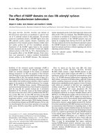
Airway responsiveness to MCh
To test the airway responsiveness of asthmatic rats in
vivo, we measured respiratory parameters induced by
MCh. Airway responsiveness of rats in the asthmatic
group increased in comparison to the control group
after induction by MCh (Figure 1).
Inflammatory cells in BAL fluid
The number of inflammatory cells in BAL fluid was
measured and compared between OVA-sensitized and
control rats. Remarkably, the total cell number in BAL
fluid recovered from OVA-sensitized/challenged rats
was significantly higher than that from PBS-treated rats.
Total cells and eosinophils in asthmatic BAL fluid sig-
nificantly i ncreased compared with control rat’s, the dif-
ference significant (P < 0.05); Total cells and eosinophils
in the treatment group significantly decreased when
compared with asthmatic group, the difference signifi-
cant (P <0.05),butdidnotsignificantlydifferfromthe
control group (P>0.05) (Table 1).
IgE measurement
Plasma t otal IgE was statistically significantly higher in
OVA-sensitized rats compared with controls (330.6 ±
97.7 ng/ml vs 282.2 ± 22.7 ng/ml, respectively; P < 0.01).
Ca
2+
concentration variations in asthmatic rat ASMC
induced by WIN62577
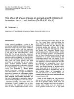
ThepurityofASMCwasconfirmedtoexceed95%by
a-actin staining (Figure 2). ASMC were loaded with the
Ca
2+
indicator Fura-2 and recorded using fluorescence
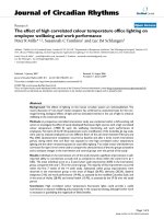
Figure 1 Airway responsiveness to MCh. Asthmatic rat inhale resistance and exhale resistance increas ed when compared with normal rat. A:
representive inhale resistance; B: representive exhale resistance.
Li et al. Journal of Inflammation 2011, 8:18
/>Page 4 of 9
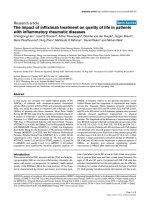
microscopy. Substance P (10
-5
M) induced [Ca
2+
]
i
to
increase in control ASMC (Figure 3A, n = 5, P < 0.05).
In contrast, [Ca
2+
]
i
decreased in asthmatic rat ASMC
exposed to WIN62577 (10
-8
M) (Figure 3B, n = 5, P <
0.05). R
340/380
in control A SMC was 0.2, but in asth-
matic rat ASMC the ratio was 1.25, suggest ing that cal-
cium concentration was higher in asthm atic ASMC than
in control cells. After substance P treatment, the R
340/
380
in control rat ASMC increased to 0.5; after
WIN62577 treatment, R
340/380
decreased to 0.4 in asth-
matic rat ASMC. These findings i ndicate that substance
P had the effect of elevating calcium concentration in
ASMC, while WIN62577 caused it to decline.
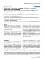
Serca2 and Ip3r mRNA expression in different groups
The equilibrium of Ca
2+
content in the SR is maintained
by SERCA pumping calcium in, while IP3R and RyR
release calcium out. SERCA and IP3R are key regulators
of Ca
2+
content in asthmatic ASMC. SERCA2 is the pre-
dominant SERCA isoform in smooth muscle. We f ound
that Serca2 mRNA decreased in asthmatic ASMC com-
pared with normal ASMC. However, after induction by
WIN62577, the expression of Serca2 mRNA in asth-
matic ASMC increased. IP3R is an SR Ca
2+
release
channel that opens upon the binding of IP3. In asth-
matic ASMC, the expression of Ip3r mRNA did not
differ from that of control ASMC. In contrast, the
expression of Ip3r mRNA decreased in asthmatic ASMC
after induction by WIN62577 (Figure 4)
The role of WIN62577 on ASMC proliferation and
migration
Because IL-13 promotes ASMC proliferation, in our
study ASMC from control rats were found to proliferate
faster after induction with IL-13, the differences among
different groups in 48 and 72 h were statistically signifi-
cant (Figure 5A, P < 0.05). Most ASMC treated with
WIN62577 and IL-13 stayed at G
1
phase compared with
those induced by IL-13 alone, with a statistically signifi-
cant difference between groups (Figure 5B, C, D, P <
0.05). The number of migrated cells significantly
decreased after WIN625 77 intervention compared with
untreated control cells (P < 0.05) (Figure 6, Table 2).
Discussion
Airway hyper-responsiveness and remodeling are impor-
tant characteristics of asthma, and both are related to
calcium levels in ASMC. In asthma, inflammatory cells
can re lease cytokines that in turn induce increased cal-
cium concentration i n ASMC, airway smooth muscle
contraction, and airway hyper-responsiveness. For exam-
ple, IL-8 has been shown to i ncrease ASMC calcium
concentration [15]. Elevation of [Ca
2+
]
i
can be caused by
Ca
2+
release f rom intracellular Ca
2+
stores or Ca
2+
influx from the extracellular space. ASMC plasma mem-
brane ion channels also contribute to changes in Ca
2+
concentration. Over a long term, increased Ca
2+
concen-
tration induces ASMC to proliferate as well as produce
and secrete pro-inflammatory factors [16].
Recently Mahn et al. reported that a SERCA2 deficiency
in ASMC contributed to their secretory and hyperproli-
ferative phenotype in asthma, suggesting that SERCA2
may play a key role in mechanisms of airway remodeling
[12]. In our study, using an asthmatic rat model we
observedthatCa
2+
homeostasis changed in asthmatic
ASMC, with increased calcium content in asthmatic rat
ASMC compared to control rat ASMC. Furthermore, sub-
stance P increased the calcium concentration of control
ASMC, and WIN62577 decreased the calcium concentra-
tion of asthmatic ASMC via increased expression of
Serca2 mRNA. How ever, WIN62577 decreased the
Table 1 Inflammation cells in different group rat’s BALF (
x
± s) ×10
4
/mL
Total eosinocyto lymphocyto granulocyto macrophage
Asthmatic group 610 ± 32* 461 ± 31* 40 ± 16* 20 ± 6.3* 88 ± 15*
Budesonide 372 ± 13
#▲
147 ± 23
#▲
19 ± 3.5
#▲
18 ± 3
#▲
56 ± 10
#▲
treatment group
normal group 172 ± 21 21 ± 7.5 8.2 ± 5.0 0.0 ± 0.0 70 ± 13
*P < 0.05, compare wi th normal group;
#
P < 0.05, compare with asthmatic group;
▲
P > 0.05, compare with normal group.
Figure 2 Immunofluorescence against a-actin suggests that
the green staining cell is ASMC.
Li et al. Journal of Inflammation 2011, 8:18
/>Page 5 of 9
expression of Ip3r mRNA in asthmatic ASMC had no dif-
ference compared with normal ASMC. Based on these
findings, we conclude that WIN62577 plays a role in
decreasing calcium concentration, which may ultimately
alleviate airway inflammation and responsiveness. As a
result, substance P antagonist WIN62577 may be an
attractive target for therapeutic approaches to asthma.
Regrettably, we were unable to examine the role of
WIN62577 in a variety of TRP channels, stretch-activated
channels, voltage-gated channels, and Ca
2+
-dependent K
+
channels, although they were involved in increased cal-
cium ion concentration.
Airway remodeling is an important cha racteristic of
asthma. The airway pathological features of asthma
include reshaping of smooth muscle cell proliferation,
hypertrophy, airway epithelium metaplasia, fibrosis,
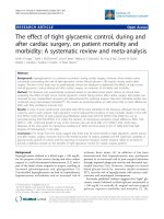
Figure 3 Effects of WIN62577 on intracellular Ca
2+
concentration ([Ca
2+
]
i
). The ratio of the intensities of emission (R
340/380
) in control group
was 0.2(Fig.3A), but in asthma group the ratio is 1.25, which suggest that the calcium concentration in asthma group was higher than the one
in control group (Fig.3B); C and D: representive the cell induced before and after substance P. calcium concentration in ASMC was increasing
intervened by substance P, the calcium concentration of asthmatic rat ASMC was decreasing intervened by substance P receptor antagonist. E
and F: representive the cell induced before and after WIN62577.
Li et al. Journal of Inflammation 2011, 8:18
/>Page 6 of 9
increased mucous cells and blood vessels, and intersti-
tial remodeling [17]. ASMC are very important effector
cells in asthma that proliferate, migrate, and contract
due to a variety of cytokines and inflammatory media-
tors, especially in asthma airway remodeling.
IL-13 is an important Th2 lymphocyte proinflamma-
tory factor [18,19] that also plays an important role in
chronic airway disease. IL-13 can change the integrity of
the airway and increase airway sensitivity [20]. Leigh et
al. demonstrated that the probability of airway hyper-
responsiveness and remodeling decreased in IL-13
knockout mice, suggesting that IL-13 played an impor-
tant role in airway remodeling [21]. IL-13 can increase
the smooth muscle cell volume and ch ange the contrac-
tile properties of smooth muscle cells and airway reac-
tivit y [22-24], as well as to promote ASMC proliferation
and participate in airway remodeling [25]. Therefore, IL-
13 was adopted in our experiment to induce ASMC
proliferation.
Figure 4 Serca2 mRNA and Ip3r mRNA express in different
group. Serca2 mRNA in asthmatic rat ASMC decreased compared
with normal ASMC (P < 0.05). But after induced by WIN62577 the
expression of Serca2 mRNA increased (P < 0.05). In asthmatic ASMC,
we found that Ip3r mRNA had no difference compared with normal
ASMC (P > 0.05). In contrast, the expression of Ip3r mRNA decreased
after induced by WIN62577 in asthmatic ASMC (P < 0.05). *P < 0.05,
control vs normal group;
#
P < 0.05, control vs asthmatic group;
▲
P >
0.05, control vs normal group. N: representive ASMC from control
group; A: representive asthmatic ASMC group untreated by
WIN62577; NK: representive asthmatic ASMC after WIN62577 treated.
Figure 5 MTT and FCM analysis the effect of NK-1R antagonist to ASMC proliferation. In IL-13 intervention group ASMC proliferate more
faster than in normal group, from 24 h to 48 h the difference become significant (P < 0.05). In NK-1R antagonist intervention group ASMC
proliferate faster than in normal group but slower than in IL-13 intervention group, especially during 48 h to 72 h (Fig.5A). Flow cytometric
analysis of ASMC cell cycle (Fig.5B, 5C, 5D). Most of ASMC stayed at G
1
stage in WIN62577 intervention group compared with in normal group.
B, C and D: representative examples of normal group, IL-13 intervention group and IL-13 with WIN62577 intervention group.
Li et al. Journal of Inflammation 2011, 8:18
/>Page 7 of 9
MTT and FCM analys is demonstrated that WIN62577
inhibited the ASMC proliferation induced by IL-13. FCM
analysis of the ASMC cell cycle suggested that most
ASMC remained at G
1
phase after WIN62577 treatment.
G
1
phase is the key to the entire cell cycle, and the cell
cycle protein D is the key protein in G
1
phase that deter-
mines transformation from G
1
to S phase. Therefore the
role of WIN 62577 on protein D and other control genes
should be studied further. In addition, IL-13 binds the IL-
13 receptor on the cell surface to activate cell receptor
protein tyrosine kinase (PTK). NK-1R is a G-protein
receptor that activates the phosphatidyl inositol bispho-
sphate (PIP2) second messenger system to promote IP3
binding to IP3R and calcium release from the SR. The
increased concentration of calcium ions could cause mem-
brane polarization and activate the PTK to achieve its bio-
logical function [26]. However, the mechanism of how
NK-1R antagonists act on the IL-13 receptor remains
unknown. Therefore, the relationship between WIN62577
and IL-13 receptor should be investigated in the future.
In asthma, eosinophils, mast cells, and other cells
secrete cytokines and inflammatory mediators that pro-
mote the development of asthma. Jonsson et al.demon-
strated that substance P induced eosinophils from
asthmatic patients to become active and demonstrate
chemotropism [27]. In this experiment, we demonstrated
tha t NK-1R antagonist WIN62577 had the effect of inhi-
biting ASMC migration in vitro, indicating that
WIN62577 may contribute to the inhibition of airway
remodeling. Taken together, our results suggest that NK-
1R antagonist WIN62577 could decrease ASMC calcium
concentration and inhibit ASMC proliferation and migra-
tion, and therefore may be useful to alleviate asthma air-
way remodeling and airway hyper-responsiveness.
Acknowledgements
This study was supported in part by a grant from the Liaoning provincial
scientific research projects (20060953).
Authors’ contributions
ML carried out the ASMC culture and participated in the Ca
2+
concentration
detecting and drafted the manuscript YY carried out the immunoassays and
ELISA detecting. YS participated in the design of the study and performed
the statistical analysis. BW participated in ASMC proliferation and migration
analysis. All authors read and approved the final manuscript.
Declaration of competing interests
The authors declare that they have no competing interest s.
Received: 12 November 2010 Accepted: 21 July 2011
Published: 21 July 2011
References
1. Global Initiative for Asthma (GINA): Global strategy for asthma
management and prevention. Bethesda MD: National Heart, Lung, and
Blood Institute; World Health Organization; 2006.
2. Ward C, Reid DW, Orside BE, Feltis B, Ryan VA, Johns DP, Walters EH: Inter-
relationships between airway inflammation, reticular basement
membrane thickening and bronchial hyper-reactivity to methacholine in
asthma; a systematic bronchoalveolar lavage and airway biopsy analysis.
Clin Exp Allergy 2005, 35:1565-1571.
3. Ammit AJ, Panettieri RA Jr: Airway smooth muscle cell hyperplasia: a
therapeutic target in airway remodeling in asthma? Prog Cell Cycle Res
2003, 5:49-57.
4. Pozzan T, Rizzuto R, Volpe P, Meldolesi J: Molecular and cellular
physiology of intracellular calcium stores. Physiol Rev 1994, 74:595-636.
5. Sathish V, Leblebici F, Kip SN, Thompson MA, Pabelick CM, Prakash YS,
Sieck GC: Regulation of sarcoplasmic reticulum Ca
2+
reuptake in porcine
airway smooth muscle. Am J Physiol Lung Cell Mol Physiol 2008,
294:787-796.
6. Parameswaran K, Janssen LJ, O’Byrne PM: Airway hyperresponsiveness and
calcium handling by smooth muscle: A “deeper look”. Chest 2002,
121:621-624.
7. Triggle DJ: Calcium, the control of smooth muscle function and
bronchial hyperreactivity. Allergy 1983, 38:1-9.
Figure 6 Transwell analyzed the effect of NK-1R ant agonist on ASMC migration. The number of ASMC in WIN62577 intervened group
compared with normal group the difference was statistically significant (P < 0.05). A and B: representive examples of normal group and
WIN62577 induced group.
Table 2 Transwell detect the role of WIN62577 to ASMC
migration
group Mean ± SD n
Normal control group 23 ± 3 15
WIN62577 intervened group 16 ± 2* 15
*P < 0.05, control vs normal group.
Li et al. Journal of Inflammation 2011, 8:18
/>Page 8 of 9
8. Mahn K, Hirst SJ, Ying S, Holt MR, Lavender P, Ojo OO, Siew L, Simcock DE,
McVicker CG, Kanabar V, Snetkov VA, O’Connor BJ, Karner C, Cousins DJ,
Macedo P, Chung KF, Corrigan CJ, Ward JP, Lee TH: Diminished sarco/
endoplasmic reticulum Ca
2+
ATPase (SERCA) expression contributes to
airway remodelling in bronchial asthma. Proc Natl Acad Sci USA 2009,
106:10775-80.
9. Groneberg DA, Harrison S, Dinh QT, Geppetti P, Fischer A: Tachykinins in
the respiratory tract. Curr Drug Targets 2006, 7:1005-1010.
10. Dinh QT, Klapp BF, Fischer A: Airway sensory nerve and tachykinins in
asthma and COPD. Pneumologie 2006, 60:80-85.
11. Patterson RN, Johnston BT, Ardill JE, Heaney LG, McGarvey LP: Increased
tachykinin levels in induced sputum from asthmatic and cough patients
with acid reflux. Thorax 2007, 62:491-495.
12. Bai TR, Zhou D, Weir T, Walker B, Hegele R, Hayashi S, McKay K, Bondy GP,
Fong T: Substance p (NK1)-and neurokinin A (NK2)-receptor gene
expression in inflammatory airway diseases. Am J physiol 1995,
269:309-317.
13. Zhou Y, Zhou X, Wang X: 1, 25-Dihydroxyvitamin D3 prevented allergic
asthma in a rat model by suppressing the expression of inducible nitric
oxide synthase. Allergy and Asthma Proceedings 2008, 29:258-267.
14. An SS, Laudadio RE, Lai J, Rogers RA, Fredberg JJ: Stiffness changes in
cultured airway smooth muscle cells. Am J Physiol Cell Physiol 2002,
283:792-801.
15. Govindaraju V, Michoud MC, Al-Chalabi M, Ferraro P, Powell WS, Martin JG:
Interleukin-8: novel roles in human airway smooth muscle cell
contraction and migration. Am J Physiol Cell Physiol 2006, 291:957-965.
16. Perez-Zoghbi JF, Karner C, Ito S, Shepherd M, Alrashdan Y, Sanderson MJ:
Ion channel regulation of intracellular calcium and airway smooth
muscle function. Pulm Pharmacol Ther 2009, 22:388-97.
17. Kondo M, Tamaoki J, Takeyama K, Nakata J, Nagai A: Interleukin-13 induces
goblet cell differentiation in primary cell culture from Guinea pig
tracheal epithelium. Am J Respir Cell Mol Biol 2002, 27:536-541.
18. O’Byrne PM, Inman MD, Adelroth E: Reassessing the Th2 cytokine basis of
asthma. Trends Pharmacol Sci 2004, 25:244-248.
19. Zimmermann N, Hershey GK, Foster PS, Rothenberg ME: Chemokines in
asthma: cooperative interaction between chemokines and IL-13. J Allergy
Clin Immunol 2003, 111:227-242.
20. Riffo-Vasquez Y, Pitchford S, Spina D: Cytokines in airway inflammation. Int
J Biochem Cell Biol 2000, 32:833-853.
21. Leigh R, Ellis R, Wattie JN, Hirota JA, Matthaei KI, Foster PS, O’Byrne PM,
Inman MD: Type 2 cytokines in the pathogenesis of sustained airway
dysfunction and airway remodeling in mice.
Am J Respir Crit Care Med
2004, 169:860-867.
22. Grünig G, Warnock M, Wakil AE, Venkaya R, Brombacher F, Rennick DM,
Sheppard D, Mohrs M, Donaldson DD, Locksley RM, Corry DB: Requirement
for IL-13 independently of IL-4 in experimental asthma. Science 1998,
282:261-263.
23. Wills-Karp M, Luyimbazi J, Xu X, Schofield B, Neben TY, Karp CL: Donaldson
DD.1998. Interleukin-13: central mediator of allergic asthma. Science
1998, 282:2258-2261.
24. Walter DM, McIntire JJ, Berry G, McKenzie AN, Donaldson DD, DeKruyff RH,
Umetsu DT: Critical role for IL-13 in the development of allergen-induced
airway hyperreactivity. J Immunol 2001, 167:4668-4675.
25. Kellner J, Gamarra F, Welsch U, Jörres RA, Huber RM, Bergner A: IL-13R-2
Reverses the Effects of IL-13 and IL-4 on Bronchial Reactivity and
Acetylcholine-Induced Ca
2+
Signaling. Int Arch Allergy Immunol 2007,
142:199-210.
26. Cascieri MA, Ber E, Fong TM, Sadowski S, Bansal A, Swain C, Seward E,
Frances B, Burns D, Strader CD: Characterization of the binding of a
potent, selective radioindinated antagonist to the human neurokinin-1
receptor. Mol Pharmacol 1992, 42:458-463.
27. Jönsson M, Norrgård O, Forsgren S: Substance P and the neurokinin-1
receptor in relation to eosinophilia in ulcerative colitis. Peptides 2005,
26:799-814.
doi:10.1186/1476-9255-8-18
Cite this article as: Li et al.: The effect of substance P on asthmatic rat
airway smooth muscle cell proliferation, migration, and cytoplasmic
calcium concentration in vitro. Journal of Inflammation 2011 8:18.
Submit your next manuscript to BioMed Central
and take full advantage of:
• Convenient online submission
• Thorough peer review
• No space constraints or color figure charges
• Immediate publication on acceptance
• Inclusion in PubMed, CAS, Scopus and Google Scholar
• Research which is freely available for redistribution
Submit your manuscript at
www.biomedcentral.com/submit
Li et al. Journal of Inflammation 2011, 8:18
/>Page 9 of 9