Báo cáo y học: "Persistent sciatica induced by quadratus femoris muscle tear and treated by surgical decompression: a case report" docx
Bạn đang xem bản rút gọn của tài liệu. Xem và tải ngay bản đầy đủ của tài liệu tại đây (1.02 MB, 4 trang )
CAS E REP O R T Open Access
Persistent sciatica induced by quadratus femoris
muscle tear and treated by surgical
decompression: a case report
Artan Bano
1
, Apostolos Karantanas
2
, Dritan Pasku
1*
, George Datseris
3
, George Tzanakakis
4
, Pavlos Katonis
1
Abstract
Introduction: Quadratus femoris tear is an uncommon injury, which is only rarely reported in the literature. In the
majority of cases the correct diagnosis is delayed due to non-specific symptoms and signs. A magnetic resonance
imaging scan is crucial in the differential diagnosis since injuries to contiguous soft tissues may present with similar
symptoms. Presentation with sciatica is not reported in the few cases existing in the English literature and the
reported treatment has always been conservative.
Case presentation: We report here on a case of quadratus femoris tear in a 22-year-old Greek woman who
presented with persistent sciatica. She was unresponsive to conservative measures and so was treate d with surgical
decompression.
Conclusion: The correct diagnosis of quadratus muscle tear is a challenge for physicians. The treatment is usually
conservative, but in cases of persistent sciatica surgical decompression is an alternative option.
Introduction
Traumatic quadratus femoris muscle tear is a clinically
unsuspected injury. The immediate and correct diagno-
sis is a challeng e because of its rarity and similarities to
other disorders that cause groin pain. Only a few cases
of partial and complete rupture of quadratus femoris
muscle in the young active population have been
reported in the literature [1,2]. In all cases, magnetic
resonance imaging (MRI) was crucial both in correct
diagnosis and gu idance of treatment. Simultane ously,
different th erapeutic techniques were used including the
injection of methylprednisolone acetate (Depo-Medrol),
transcutaneous neurostimulation, ultrasound and physi-
cal rehabilitation techniques [1]. We present a rare case
of quadratus femoris muscle rupture assoc iated with
persistent sciatica, which was treated with surgical
decompression.
Case presentation
A 22-year-old Greek woman sustained a direct injury to
the right buttock following a fall down the stairs. After
the injury she had an antalgic gait due to pain in the
right inferior g luteal area with radiati on to the proximal
posterior thigh. Pain was aggravated by sitting and
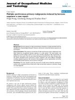
squatting. MRI examination at that time revealed an
extensive hematoma extending to both the quadratus
femoris and obturator externus muscles, in keepin g with
strain grade II (Figure 1). She was treated with non-ster-
oidal anti-inflammatory drugs (NSAID) without
improvement.
Six months after the injury, she was referred to our
tert iary health care hospital for consultation due to per-
sistent sc iatica. Physical examination revealed an active
young woman with healthy muscular development.
There were no abnormalities on examination, such as
soft tissue swelling, ecchymosis or erythema of the right
gluteus and lower leg. There was tenderness upon pal-
pation at the right ischial tuberosity associated with
reduced muscular strength at right hip external rotators.
Right straight leg rising (SLR) reproduced symptoms at
30° and her Lasegue test was positive. Passi ve hip inter-
nal rotation also reproduced pain in the proximal pos-
terior thigh, with positive Freiberg and flexion,
adduction, internal rotation (FADIR) tests. Her vascu lar
clinical tests and the lumbar spine examination were
* Correspondence:
1
Department of Orthopaedic and Traumatology, University Hospital of
Heraklion, 71110, Crete, Greece
Bano et al. Journal of Medical Case Reports 2010, 4:236
/>JOURNAL OF MEDICAL
CASE REPORTS
© 2010 Bano et al; licensee BioMed Central Ltd. This is an Open Access article distributed under the terms of the Creative Commons
Attribution License (http://creativecommons .org/licenses/by/2.0), which permits unrestricted use, distributio n, and reproduction in
any medium, provided the original work is properly cited.
normal. Standard hip, lumbar spine and pelvis radio-
graphs were unremarkable. The complete laboratory
work-up did not reveal any indication for infection or
coagulopathy. Based on the above, the initial clinical
impression was piriformis syndrome. A new MRI exami-
nation of our patient was requested for confirmation.
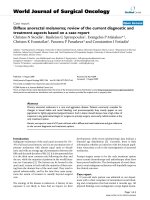
This show ed that the previous muscular strain s howed
only a minor degree of hematoma absorption compared
to the previous study (Figure 2). Thus, hematoma for-
mation was thought to be responsible for her persistent
sciat ica. A conservative approach, by means of strength-
ening of our patient’s external rotators muscle s, did not
show any improvement for one month. The lack of
obvious fluid effusion did not allow computed tomogra-
phy (CT)-guided drainage. A surgical exploration of our
patient was then performed through a posterolateral
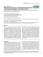
approach of the right hip. Intra-operatively, an atrophic
quadratus femoris muscle was found, with complete
detachment at the tendon-bone junction from the quad-
rate tubercle of the femur (grade III strain) (Figure 3).
An associated solid mass (5 cm × 2 cm × 3 cm), repre-
senting chronic hematoma and fibrosis, was attached to
and compressed the sciatic nerve. After decompressing
the sciatic nerve from the fibrotic and granulation tissue,
the newly formed mass was evacuated. The greater tro-
chanteric bursa and contig uous structures were noted to
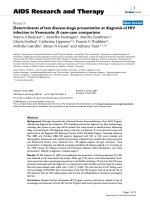
be normal. The histological findings were compatible with
degenerative muscular changes including f ibrotic tissue,
significant atrophy and fatty replacement (Figure 4).
Post-operatively, management consisted of physical
rehabilitation with empha sis on the strengthening of the
external rotator muscles with pain-free isometric pro-
gressive exercises. One month after surgery, our patient
was free of symptoms and returned to work. At the
one-year follow-up, she had no abnormal symptoms or
signs.
Discussion
Post-traumat ic pain located in the buttock area may
develop following a pelvic or coccygeal fracture or a
muscle strain, with hematoma resulting in sciatic nerve
compression. A traumatic lumbar disc herniation may
be found in patients with radicular pain. For a correct
clinical evaluation it is essential to assess the osseous
structures and the muscles around the hip joint. The
quadratus femoris muscle is a flat quadrilateral muscle
that arises from the upper external border of the ischial
tuberosity and inserts at the quadrate tubercle of the
femur [3]. It acts as a hip external rotator and assists
adduction [3,4]. The quadratus femoris muscle is inner-
vated by the quadratus f emoris nerve which rises from
the ventral roots of the L4, L5 and S1 nerves in 79.4%
of p atients [5]. In adults, the myotendinous junction is
the most vulnerable location for injury [6,7]. The tendon
insertion in the bone may also be affected.
Only a few cases describing a quadratus femoris muscle
injury have been reported in the literature. The incidence
of the quadratus femoris muscle tear is unknown.
O’ Brien and Bui-Mansfield presented a review of seven
cases [1]. In this study, this type of injury occurs predo-
minantly in women (as in our case) with a female to male
ratioof6:1.Theageofpatientsrangesfrom17to43
years with an average a ge of 29.6 years. The symptoms
were hip pain in three patients, gr oin pain in one pati ent
and deep posterior thigh or gluteal pain in three patie nts.
In none of the cases reported was there a correct clinical
diagnosis of quadratus femoris muscle tear. Diagnosis
was confused with a hamstring injury, snapping hip syn-
drome or lumbar radiculopathy. The delay from time of
injury to correct diagnosis varied from one day to five
months [1]. In one case the injury was located at the ten-
don insertion and in the rest at the musculotendinous
part. All cases were evaluated by MRI examination.
Figure 1 MRI performed a few days after injury. (a) The transverse fat suppressed proton density turbo spin echo (TSE) and (b) the coronal
short tau inversion recovery (STIR) images, show the hematoma formation in the quadratus femoris muscle (arrows) extending to the obturator
internus muscle (open arrows).
Bano et al. Journal of Medical Case Reports 2010, 4:236
/>Page 2 of 4
Figure 2 The follow-up MRI examination was performed six months later. (a) The transverse fat suppressed proton density (PD)-weighted
TSE image, shows persistent dimensions of the hematoma-like lesion in the quadratus femoris (arrow) and the obturator internus (open arrow)
muscles. The corresponding T1-weighted spin echo (SE) images show the high signal intensity on the bone-tendinous junction of the quadratus
femoris (arrows in b) and obturator internus (arrow in c). These areas histologically turned out to correspond to a mixture of chronic hematoma,
fibrosis, granulation tissue and fatty infiltration.
Figure 3 Intra-operative picture showing the sciatic nerve
(white arrow) and the ruptured quadratus muscle (black
arrow).
Figure 4 Hematoxylin and eosin stain, magnification ×400
(×400, H&E). Histopathological examination of the removed mass
showing a significant quantity of fibrotic tissue and atrophy of
muscles bundles.
Bano et al. Journal of Medical Case Reports 2010, 4:236
/>Page 3 of 4
The exact mechanism of this injury i s unknown. In
tennis players it may result from a strong eccentric
stress upon the quadratus femoris muscle in an attempt
to control hip internal rotation during the follow-
through phase of serving [2]. On the other hand, a con-
genitally smaller distance between the lesser trochanter
and the ischial tuberosity is a predisposing factor for
impingement of the quadratus femoris muscle [8].
The treatment of muscular strain consists of a carefully
planned physical rehabilitation programme. In the first
days after injury, progressive flexibility and pain-free
strengthening exercises for the external hip rotator muscle
should be performed. Then, the strengthening exercises are
progressed to e ccentric loading, as symptoms subside [ 2].
MRI has an important role in confirming the clinical
suspicion, ruling out other soft tissue injuries and aiding
prognosis [1,9,10]. Publis hed case reports have shown
the correlation of quad ratus femoris tendinitis with
groin pain [11] and muscle tear with hip pain [1].
According to O’ Br ien and Bui-Mansfield, axial T2-
weighted fat-suppressed magnetic resonance (MR)
images have demonstrated the presence of edema
between the lesser t rochanter and ischial tuberosity. On
sagittal T2-weighted fat-suppressed images the edema is
localized posterior to the lesser trochanter [1].
We suggest that in our case the grade III, quadratus
femoris strain at the tendon-bone junction resulted in
an organized mass which compressed the sciatic nerve,
simulating piriformis syndrome. To our knowledge, this
is the first case of quadratus femoris tear treated by
open surgical decompression due to persistent sciatica.
Conclusions
The primary symptoms of a severe quadratus femoris
strain are buttock pain with posterior thigh pain, which
is a ggravated by sitting or activity, and reproduction of
buttock pain on prolonged hip flexion, adduction and
internal rotation. MRI is crucial in identifying this unu-
sual injury and in excluding damage to neighbouring
structures. However, due to the presence of e xtensive
hematoma, imaging may downstage the degree of st rain.
The above injury should be considered in the differential
diagnosis of any patient presenting with proximal thigh
pain after injury. The therapy is usually conservative
consisting of rehabilitation but, in the case o f persisting
symptoms, open sciatic nerve decompression should be
an alternative approach.
Consent
Written informed consent was obtained from the patient
for publication of this case report and any accompany-
ing images. A copy of the written consent is available
for review by the Editor-in-Chief of this journal.
Author details
1
Department of Orthopaedic and Traumatology, University Hospital of
Heraklion, 71110, Crete, Greece.
2
Department of Radiology, University
Hospital of Heraklion, 71110, Crete, Greece.
3
Department of Pathology,
Medical School, University of Crete, Heraklion, 71003, Greece.
4
Department of
Histology, Medical School, University of Crete, Heraklion, 71003, Greece.
Authors’ contributions
DP, AB and PV initiated and co-wrote the paper and performed the surgical
treatment of the muscle rupture. AK analyzed the MR images, prepared the
illustrations, and performed the proof editing. GD and GT examined the
specimen and prepared the histological illustrations of the excised mass. All
authors have read and approved the final manuscript.
Competing interests
The authors declare that they have no competing interests.
Received: 4 November 2009 Accepted: 2 August 2010
Published: 2 August 2010
References
1. O’Brien SD, Bui-Mansfield LT: MRI of quadratus femoris muscle tear:
another cause of hip pain. AJR Am J of Roentgenol 2007, 189:1185-1189.
2. Willick SE, Lazarus M, Press JM: Quadratus femoris strain. Clin J Sport Med
2002, 12:130-131.
3. Gray H: Anatomy of the human body Philadelphia, PA: Lea & Febiger, 30
1985, 570.
4. Kendall FP, McCreary EK: Muscles testing and function Baltimore: Williams &
Wilkins, 4 1993, 232.
5. Aung HH, Sakamoto H, Akita K, Sato T: Anatomical study of the obturator
internus, gemelli and quadratus femoris muscles with special reference
to their innervation. Anat Rec 2001, 263:41-52.
6. Taylor DC, Dalton JD Jr, Seaber AV, Garret WE Jr: Experimental muscle
strain injury. Early functional and structural deficits and the increased
risk for reinjury. Am J Sports Med 1993, 21:190-194.
7. Tidball JG, Salem G, Zernicke R: Site and mechanical conditions for failure
of skeletal muscle in experimental strain injuries. J Appl Physiol 1993,
74:1280-1286.
8. Kassarijian A: Signal abnormalities in the quadratus femoris muscle: tear
or impingement. AJR Am J Roentgenol 2008, 190:380-381.
9. Kujula UM, Orava S, Jarvinen M: Hamstring injuries. Current trends in
treatment and prevention. Sports Med 1997, 23:397-404.
10. Speer KP, Lohnes J, Garret WE: Radiographic imaging of muscle strain
injury. Am J Sports Med 1993, 21:89-96.
11. Klinkert P Jr, Porte RJ, de Rooij TP, de Vries AC: Quadratus femoris
tendinitis as a cause of groin pain. Br J Sports Med 1997, 31:348-349.
doi:10.1186/1752-1947-4-236
Cite this article as: Bano et al.: Persistent sciatica induced by quadratus
femoris muscle tear and treated by surgical decompression: a case
report. Journal of Medical Case Reports 2010 4:236.
Submit your next manuscript to BioMed Central
and take full advantage of:
• Convenient online submission
• Thorough peer review
• No space constraints or color figure charges
• Immediate publication on acceptance
• Inclusion in PubMed, CAS, Scopus and Google Scholar
• Research which is freely available for redistribution
Submit your manuscript at
www.biomedcentral.com/submit
Bano et al. Journal of Medical Case Reports 2010, 4:236
/>Page 4 of 4