Báo cáo y học: "Recurrent lower gastrointestinal bleeding from idiopathic ileocolonic varices: a case report" ppsx
Bạn đang xem bản rút gọn của tài liệu. Xem và tải ngay bản đầy đủ của tài liệu tại đây (630.63 KB, 4 trang )
CAS E REP O R T Open Access
Recurrent lower gastrointestinal bleeding from
idiopathic ileocolonic varices: a case report
Ravula Phani Krishna
1
, Rajneesh Kumar Singh
1*
, Uday C Ghoshal
2
Abstract
Introduction: Varices of the colon are a rare cause of lower gastrointestinal bleeding, usually associated with portal
hypertension due to liver cirrhosis or other causes of portal venous obstruction. Idiopathic colonic varices are
extremely rare. Recognition of this condition is important as idiopathic colonic varices may be a cause of recurrent
lower gastrointestinal bleeding.
Case presentation: We report the case of a 21-year-old Asian man from north India who presented with recurrent
episodes of lower gastrointestinal bleeding. Colonoscopy revealed varices involving the terminal ileum and colon
to the sigmoid. Thorough evaluation was undertaken to rule out any underlying portal hypertension. Our patient
underwent subtotal colectomy including resection of involved terminal ileum and an ileorectal anastomosis.
Conclusion: Colonic varices are an uncommon cause of lower gastrointestinal bleeding. Idiopathic colonic varices
are diagnosed after excluding underlying liver disease and portal hypertension. Recognition of this condition is
important as prognosis is good in the absence of liver disease and is curable by resection of the involved bowel.
Introduction
Varices of the colon are a rare cause of lower gastroin-
testinal bleed, usually associated with portal hyperten-
sion due to liver cirrhosis or other causes of portal
venous obstruction. Idiopathic colonic varices are extre-
mely rare. Recognition of this condition is important as
idiopathic colonic varices may be a cause of recurrent
lower gastrointestinal bleed.
Case report
A 21-year-old Asian man from north India presented with
history of recurrent episodes of lower gastrointestinal
bleedi ng over the past six years. He had intermittent epi-
sodes, one to two per year, of passing bloody maroonish
stools with occasional hematochezia. Episodes were self
limited lasting two to three days,butrequiredrepeated
hospital admissions with multiple bl ood transfusions. He
had no significant past medical or family history of similar
complaints. Physical examination was unremarkable. He
was admitted with a fresh episode of lower gastrointestinal
bleeding, which subsided spontaneously. Coagulation
profile, liver function tests and hepatitis serology were nor-
mal. Upper gastrointestinal endoscopy to the third part of
the duodenum did not reveal findings suggestive of portal
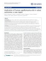
hypertension or any other bleeding source. Colonoscopy
revealed several large dilated tortuous sub-mucosal varices
extending from the upper rectum, sigmoid, the entire
colon extending into the terminal ileum (Figure 1). Our
patient was examined for portal hypertension. Doppler
ultrasound revealed normal liver size and echotexture,
portal vein 10 mm, splenic vein 7 mm with normal hepa-
topetal flow and no evidence of collaterals. Magnetic reso-
nance portovenogram revealed no evidence of cirrhosis or
portal hypertension. Selective mesenteric angiography was
carried out to search for any other vascular lesions in the
gastrointestinal tract. All vascular territories were found to
be normal and colonic lesions were undetected on an
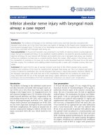
angiogram . Small bowel evaluation with enteroclysis was
normal. However, capsule enteroscopy revealed evidence
of tortuous dilated vessels in distal ileum (Figure 2).
At surgery his liver was normal and there was no evi-
dence of portal hypertension. Intra-operative ly, portal
pressure measured by cannulating mesenteric veins was
normal. Small bowel was no rmal except for the terminal
15 cm, which showed evidence of dilated tortuous sub-
serosal vessels with a clear demarcation from rest of
* Correspondence:
1
Department of Surgical Gastroenterology, Sanjay Gandhi Post-graduate
Institute of Medical Sciences, Lucknow 226014, India
Full list of author information is available at the end of the article
Krishna et al. Journal of Medical Case Reports 2010, 4:257
/>JOURNAL OF MEDICAL
CASE REPORTS
© 2010 Krishna et al; licensee BioMed Central Ltd. This is an Op en Access artic le distr ibuted unde r the terms of the Creat ive Commons
Attribution License ( which permits unres trict ed use, distribution, and reproduction in
any medium, provided the original work is properly cited.
Figure 1 Colonoscopy shows dilated tortuous veins extending throughout the colon.
Figure 2 Capsule endoscopy shows dilated tortuous veins in terminal ileum.
Krishna et al. Journal of Medical Case Reports 2010, 4:257
/>Page 2 of 4
small bowel marked by a meandering dilated mesenteric
vein (Figure 3a), confirmed by intra-operative entero-
scopy. Serosal aspect of colon was normal except few
dilated veins at sigmoid (Fig ure 3a). There were no col-
laterals in the colonic mesentery or retroperitoneum.
Sub-total colectomy including the terminal ileum w as
performed with an ileorectal anastomosis. The rectum
was relatively spared of varices. The colectomy specimen
revealed multiple dilated tortuous sub-mucosal vessels
in the colon (Figure 3b). Histology revealed large dilated
thin walled vascular channels in the submucosa.
Discussion
Lower gastrointestinal bleeding is a frequent cause of
hospital admissions. Common causes of lower gastroin-
testinal bleeding are diverticulae, vascular ectasia, colitis,
non-specific caecal ulcers, neoplasia and proctal lesions
[1]. Varices of the colon are a rare cause of lower gas-
trointestinal bleed. Incidence in one autopsy se ries was
0.007% [2]. Varices are usually associated with portal
hypertension with the most common locations being the
rectosigmoid and c eacum. In one study a mong cirrho-
tics, colonic varices were present in 31%, but bleeding
from colonic varices was seen in only 1% [3]. Other less
common causes of colonic varices are congestive heart
failure, mesenteric vein thrombosis, pancreatitis with
splenic vein thrombosis and post-operative adhesions
[1]. Idiopathic varices are rare. Establishing a diagnosis
of idiopathic colonic varices needs exclusion of other
etiologies such a s cirrhosis and portal venous obstruc-
tion by thorough evaluation. Several reported cases have
shown an associated familial aggregation. To date only
10 cases of familial colonic varices and an additional 10
cases with no other family members affected have been
reported [4-11]. Most patients present before their third
decade. This strongly suggests that these varices may be
congenital and represent an inbred vascular anomaly. In
patients with familial involvement the numbers are too
small to draw any conclusion on possible modes of
inheritance. Some authors have suggested a possible
autosomal recessive mode of inheritance [9] . Even in
patients with late presentation, after their fifties, it is
unlikely to be vascular degenerative ectasia. It is impor-
tant to note that even in cirrhotics and non-cirrhotic
patients it is unusual for varices to extend beyond the
anorectal area, in contrast with these patients. In
approxim ately half the cases the whole colon is involved
and in cases with segmental lesi ons right and left colon
are equally involved [5]. Thus it is probably more likely
that all these cases represent a significant inborn vascu-
lar anomaly [9].
Usual presentation is recurrent massive bleed. The age
at presentation has ranged from 18 years to 75 years with
no sexual predilection [4-9]. Bleeding is usually painless,
but may occasionally be associated with crampy abdom-
inal pain. Colonoscopy is the investigation of choice and
varices can be visualized as dilated tortuous venous chan-
nels. However, varices may occasionally be mistaken for
polyposis or tumor. Colonoscopy during a period of hypo-
tension, along with compression of varices due to insuffla-
tion, may cause them to be missed [2]. Barium enema is
unreliable and varices may be missed or mistaken for
polyps [5]. Mesenteric angiography is a useful diagnostic
tool, but diagnosis of colonic varices may be missed on
angiography as seen in our case and other case reports
[8,10]. Varices are detected in the venous phase of
Figure 3 (A) Terminal ileum shows evidence of tortuous dilated veins on surface (arrows) with clear demarcation from proximal small
bowel. Colon normal except for few dilated veins over surface of sigmoid (arrowhead). (B) Cut specimen of colon shows prominent tortuous
sub-mucosal vessels (arrows).
Krishna et al. Journal of Medical Case Reports 2010, 4:257
/>Page 3 of 4
angiography and the volume of contrast used may not be
sufficient to demonstrate varices [8]. Before concluding
that true idiopathic varices are present, underlying cirrho-
sis and portal venous obstruction should be excluded.
Liver function tests, hepatitis serology, Doppler studies for
portal venous system, portovenogram and wedge hepatic
venous pressure measurements should be done to rule out
liver disease or portal venous obstruction. In patients with
involvement of the entire colon, extension into the ileum
has been reported [4], as seen in our case. Therefore these
patients also need pre-operative evaluation of small bowel
as well as careful intra-operative assessment by inspection
and intra-operative endoscopy as needed to d elineate
extent of small bowel involvement.
Treatment options are conservative management and
surgical resection. Conservative treatment alone has
been attempted [3,9]; however, surgical resection of the
involved colon is the treatment of choice in view of the
risk of recurrent bleed [4-8,10]. Unlike patients with
portal hypertension and hepatocellular disease, colonic
resection can be performed with low morbidity and
mortality in this group of patients [4,8]. For patients
who are poor surgical candidates, due to advanced age
or co-morbidities , a conservative approach may be justi-
fied [3,9]. Prognosis of idiopathic colonic varices is good
at all ages compared with cirrhosis due to the low pres-
sure within varices and absence of associated liver
disease.
Our case highlights a rare cause of lower gastrointest-
inal bleed of which fewer than 20 cases have been
reported in literature. Knowledge of this entity is impor-
tant for the treating physicians as it is commonly con-
fused with portal hypertensive varices. Identification of
true idiopathic varices in such cases by a thorough
workup has important prognostic and therapeutic
implications.
Conclusions
Colonic varices are an uncommon cause of lower gas-
trointestinal bleed. Idiopathic colonic varices are diag-
nosed after excluding underlying liver disease and portal
hypertension. Family history of similar problem may be
seen in some, but not all, cases. Recognition of this con-
dition is important as prognosis is good in the absence
of liver disease and is curable by resection of involved
bowel.
Consent
Written informed consent was obtained from the patient
for publication of this case report and any accompany-
ing images. A copy of the written consent is available
for review by the journal’s Editor-in-Chief.
Author details
1
Department of Surgical Gastroenterology, Sanjay Gandhi Post-graduate
Institute of Medical Sciences, Lucknow 226014, India.
2
Department of
Medical Gastroenterology, Sanj ay Gandhi Post-graduate Institute of Medical
Sciences, Lucknow 226014, India.
Authors’ contributions
RPK and RKS were involved in conceiving the study and drafting the
manuscript. UCG was involved in revision of draft. All authors were involved
the management of the patient, contributed intellectual content and have
read and approved the final manuscript.
Competing interests
The authors declare that they have no competing interests.
Received: 22 October 2009 Accepted: 10 August 2010
Published: 10 August 2010
References
1. Miller LS, Barbarevech C, Friedman LS: Less frequent causes of lower
gastrointestinal bleeding. Gastroenterol Clin North Am 1994, 23:21-52.
2. Feldman MS, Smith VM, Warner CG: Varices of colon. Report of three
cases. JAMA 1962, 179:729-730.
3. Bresci G, Parisi G, Capria A: Clinical relevance of colonic lesions in cirrhotic
patients with portal hypertension. Endoscopy 2006, 38:830-835.
4. Lopes LM, Ramada JM, Certo MG, Perriera RP, Soares JM, Ribiero M: Massive
lower gastrointestinal bleeding from idiopathic ileocolonic varix. Dis
Colon Rectum 2006, 49:524-526.
5. Han JH, Jeon W, Chae HA, Park S, Youn SJ, Kim SH, Bae IH, Lee SJ: Acase
of idiopathic colonic varices: a rare cause of hematochezia misconceived
as tumor. World J Gastroenterol 2006, 12:2629-2632.
6. Mehta R, Deepak S, John A, Balakrishnan V: Idiopathic colonic varices.
Indian J Gastroenterol 2004, 23:30-31.
7. Villareal HA, Marts BC, Longo WE, Ure T, Verana AM, Joshi S: Congenital
colonic varices in the adult. Dis Colon Rectum 1995, 38:990-992.
8. Shreshta R, Dunkelberg JC, Shaefer JW: Idiopathic colonic varices: An
unusual cause of massive lower gastrointestinal hemorrhage. Am J
Gastroenterol 1995, 90:496-497.
9. Iredale JP, Ridings P, McGinn FP, Aurthur MJP: Familial and idiopathic
colonic varices: an unusual cause of lower gastrointestinal hemorrhage.
Gut 1992, 33:1285-1288.
10. Isbiter WH, Pease CW, Delahunt B: Colonic varices - report of a case. Dis
Colon Rectum 1989, 32:524-527.
11. Zaman L, Bebb JR, Dunlop SP, Jobling JC, Teahon K: Familial colonic
varices–a cause of “polyposis” on barium enema. Br J Radiol 2008,
81(961):e17-9.
doi:10.1186/1752-1947-4-257
Cite this article as: Krishna et al.: Recurrent lower gastrointestinal
bleeding from idiopathic ileocolonic varices: a case report. Journal of
Medical Case Reports 2010 4:257.
Submit your next manuscript to BioMed Central
and take full advantage of:
• Convenient online submission
• Thorough peer review
• No space constraints or color figure charges
• Immediate publication on acceptance
• Inclusion in PubMed, CAS, Scopus and Google Scholar
• Research which is freely available for redistribution
Submit your manuscript at
www.biomedcentral.com/submit
Krishna et al. Journal of Medical Case Reports 2010, 4:257
/>Page 4 of 4