Báo cáo y học: " Complete removal of heart-compressing large mediastinal lipoma : a case report" pps
Bạn đang xem bản rút gọn của tài liệu. Xem và tải ngay bản đầy đủ của tài liệu tại đây (1.35 MB, 3 trang )
Minematsu et al. Journal of Cardiothoracic Surgery 2010, 5:48
/>Open Access
CASE REPORT
© 2010 Minematsu et al; licensee BioMed Central Ltd. This is an Open Access article distributed under the terms of the Creative Com-
mons Attribution License ( which permits unrestricted use, distribution, and reproduc-
tion in any medium, provided the original work is properly cited.
Case report
Complete removal of heart-compressing large
mediastinal lipoma : a case report
Noritoshi Minematsu*
1
, Naoki Minato
1,2
, Keiji Kamohara
1
and Takeshi Hakuba
1
Abstract
An 83-year-old man presented with worsening of respiratory discomfort and underwent close examination, which
revealed a large mediastinal lipoma measuring 15 × 10 cm. The patient showed heart failure symptoms due to heart
compression by tumor. The tumor was completely removed safely and reliably by cutting the ascending aorta, main
pulmonary artery and superior vena cava. Although preoperative examination could not determine whether the
tumor was lipoma or liposarcoma, we selected an invasive surgical therapy because neither radiation therapy nor
chemotherapy was considered effective for either type of tumor. We report here a very rare case of heart-compressing
mediastinal tumor.
Introduction
Mediastinal lipoma is a rare tumor of the mediastinum
and causes few clinical symptoms [1]. Even if diagnosed
pathologically as benign, mediastinal lipoma causing clin-
ical symptoms is considered clinically malignant. We
achieved complete removal of a 15 × 10 cm large medi-
astinal lipoma by cutting the ascending aorta, main pul-
monary artery and superior vena cava in a patient who
developed heart failure due to heart compression by the
tumor. Tumor removal using this approach is rare and is
therefore reported here.
Case report
An 83-year-old man showed sinus rhythm on echocar-
diography, but occasionally showed paroxysmal atrial
fibrillation suspected to be due to left atrial compression.
With respiratory discomfort worsened to NYHA III, the
patient underwent close examination by computed
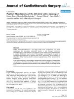
tomography and was found to have a large tumor (15 × 10
cm) behind the ascending aorta and pulmonary artery
and compressing the right and left atria (Fig 1). The
tumor was homogenous, immediately enhanced with
contrast medium, rich in fat and showed no apparent
invasion into surrounding tissues, suggesting that the
tumor was lipoma. All tumor markers, except SCC with a
high value of 9.3, showed normal values. Ultrasound car-
diography (UCG) demonstrated favorable cardiac func-
tion (EF: 70.0%) without asynergy, but revealed mitral
regurgitation of III degree due to deformation of the left
atrium caused by the tumor. No apparent tumor invasion
into the heart was observed on UCG. Cardiac catheter-
ization revealed compression of the left main trunk by the
tumor, but no significant coronary stenosis due to arte-
riosclerosis. A feeding vessel extending from the sinus
node artery into the tumor was observed.
Due to advanced age, the patient was considered at
high risk, with a EuroSCORE of 8. However, since preop-
erative imaging suggested a benign tumor with no inva-
sion to surrounding tissues, we judged that surgical
resection would improve clinical symptoms and consid-
ered the patient eligible for surgery.
Surgery
The surgical approach was achieved by median sterno-
tomy. Accumulation of about 40 ml of serous pericardial
fluid was found; cytology of the accumulated pericardial
fluid showed no tumorous change. The right end of the
tumor compressed the superior vena cava from its poste-
rior aspect. The tumor was then detached from the supe-
rior vena cava. With no invasion into surrounding tissues,
the tumor was considered detachable and removable.
Since the tumor was located behind the superior vena
cava, ascending aorta and main pulmonary artery, we
judged it necessary to cut these vessels to obtain a good
surgical field. Extracorporeal circulation was started by
transmitting blood to the right femoral artery and drain-
* Correspondence:
1
Department of Thoracic and Cardiovascular Surgery, Fukuoka Tokushukai
Hospital, 4-5 Sukukita, Kasuga City, Fukuoka, 816-0864, Japan
Full list of author information is available at the end of the article
Minematsu et al. Journal of Cardiothoracic Surgery 2010, 5:48
/>Page 2 of 3
ing blood from the superior and inferior vena cava,
ascending aorta and main pulmonary artery were cut.
The tumor was encapsulated with a thin membrane and
showed no invasion into surrounding tissues. A tight
adhesion was observed in an extra-pericardial mediasti-
nal fat tissue in the superior-posterior portion of the
bifurcation of the right pulmonary artery. Except this
portion, the tumor was relatively easily detached from
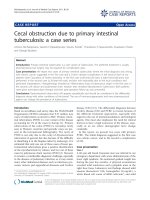
surrounding tissues. A solid tumor encapsulated with a
thin membrane was completely removed as a mass (Fig
2A). The tumor weighed 500 g (Fig 2B). The cut superior
vena cava, ascending aorta and main pulmonary artery
were reanastomosed. Extracorporeal circulation was dis-
continued without difficulty.
Pathology
Macroscopic findings
The tumor was mature, 10 × 15 cm in size, filled with fat,
and showed a clear margin. Its surface was smooth and
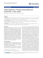
covered by a thin membrane. The cut surface of the
tumor showed a multilocular structure with fibrous septa
(Fig 3A).
Microscopic findings
Some tumor cells resembled liposarcoma cells and were
variable in size. Unlike liposarcoma, however, the major-
ity of cells were differentiated with low cellular density
and poor vascular proliferation (Fig 3B). With the
absence of lipoblast characteristic of liposarcoma and
abundance of mature fat droplets, the tumor was diag-
nosed as lipoma.
Postoperative course
Although the patient was in advance age and thus was
hospitalized for a relatively long period of time, he recov-
ered and was discharged from hospital with no serious
complications such as quadriplegia.
Discussion and Conclusions
Lipoma can occur in any soft tissues but is uncommon in
the mediastinum, only accounting for 1.6-2.3% of all pri-
mary mediastinal tumors [2]. Most cases of mediastinal
lipoma occur in the anterior mediastinum. Mediastinal
lipoma causes superior vena cava syndrome or Horner's
syndrome due to tumor mass effect, spinal nerve paraly-
sis, swallowing disorder due to esophageal compression,
respiratory discomfort and arrhythmia [3]. Therefore,
although lipoma is histologically diagnosed as a benign
tumor and grows slowly, those growing and causing clini-
cal symptoms should be considered clinically malignant
Figure 1 Computed tomography showed a large tumor behind
the ascending aorta and pulmonary artery and compressing the
right and left atria.
Figure 2 (A) Intraoperative photograph and (B) surgical specimen showed a giant tumor located within the middle and posterior mediasti-
num.
Minematsu et al. Journal of Cardiothoracic Surgery 2010, 5:48
/>Page 3 of 3
and subjected to surgical procedures for complete
removal.
Although the patient was in advanced age and consid-
ered at high risk, we judged that the patient was eligible
for surgical therapy because he showed heart failure
symptoms due to heart compression by tumor. We also
considered radiation therapy and chemotherapy as alter-
native options; however, we found a number of reports
skeptical about the effectiveness of these therapies and
thus considered them to be less effective.
For the surgical approach, thoracoscopic tumor exci-
sion has recently been reported. However, since the
tumor was large (10 × 15 cm) and adjacent to the poste-
rior aspect of the left atrium, we considered it difficult to
perform thoracoscopic surgery due to high risk of bleed-
ing [4]. There also are several case reports of liposarcoma
developing after resection of mediastinal lipoma. How-
ever, in these cases, although the tumors were pathologi-
cally diagnosed as lipoma at the initial surgery, they
actually might have been well-differentiated liposarcoma
with histological characteristics similar to those of
lipoma. In the present case, we made a preoperative diag-
nosis of lipoma, but could not rule out the possibility of
liposarcoma. Well-differentiated liposarcoma has a high
local recurrence rate (53%) and a strong tendency toward
local recurrence following incomplete resection (local
recurrence rate following complete resection: 30%) [5].
Therefore, complete tumor removal was essential, and
the thoracoscopic approach was considered inadequate.
Although an invasive approach, we chose to perform sur-
gery under direct vision and extracorporeal circulation in
consideration of safety and reliability.
We encountered an aged patient with heart failure
symptoms of NYHA III who was found to have a large
mediastinal tumor and obtained a favorable outcome
through complete removal of the tumor after cutting of
the ascending aorta, main pulmonary artery and superior
vena cava. Although we were aware of the high risk of
surgery, we selected an invasive treatment option in con-
sideration of the need for a good surgical field due to the
large tumor and for complete removal of the tumor due
to high possibility of recurrence. The selected treatment
strate allowed the patient to return to his daily activities
without recurrence for at least 18 mon postoperation.
Consent
Written informed consent was obtained from the patient
for publication of this case report and any accompanying
images. A copy of the written consent is available for
review by the Editor-in-Chief of this journal.
Competing interests
The authors declare that they have no competing interests.
Authors' contributions
MN was the primary caregiver for this patient and reviewed the manuscript. KK
and TH also cared for this patient. NM performed data collection and drafted
the manuscript. All authors read and approved the final manuscript.
Author Details
1
Department of Thoracic and Cardiovascular Surgery, Fukuoka Tokushukai
Hospital, 4-5 Sukukita, Kasuga City, Fukuoka, 816-0864, Japan and
2
Department of Thoracic and Cardiovascular Surgery, Kansai Medical
University, Japan
References
1. Jui-Sheng Hsu, Wan-Yi Kang, Gin-Chung Liu, Eing-Long Kao, Ming-Tsung
Chuang, Shah-Hwa Chou: Giant fibrolipoma in the mediastinum: An
unusual case. Ann Thorac Surg 2005, 80:e10-2.
2. Scott Gaerte C, Cristopher Meyer A, Helen Winer-Muram T, Robert Tarver
D, Dewey Conces J Jr: Fat-containing lesions of the chest. Radiographics
2002, 22:S61-78.
3. Schweitzer DL, Aguam AS: Primary liposarcoma of the mediastinum.
Thorac Cardiovasc Surg 1977, 74:83-97.
4. Aubert A, Chaffanjon P, Peoc'h M, Brichon PY: Chest wall implantation of
a mediastinal liposarcoma after thoracoscopy. Ann Thorac Surg 2000,
69:1579-80.
5. Enzinger FM, Winslow DJ: Liposarcoma: a study of 103 cases. Virchows
Arch Patho1 Anat Physiol Klin Med 1962, 335:367-388.
doi: 10.1186/1749-8090-5-48
Cite this article as: Minematsu et al., Complete removal of heart-compress-
ing large mediastinal lipoma : a case report Journal of Cardiothoracic Surgery
2010, 5:48
Received: 23 March 2010 Accepted: 3 June 2010
Published: 3 June 2010
This article is available from: 2010 Minematsu et al; licensee BioMed Central Ltd. This is an Open Access article distributed under the terms of the Creative Commons Attribution License ( which permits unrestricted use, distribution, and reproduction in any medium, provided the original work is properly cited.Journal of Cardiothoracic Surgery 2010, 5:48
Figure 3 (A) The tumor showed a multilocular structure with fi-
brous septa and (B) the majority of cells were differentiated with
low cellular density and poor vascular proliferation.