Báo cáo y học: "Perforated Meckel’s diverticulum presenting with combined bowel and urinary obstruction and mimicking Crohn’s disease: a case report" pps
Bạn đang xem bản rút gọn của tài liệu. Xem và tải ngay bản đầy đủ của tài liệu tại đây (624.71 KB, 5 trang )
CAS E REP O R T Open Access
Perforated Meckel’s diverticulum presenting with
combined bowel and urinary obstruction and
mimicking Crohn’s disease: a case report
Banny S Wong
1
, David W Larson
2
, Thomas C Smyrk
3
, Amy S Oxentenko
1*
Abstract
Introduction: Meckel’s diverticulum is a common congenital anomaly of the gastrointestinal tract, but is an
uncommon cause of serious complications in adults. Although cases of patients with hemorrhage, bowel
obstruction or perforation associated with Meckel’s diverticulum have been reported, there have been no prior
reports of patients with combined urinary and bowel obstruction due to abscess formation.
Case presentation: We describe the case of a 21-year-old man with a history of recurrent papillary thyroid can cer,
but no prior abdominal surgeries, who presented with a one-month history of rectal pain and new-onset
obstipation with urinary retention. He reported night sweats and weight loss, and had a second-degree relative
with known Crohn’s disease. A digital rectal examination was notable and revealed marked tenderness with
proximal induration. A computed tomography scan of the patient’s abdomen revealed a large, complex,
circumferential perirectal ab scess compressing the rectal lumen and base of the urinary bladder, associated with
terminal ileal thickening and an ileocecal fistula. A flexible sigmoidoscopy with an endorectal ultrasound scan
displayed a complex abscess with extensive mucosal and surrounding inflammation. An exploratory laparotomy
revealed a Meckel’s diverticulum with a large perforation at its base, positioned near the ileocecal fistula and
immediately superior to the perirectal abscess. The section of small bowel containing the Meckel’s diverticulum,
the terminal ileum, and the cecum, were all resected, and the abscess was debrided.
Conclusions: Pre-operative diagnosis of Meckel’s diverticulum can be difficult. If the nature of the complication
makes ultimate surgical management likely, an early laparoscopic or open exploration should be performed to
prevent the morbidity and mortality associated with late complications.
Introduction
Meckel’s diverticulum is a congenital anomaly found in
approximately 2% of the general population. Complica-
tions develop in only 4% of patients with this malforma-
tion, with most cases presenting in childhood [1].
Complications of Meckel’s diverticulum include hemor-
rhage, bowel obstruction, inflammation, and perforation.
All of these complications can be challenging to diag-
nose because patients may present with non-specific
symptoms, which produce a clinical picture that can
mimic other more common gastrointestinal disorders
[2]. We report an unusual case of a patient with a
perforated Meckel’s diverticulum and secondary perirec-
tal abscess formation who presented with rectal pain,
obstipation, and urinary retention. Clinical considera-
tions included Crohn’s disease and malignancy. A defini-
tive diagnosis and treatment for this patient co uld not
have been achieved without a surgical approach.
Case presentation
A 21-year-old Caucasian man was transferred to the
Clinic for an evaluation of a complex perirectal abscess.
The patient had experienced rectal pain with defecation
for a month prior to presentation, and was initially trea-
ted conservatively for presumed hemorrhoidal disease.
His symptoms progressed so that passing flatus alone
caused him significant discomfort. He then developed
worsening constipat ion. A r ectal examination was
* Correspondence:
1
Division of Gastroenterology and Hepatology, Department of Internal
Medicine, Mayo Clinic, 200 First Street SW, Rochester, Minnesota 55905, USA
Full list of author information is available at the end of the article
Wong et al. Journal of Medical Case Reports 2010, 4:264
/>JOURNAL OF MEDICAL
CASE REPORTS
© 2010 Wong et al; licensee BioMed Central Ltd. This is an Open Access article distributed under the terms of the Creative Commons
Attribution Lice nse (http://c reativecommons.org/licenses/by/2.0), which p ermits unrestricted use, distribution, and reprodu ction in
any medium, provided the original work is properly cited.
notable and revealed marked tenderness and induration.
Imaging and a proctoscopic examination under anesthe-
sia were performed at an outside hospital before admis-
sion to our clinic, and a complex perirectal fluid was
collected but could not be adequately drained. The
patient then developed increasing difficulty urinating
and a four-day history of obst ipation, prompting
transfer.
The patient’s past medical history was notable for the
occurrence of a papillary thyroid carcinoma with cervi-
cal lymph node metastases that was diagnosed one year
before and treated with a total thyroidectomy and post-
operative radioiodine therapy. He had a recent lymph
node recurrence prompting a modified radical neck dis-
section at an outside hospital one week before transfer
to our clinic. He had no history of prior abdominal or
pelvic surgeries. The only med ication he was taking was
levothyroxine. He was single, and denied tobacco, alco-
hol, or dru g use. He also denied any rectal instrumenta-
tion or anal intercourse. His family history was notable
for a paternal uncle with Crohn’s disease, and a paternal
grandmother with a history of resected thyroid cancer.
Areviewofthepatient’ s systems was positive for a 10
kg weight loss in the past month, anorexia, a decrease
in stool caliber, fatigue, and painful u rination, in addi-
tion to the presenting complaints. He denied having a
fever, but did complain of night sweats.
A physical examination revealed a tall, thin man in no
acute distress. His maximum body temperature was 37°
C, with a blood pressure of 100/60 mmHg and a heart
rate of 88 beats per minute, with normal respiration and
oxygen saturation. His abdomen was soft, with normal
bowel sounds, no distension, and no palpable masses.
He had mild tenderness in his right and left lower quad-
rantsaswellashissuprapubicregion,butnorebound
or guarding. A Foley catheter was put into position. A
perianal inspection was negative for fistulae, fissures, or
external hemorrhoids. A digital rectal examination could
not be completed because of significant tenderness.
On the day of admission, laboratory tests showed the
patient had mild microcytic anemia (hematocrit of 10.5
g/dL and mean cell volume of 79.7 fL) and hy poalbumi-
nemia (3.3 g/dL), with the remainder of the complete
blood count, electrolyte level and liver biochemistry
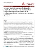
being within normal limits. A computed tomography
(CT) scan of the patient’s abdomen and pelvis demon-
strated an extensive, locula ted fluid collection encircling
the distal rectum, with a large amount of surrounding
inflammation. Compression of the patient’s distal rectal
lumen and bladder neck by the perirectal collection was
seen, but adequate bladder decompress ion was obtained
using a Foley catheter. Wall thickening of the terminal
ileum and a fistulous tract from the distal ileum to the
cecum were noted (Figure 1).
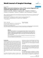
A flexible sigmoidoscop y on day two demonstrated an
erythematous, edematous rectosigmoid colon with mul-
tiple areas of extrinsic compression (Figure 2). Although
a colonoscopy was attempted for cecal and ileal inspec-
tion and tissue sampling, the patient could not tolerate
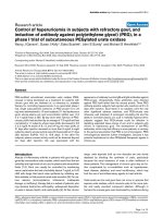
further advancement of the endoscope. An endorectal
ultrasound showed a complex solid and cystic structure
surrounding the rectosigmoid area, with mobile fluid,
solid debris, and significant surrounding inflammation
(Figure 3). The process abutted the sphincteric complex,
prostate gland, and bladder. A biopsy of the patient’s
rectum revealed focal acute inflammation with a poorly
formed mucosal granuloma, but no chronic architectural
changes. A fine-needle aspirate of the cy stic structure
produced a yellow, turbid fluid containing many leuko-
cytes and mixed bacterial flora, but no malignant cells.
On day six of his hospitalization, the patient had an
exploratory laparoscopy and the terminal ileum and
cecum were found to be densely adhered to the pelvic
side wall, with an apparent fistulous tract further fixing
the distal ileum to the cecum. Laparoscopic mobiliza-
tion of the involved structures was not possible, and
conversion to laparotomy with a low midline incision
was performed. The ileum and cecum were mobilized
to expose a large, perirectal and pelvic lateral sidewall
abscess, which was thoroughly debrided. A Meckel’ s
diverticulum with a large perforation at its ba se was
densely adherent to an ileocecal fistula, with surround-
ing inflammation and fibrosis. Given the inability to
repair the defect in the terminal ileum from the fistu-
lous opening, resection of a segment of the terminal
ileum and cecum was performed with a side-to-side,
functional end anastomosis. The portion of small
bowel containing the Meckel’s diverticulum was also
resected, with a primary anastomosis accomplished.
On abdominal exploration, no other obvious abnorm-
alities were seen to support a diagnosis of Crohn’sdis-
ease or malignancy. Fecal diversion was deemed
unnecessary because the perforated Meckel’sdiverticu-
lum was felt to be the underlying source of the
patient’ s symptoms, and resection and debridement
with single intraabdominal drain placement had
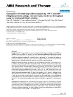
allowed for decompression. Gross pathologic examina-
tion of the surgical specimen confirmed a perforated
Meckel’s diver ticulum (Figure 4) with extensive acute
inflammation an d fibrosis of the adj acent small and
largebowels.AhistologicexaminationoftheMeckel’ s
diverticulum did not reveal gastric- or pancreatic-type
mucosa. There was no histologic evidence to support a
diagnosis of Crohn’s disease. Specifically, there was no
chronic inflammation, crypt architectural distortion or
additional granulomas seen in the area surrounding
the Meckel’s diverticulum, in the ileum or the cecum.
In addition, no evidence of malignancy was seen.
Wong et al. Journal of Medical Case Reports 2010, 4:264
/>Page 2 of 5
Postoperatively, the patient recovered uneventfully. He
initially received three days of total parenteral nutrition
but was subsequently advanced to a normal diet without
difficulty prior to hospital discharge on day 14. He
regained normal bowel and bladder function before dis-
missal. Clinical follow up over the next thre e years was
unremarkable, with no evide nce of inf lammatory bowel
disease or long-term bowel or bladder sequelae.
Discussion
We report a complicated and unusual case of a patient
with a pe rforated Meckel’s diverticulum who presented
with obstipation and urinary retention. The patient
required an open laparotomy for definitive diagnosis
and management.
Figure 1 A computed tomography scan of the patient’s abdomen and pelvis revealed an extensive, loculated fluid collection (arrow)
encircling the distal rectum (*) with surrounding inflammation, consistent with a perirectal abscess. Additionally, a thickening of the wall
of the terminal ileum with a fistulous tract from the distal terminal ileum to cecum was noted but is not shown in this figure.
Figure 2 Endoscopic view of the rectosigmoid mucosa
demonstrates erythema and edema, with luminal narrowing
due to multiple areas of extrinsic compression from the
abscess.
Figure 3 Endorectal ultrasound image showing a loculated
structure, with mobile fluid, solid debris, and significant
surrounding inflammation, around the rectosigmoid area.
Wong et al. Journal of Medical Case Reports 2010, 4:264
/>Page 3 of 5
Complications in pa tients with Meckel’s diverticulum
are rare; most patients remain asymptomatic for l ife
[3]. In both adults and children, intestinal obstruction
and bleeding have been reported to be two o f the most
common complications of a Meckel’ s diverticulum
[3-6]. Small bowel obstructions related to Meckel’ s
diverticulum have been reported due to intussuscep-
tion, incarceration in a hernia sac, or entrapment by
an adhesive band, or as being secondary to neoplasm
[7]. The pre-operative diagnosis of a patient with
Meckel’s diverticulum often presents a challenge to the
clinician in both children and adults, because present-
ing symptoms can be non-specific and the differential
diagnosis broad [4].
The perforation of a Meckel’s diverticulum may mimic
acute appendicitis and present as an acute abdomen [6].
In our case, perforation did not produce peritonitis, but
presumably led to the formation of an ileocecal fistula
and a pelvic abscess via local inflammation, which
remained relatively asymptomatic until the abscess
became large enough to cause external compression of
both the rectum and the bladder neck.
The presence of ectopic gastric mucosa is common in
complicated and symptomatic cases of Meckel’sdiverti-
culum, including patients with bleeding, inflammation,
or perforation [3,4,6]. Interestingly, our patient’s Meck-
el’s diverticulum did not contain ectopic gastric or pan-
creaticmucosaonhistologicexamination.Other
reported etiologies in patients with perforated Meckel’s
diverticulum include trauma [8], ingested sharp foreign
bodies such as a tooth pick [9] or fish bone [3], and
tumors such as leiomyosarcoma within the div erticulum
[3,10]. In addition, obstruction of the diverticular lumen
or diverticular torsion may lead to diverticulitis with
inflammation severe enough to lead to perforation, simi-
lar to some cases of appendicitis. A Meckel’s diverticuli-
tis may conceivably be the source of our patient’ s
perforation given the l ack of trauma, foreign body, or
neoplasm found on surgical exploration and the histolo-
gic examination of the resected specimen.
The initial differential diagnoses for this patient
included inflammatory bowel disease (IBD), malignancy,
and perforated appendicitis. CT imaging failed to visua-
lize the Meckel’s diverticulum, partly because adminis-
tration of intraluminal rectal contrast was
contraindicated with bowel obstruction and a high risk
of perforation. A recent study found that CT imaging
can be helpful in the diagnosi s of patients with Meckel’s
diverticulitis, but confirmed that bowel obstruction pre-
sents a greater diagnostic challenge due to a decreased
Figure 4 Gross pathology specimen of the patient’s resected bowel reveals a Meckel’s diverticulum (arrow) with perforation at its tip,
attached to the small intestine.
Wong et al. Journal of Medical Case Reports 2010, 4:264
/>Page 4 of 5
sensitivity without intraluminal opacification [11]. In our
patient, the CT findings of terminal ileal thickening, an
ileocecal fistula, and a pelvic abscess increased the suspi-
cion for Crohn’s disease.
Endorectal ultrasound findings were more consistent
with the appearance of a complicated abscess rather than
malignancy. A fine-needle aspirate of the abscess fluid
also lacked malignant cytology. Endoscopicall y, there
were no ulcerations or gross findings to support a diag-
nosis of IBD, and rectal biopsy specimens did not show
chronic inflammatory changes. However , difficulty in
advancing the colonoscope precluded biopsy of the term-
inal ileum pre-operatively to rule out Crohn’ sdisease.
Ultimately, laparotomy was required both to diagnose
and to treat this patient definitively. Surgical pathology
showed no evidence of Crohn’sdiseaseormalignancy,
and the patient continues to do well more than three
years post-operatively. Interestingly, reports of patients
with Meckel’s diverticulum masquerading as Crohn’s dis-
ease are rare [12], but cases of patients with Meckel’ s
diverticulum associated with confirmed Crohn’s disease
are not uncommon. However, it is not clear whether the
prevalence of Meckel’ s diverticulum is increased in
patients with diagnosed Crohn’s disease [13].
In summary, our case illustrates the difficulty in diagnos-
ing a complex case of a patient with a perforated Meckel’s
diverticulum. Both CT and endorectal ultrasound failed to
achieve the diagnosis. Nuclear imaging with a ‘Meckel’s
scan’ was not pursued because of the complicated nature
of the case. Due to the lack of ectopic gastric mucosa in
the resected specimen, the scan would not have assisted in
diagnosis even if performed. Laparoscopy did not lead to
the diagnosis due to inflammatory adhesions precluding
adequate exposure, and therefore, a laparotomy was una-
voidable and proved definitive in facilitating both diagnosis
and management in our patient.
Conclusions
As illustrated in our case and supported by other
reports, pre-operative diagnosis of patients with Meck-
el’s diverticulum can be challenging. Nuclear imaging
using technetium-99 m pertechnetate can be consid-
ered for detection of ectopic gastric mucosa associated
with many of the complications of Meckel’sdiverticu-
lum. However, if the nature of the complication is
likely to require surgical management, an early lapar o-
scopic or open exploration should be performed to
prevent the morbidity a nd mortality associated with
late complications.
Consent
Written informed consent was obtained from the patient for publication of
this case report and any accompanying images. A copy of the written
consent is available for review by the Editor-in-Chief of this journal.
Competing interests
The authors declare that they have no competing interests.
Authors’ contributions
BSW and ASO wrote the manuscript. DWL reviewed the manuscript and
provided surgical specimens and expertise. TCS reviewed the manuscript
and provided pathology expertise. All authors read and approved the final
manuscript.
Author details
1
Division of Gastroenterology and Hepatology, Department of Internal
Medicine, Mayo Clinic, 200 First Street SW, Rochester, Minnesota 55905, USA.
2
Division of Colon and Rectal Surgery, Department of Surgery, Mayo Clinic,
200 First Street SW, Rochester, Minnesota 55905, USA.
3
Department of
Anatomic Pathology, Mayo Clinic, 200 First Street SW, Rochester, Minnesota
55905, USA.
Received: 21 September 2009 Accepted: 11 August 2010
Published: 11 August 2010
References
1. Turgeon DK, Barnett JL: Meckel’s diverticulum. Am J Gastroenterol 1990,
85:777-781.
2. Yahchouchy EK, Marano AF, Etienne JC, Fingerhut AL: Meckel’s
diverticulum. J Am Coll Surg 2001, 192:658-662.
3. Park JJ, Wolff BG, Tollefson MK, Walsh EE, Larson DR: Meckel diverticulum:
the Mayo Clinic experience with 1476 patients (1950-2002). Ann Surg
2005, 241:529-533.
4. Bemelman WA, Hugenholtz E, Heij HA, Wiersma PH, Obertop H: Meckel’s
diverticulum in Amsterdam: experience in 136 patients. World J Surg
1995, 19:734-737.
5. Brown RL, Azizkhan RG: Gastrointestinal bleeding in infants and children:
Meckel’s diverticulum and intestinal duplication. Semin Pediatr Surg 1999,
8:202-209.
6. Bani-Hani KE, Shatnawi NJ: Meckel’s diverticulum: comparison of
incidental and symptomatic cases. World J Surg 2004, 28:917-920.
7. Leijonmarck CE, Bonman-Sandelin K, Frisell J, Raf L: Meckel’s diverticulum
in the adult. Br J Surg 1986, 73:146-149.
8. Kazemi K, Jalaeian H, Fattahi MR, Hosseini SV, Shafiee M, Roshan N:
Ruptured Meckel’s mesodiverticulum and Meckel’s diverticulum
following blunt abdominal trauma. Med Princ Pract 2008, 17:161-163.
9. Greenspan L, Abramovitch A, Tomarken J, Cohen Z: Perforation of a
Meckel’s diverticulum by a foreign body. Can J Surg 1983, 26:184-185.
10. De Mulder RM, Verschave JG: Perforated leiomyosarcoma of Meckel’s
diverticulum. Case report. Eur J Surg 1991, 157:69-70.
11. Bennett GL, Birnbaum BA, Balthazar EJ: CT of Meckel’s diverticulitis in 11
patients. AJR Am J Roentgenol 2004, 182:625-629.
12. Henneberg Holmboe C, Thorlacius-Ussing O, Teglbjaerg PS, Vinter-Jensen L:
Inverted Meckel’
s diverticulum masquerading Crohn disease in the small
intestine. Scand J Gastroenterol 2003, 38:225-227.
13. Andreyev HJ, Owen RA, Thompson I, Forbes A: Association between
Meckel’s diverticulum and Crohn’s disease: a retrospective review. Gut
1994, 35:788-790.
doi:10.1186/1752-1947-4-264
Cite this article as: Wong et al.: Perforat ed Meckel’s diverticulum
presenting with combined bowel and urinary obstruction and
mimicking Crohn’s disease: a case report. Journal of Medical Case Reports
2010 4:264.
Wong et al. Journal of Medical Case Reports 2010, 4:264
/>Page 5 of 5