Báo cáo y học: "Aplastic anemia associated with interferon alpha 2a in a patient with chronic hepatitis C virus infection: a case report" pptx
Bạn đang xem bản rút gọn của tài liệu. Xem và tải ngay bản đầy đủ của tài liệu tại đây (382.67 KB, 5 trang )
CAS E REP O R T Open Access
Aplastic anemia associated with interferon alpha
2a in a patient with chronic hepatitis C virus
infection: a case report
Savvas Ioannou, Gregorios Hatzis, Ioanna Vlahadami, Michael Voulgarelis
*
Abstract
Introduction: Hepatitis-associated aplastic anemia is a common syndrome in patients with bone marrow failure.
However, hepatitis-associated aplastic anemia is an immune-mediated disease that does not appear to be caused
by any of the known hepatitis viruses including hepatitis C virus. In addition, to the best of our knowledge there
are no reported cases of patients with chronic hepatitis C virus infection developing aplastic anemia associated
with pegylated interferon alpha 2a treatment.
Case presentation: We report the case of a 46-year-old Greek man who developed severe aplastic anemia during
treatment with pegylated interferon alpha 2a for chronic hepatitis C virus infection. He presented with generalized
purpura and bruising, as well as pallor of the skin and mucous membranes. His blood tests showed pancytopenia.
He underwent allogeneic bone marrow transplantation after compl eting two courses of immunosuppressive
therapy with antithymocyte globulin and cyclosporin A.
Conclusions: The combination of a specific environmental precipitant represented by the hepatitis C virus
infection, an altered metabolic detoxification pathway due to treatment with pegylated interferon alpha 2a and a
facilitating genetic backgroun d such as polymorphism in metabolic detoxification pathways and specific human
leukocyte antigen genes possibly conspired synergistically in the development of aplastic anemia in this patient.
Our case clearly shows that the causative role of pegylated interferon alpha 2a in the development of aplastic
anemia must not be ignored.
Introduction
HepatitisCvirus(HCV)infectionisamajorpublic
health issue. In developed countries, HCV accounts for
20% of cases of acute hepatitis, 70% of cases of chronic
hepatitis, 40% of cases of end-stage cirrhosis, 60% of
cases of hepatocell ular carcinoma, and 30% of liver
transplants [1]. Moreover, extrahepati c manifestations of
chronic HCV i nfection are clinically present in almost
40% of infected patients. These manifestations include
essential mixed cryoglobulinemia, sicca syndrome, mem-
branoproliferative glomerulonephritis, thrombocyto-
penia, and autoimmune hemolytic anemia (AIHA) [2].
Hepatitis-associated aplastic anemia (HAA) is a not
uncommon syndrome in patients with bone marrow
failure, with hepatitis documented in 2 to 5% of cases of
aplastic anemia (AA) occurring in t he West [3,4] and 4
to 10% in the Far East [5]. Characteristically, the HAA
syndrome is more prevalent among young men. The
hepatitis general ly follows a benign course, but the
onset of AA two to three months later is usually fatal if
left untreated. HAA may be induced by the presence of
HCV or hepatitis B virus infection, and also by infec-
tions with other viruses such as human immunodefi-
ciency virus (HIV), Epstein-Barr virus (EBV),
transfusion-transmitted virus and echovirus [6]. How-
ever, most cases of HAA are seronegative for the known
hepa titis viruses, includ ing hepatitis A, B, C, and G (GB
virus C) [7]. T he clinical features of the syndrome and
the patient’s response to immunosuppressive treatment
strongly indicate that the liver and marrow abnormal-
ities in patients with HAA are immune-mediated [8,9].
Pegylated interferon alpha 2a (PEG-IFN-a 2a) or 2b
plus ribavirin is currently the standard regimen for
* Correspondence:
Department of Pathophysiology, Medical School, National University of
Athens, Athens, Greece
Ioannou et al. Journal of Medical Case Reports 2010, 4:268
/>JOURNAL OF MEDICAL
CASE REPORTS
© 2010 Ioannou et al; licensee BioMed Central Ltd. This is an Open Access article distributed under the terms of the Creative Commons
Attribu tion License (h ttp://creativecommons.org/licenses/by/2.0), which permits unrestricted use, distributio n, and reproduction in
any medium, provided the original work is prope rly cited.
patients with HCV infection. A wide range of adverse
reactions, including flu-like symptoms, nausea, anorexia,
diarrhea, psychiatric symptoms, alopecia, injection-site
reactions, leukopenia, thrombocytopenia, hemolytic ane-
mia, cough, dyspnea , rash, pruritus, insomnia, and
ataxia, have been associated with PEG-IFN-a 2a plus
ribavirin treatment. Treatment wit h interferon (IFN) -a
has also been reported to trigger autoimmune phenom-
ena in up to 3% of cases, with AIHA being the most
prevalent and most significant phenomena seen in clini-
cal practice [10]. Furthermore, due to its inhibition of
cellular growth, interference with oncogene expression
and augmentation of lymphocyte cytotoxicity for target
cells, IFN-a may cause bone marrow suppression,
including potentially severe cytopenias and, very rarely,
AA [11].
The primary observed serious adverse side effect of riba-
virin treatment is hemolytic anemia. Ribavirin is an
antiviral nucleoside analogue; the mechanism of ribavirin-
induced h emolytic anemia has not been clearly estab-
lished. Anemia is most likely related to extensive ribavirin
accumulation in erythrocytes subsequent to active unidir-
ectional transmembraneous transport. Ribavirin exerts its
toxicity through an inhibition of intracellular energy meta-
bolism and oxidative m embrane damage, leading to an
accelerated extravascular hemolysis by the reticulo-
endothelial system [12]. Lau et al. describe how ribavirin,
following uptake into cells, is phosphorylated and con-
verted to ribavirin triphosphates, which then must be
dephosphorylated for elimination from the cells [13].
However, becaus e red blood cells lack dephosphorylation
enzymes, ribavirin accumulates in cells and destroys them,
causing hemolytic anemia. Severe anemia develops in
about 10% of patients treated with ribavirin, and they
require close monitoring of hemoglobin (Hb) levels and
often ribavirin dose reduction, which may compromise
sustained virologic response.
Herein, we report the development of AA in a patient
with chronic HCV infection following treatment with
PEG-IFN-a 2a plus ribavirin. By reviewi ng the literature
on the subject and the course of the patient’ sdisease,
we have come to the conclusion that, on balance, the
development of AA was a side effect of the patient’s
treatment with PEG-IFN-a 2a within a facilitating
genetic and environmental background.
Case presentation
A 46-year-old Greek man was diagnosed with HCV
infection (genotype 4 h) and a combination treatment of
PEG-IFN-a 2a ( 180 μg, weekly) and ribavirin (1200 mg/
day) was commenced for a period of 48 weeks. Before
starting the c ombination treatment his blood tests were
normal with a platelet count of 250,000 cells/mm
3
,Hb
of 16.3 g/dl, and a white blood cell (WBC) count of
6300 cells/mm
3
. The treatment was well tolerated by
the patient with a normalization of his liver function
tests. Four mo nths later he was referred to the depart-
ment of pathophysiolo gy with a bleeding tendency and
unexplained fatigue of recent onset. No contact with a
benzene or pesticide was mentioned by the patient. A
physical examination revealed generalized purpura and
bruising, and pallor of the skin and mucous membranes.
The patient’ s liver, spleen and lymph nodes were not
enlarged. Routine blood work showed severe pancyto-
penia with a platelet count of 20,000 cells/mm
3
,Hb
of 7.9 g/dl, reticulocytes at 0% and a WBC count of
600 cells/mm
3
with an absolute neutrophil count of
180 cells/mm
3
. Further investigation showed the patient
had a normal liver function test and normal prothrom-
bin time. On admission, his serum HCV ribonucleic
acid (RNA) levels were more than 1 × 10
6
units/ml. Ser-
ology for HIV, and hepatitis A and B viruses was nega-
tive, as were immunoglobulin (Ig) M antibodies against
cytomegalovirus, parvovirus B19, herpes simplex viruses
1 and 2, and EBV. Further investigations showed the fol-
lowing: urea 20 mg/dl (normal range 17 to 50 mg/dl),
creatinine 1.0 mg/dl (normal range 0.7 to 1.4 mg/dl),
sodium 139 mMol/L (normal range 136 to 145 mMol/L),
potassium 3.8 mMol/L (normal range 3.5 to 5.0 mMol/
L), glucose 99 mg/dl (normal range 74 to 115 mg/dl),
calcium 8.8 mg/dl (normal range 8.6 to 10.2 mg/dl), amy-
lase 48 U/L (normal range 20 to 104 U/L), creatine phos-
phokinase 200 U/L (normal range 20 to 190 U/L), lactate
dehydrogenase 296 U/L (normal range 200 to 460 U/L),
uric acid 4.6 mg/dl (normal range 3.5 to 7.2 mg/dl), ery-
throcyte sedimentation rate 34 mm in the first hour (nor-
mal range 0 to 20 mm), and C-reactive protein 37.4 mg/L
(normal range 0 to 5 mg/L). Screening for several autoan-
tibodies was negative. Thyroid function tests and comple-
ment serum levels were normal. A serum protein
electrophoresis showed no hypogammaglobulinemia or
abnormal bands. A computed tomography examination
of the patient’ s a bdomen and t horax was unremark-
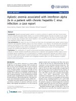
able. The patient’s bone marrow biopsy was profoundly
hypocellular with a decrease in all haematopoietic cells
(Figure 1); the bone marrow space was composed mostly
of fat cells and marrow stroma. The CD34 cell popula-
tion was more than 1%. Malignant infiltrates or fibrosis
were absent. Fluorescence-activated cell s orting analysis
of the patient’s bone marrow showed decreased marrow
elements with normal lymphocyte gate. A cytogenetic
examination showed the patient had a normal karyotype.
The pres ence of paroxysmal nocturn al hemoglobulinuria
was excluded by flow cytometry with the use of anti-
CD55 and anti-CD59 antibodies. Human leukocyte ant i-
gen (HLA) typing revealed thepresenceofDRB1*0701
and DRB1*1501 alleles. HLA matching identified a sister
with an identical HLA type.
Ioannou et al. Journal of Medical Case Reports 2010, 4:268
/>Page 2 of 5
The diagnosis of severe AA was made in the patient.
Treatment with PEG-IFN-a 2a and ribavirin were discon-
tinued. Ho wever, after two weeks, the pancytopenia did
not resolve and the patient was started on immunosup-
pressive therapy with rabbit antithymocyte globulin
(Thymo-globulin, Genzyme; 15 mg/kg/day, for five conse-
cutive days) and cyclosporin A (6 mg/kg/da y, in divided
doses every 12 hours). Prophylaxis against serum sickness
was instituted with methylprednisolone (2 mg/kg/day) for
five days with subsequent halving of the dose every week
until discontinuation on day 28. The patient had a partial
response that was noted on day 60 with a platelet count
of 27,000 cells/mm
3
, Hb of 9.3 g/dl and WBC of 5000
cells/mm
3
. The patient was dependent on red blood cell
and platelet transfusions and was on granulocyte colony
stimulating factor (400 μg/m
2
/day, three times a week).
Therefore, on day 120 a second course of antithymocyte
globulin therapy was given. The patient received a full
cyclosporin A dose for six months, after which cyclos-
porin A was tapered off slowly (0.5 mg/kg/month).
During the period of aplasia, the patient was persistently
pyrexial and broadspectrum antibiotics in the form of
an antipseudomonal penicillin (piperacillin/tazobactam)
and a carbapenem (meropenem) were administered con-
secutively, as well as an antifungal agent (liposomal
amphotericin B).
Eight months after the first course of immunosuppres-
sive treatment, the patient’ s Hb was 10.6 g/dl, platelet
count was 32,000 cells/mm
3
and WBC wa s 3590 cells/
mm
3
with an absolute neutrophil count of 2261 cells/
mm
3
.Atthattimethepatientwasstillreceivingblood
and platelet transfusions. His serum HCV RNA levels
were more than 1 × 10
6
units/ml indicating that the
patient was continuously viremic. His liver function
tests remained normal during follow up. The patient
underwent allogeneic bone marrow transplantation. He
experienced a hemorrhagic stroke due to prolonged
thrombocytopenia and died during the recovery phase.
Discussion
AA is characterized by a diminished number of or
absent bone marrow precursor cells and peripheral cyto-
pen ias. The disease is estimated to occur in two to four
people per million per year [14,15]. Numerous studies
have shown that AA behaves as an immune-mediated
disease. Cytotoxic T cells expressing T-helper 1 cyto-
kines, especially IFN-g, have been implicated in the
pathophysiolo gy of T cell-induced, Fas-mediated stem
cell apoptosis of CD34 target stem cells [16]. Why
T cells are activated in patients with AA is unclear.
A number of reports have documented a significantly
increased incidence of HLA-DR15 in patients with AA
[17]. Additionally, in a recent study, HLA-DRB1 gene
analysis showed an increased prevalence of DRB1*07 in
patients with AA compared with the normal population,
at 15.7% and 8.3%, respectively. This raises the possibi-
lity that HLA-DRB1*07 plays a significant role in the
development of AA [18]. Our patient had both
DRB1*0701 and DRB1*1501 alleles, which may indicate
that their presence is likely to allow for preferenti al pre-
sentation of peptides, such as viruses or drugs, to speci-
fic T cells, driving the autoimmune T cell-mediated
destruction of the patient’ s hematopoietic cells. This
process might have been further enhanced both in
quantity and quality by the action of the IFN-a treat-
ment that the patient received.
However, the association of AA and chronic HCV
infection remains ill-defined. In a recent report, there
were two cases of patients with AA, unrelated to IFN -a
therapy, among 35 patients with chronic HCV infection
[19]. Another case of a pat ient with severe AA associated
with HCV infection has also been reported [20]. Several
other studies have shown that the prevalence of anti-
HCV antibodies in patients with HAA receiving blood
transfusions increases with the duration and number of
transfusions, and is therefore probably transfusion related
[21]. Taking these data into account and considering our
patient’ s clinical course (normal liver function tests at
presentation, late onset of AA) , it is unlikely that the
HCV infection alone was the cause of his ensuing AA.
Bone marrow aplasia may also occur as an idiosyn-
cratic drug reaction, with a sudden onset after several
months of therapy, and it is usually irreversible. In this
regard, two cases of patients with bone marrow hypopla-
sia and fibrosis following IFN-a treatment have been
reported in the literature [22]. A severe and persistent
pancytopenia has also been described in a 42-year-old
woman with a non-Hodgkin’slymphomafollowinga
course o f 10 days of i ntramuscular leukocyte IFN-a [23].
Figure 1 A bone marrow biopsy showing the absence of
hematopoietic tissue and its replacement with fat. Hematoxylin
& eosin staining. 20× magnification.
Ioannou et al. Journal of Medical Case Reports 2010, 4:268
/>Page 3 of 5
Aslam and Singh reported a case of AA with IFN-b 1a in
a patient with multiple sclerosis [24]. However, to date,
there have been no reports of patients with severe AA
associated with PEG-IFN-a 2a in chronic HCV infection.
Some reports have suggested a genetic predisposition
to bone marrow injury in patients with an idiosyncratic
drug reaction. In such cases, direct toxicity may occur,
possibly due to genetically determined differences in
metabolic detoxification pathways [25,26]. Interestingly,
themostcommonlyuseddoseofIFN-a in humans
inhibits cytochrome P450, thus decreasing the hepatic
clearance of some drugs, and this inhibition persists
during IFN-a therapy leading to various forms of hepa-
tic and extrahepatic toxicity [27].
On the other hand, clinical characteristics and circum-
stantial evidence suggest that idiosyncratic drug reac-
tions are caused by reactive metabolites and are
immune-mediated. The possible mechanisms of stem
cell damage by drug-mediated immune damage have
not been clearly defined. One suggestion mechanism is
the ‘ spoiled membrane hypothesis’ ,whichenvisages
aberrant stem cell antigens as a result of drug action
[28]. Another possibility that has not been widely
explored is that drug-induced AA is uncommon because
it requires a coincident event at or near the time the
drug is given. We could speculate that such an event
might be a virus infection such as with HCV. Therefore,
we suggest that the combination of a specific environ-
mental precipitant repr esented by the HCV infection, an
aberrant expression of cellular proteins in the patient’s
bonemarrowcellscausedbyadisturbedPEG-IFN-a
2a-associated drug metabolic detoxification p athway,
and a facilitating genetic background (specific HLA
genes) offering a more effective presentation of viral and
drug metabolites to the T cells conspired, possibly
synergistically, in the initiation of the destructive
immune attack towards the patient’s bone marrow cells
and the development of severe AA in our patient.
The approach to treating a patient with medication-
induced AA entails stopping the offending drug while
supporting the patient during the period of pancytope-
nia. The therapeutic issue revolves around the dilemma
of a period of initial observation versus aggressive ther-
apy, such as immunosuppression or bone marrow trans-
plantation. Waiting for a week and then conducting a
repeat bone marrow biopsy may avoid potential side
effects associated w ith the therapy without foreclosing
on a definitive treatment, which is to be promptly insti-
tuted in the absence of signs of recovery.
Conclusions
We present a case of a 46-year-old man who developed
severe AA while being treated with PEG-IFN-a 2a for
chronic HCV infection. To the best of our knowledge, this
is the first report of a patient with this complication asso-
ciated with PEG-IFN-a 2a in the growing body of liter a-
ture. As health care providers, physicians should be aware
of this rare but life-threatening complication of PEG-IFN-
a 2a treatment.
Abbreviations
AA: aplastic anemia; AIHA: autoimmune hemolytic anemia; EBV: Epstein-Barr
virus; HAA: hepatitis-associated aplastic anemia; Hb: hemoglobin; HCV:
hepatitis C virus; HIV: human immunodeficiency virus; HLA: human leukocyte
antigen; IFN: interferon; Ig: immunoglobulin; PEG-IFN-a 2a: pegylated
interferon alpha 2a; RNA: ribonucleic acid; WBC: white blood cells.
Consent
Written informed consent was obtained from the patient for publication of
this case report and any accompanying images. A copy of the written
consent is available for review by the Editor-in-Chief of this journal.
Competing interests
The authors declare that they have no competing interests.
Authors’ contributions
SI and MV were responsible for writing the manuscript. IV and GH provided
clinical details and contributed to the final manuscript. All authors read and
approved the final manuscript.
Received: 11 December 2009 Accepted: 12 August 2010
Published: 12 August 2010
References
1. EASL International Consensus Conference on Hepatitis C: Consensus
statement. J Hepatol 1999, 30:956-961.
2. Palekar NA, Harrison SA: Extrahepatic manifestations of hepatitis C. South
Med J 2005, 98:1019-1023.
3. Bottiger LE, Westerholm B: Aplastic anaemia. III. Aplastic anaemia and
infectious hepatitis. Acta Med Scand 1972, 192:323-6.
4. Mary JY, Baumelou E, Guiguet M: Epidemiology of aplastic anemia in
France: A prospective multicentric study. Blood 1990, 75:1646-53.
5. Young NS, Issaragrasil S, Chieh CW, Takaku F: Aplastic anaemia in the
Orient. Br J Haematol 1986, 62:1-6.
6. Gonzalez-Casas R, Jones EA, Moreno-Otero R: Spectrum of anemia associated
with chronic liver disease. World J Gastroenterol 2009, 15:4653-4658.
7. Brown KE, Tisdale J, Barrett AJ, Dunbar CE, Young NS: Hepatitis-associated
aplastic anemia. N Engl J Med 1997, 336:1059-1064.
8. Gonzalez-Casas R, Garcia-Buey L, Jones EA, Gisbert JP, Moreno-Otero R:
Systematic review: hepatitis-associated aplastic anaemia-a syndrome
associated with abnormal immunological function. Aliment Pharmacol
Ther 2009, 30:436-443.
9. Lu J, Basu A, Melenhorst JJ, Young NS, Brown KE: Analysis of T-cell
repertoire in hepatitis-associated aplastic anemia. Blood 2004,
103:4588-4593.
10. Conrad B: Potential mechanisms of interferon-alpha induced
autoimmunity. Autoimmunity 2003, 36:519-523.
11. Platanias LC, Fish EN: Signaling pathways activated by interferons. Exp
Hematol 1999, 27:1583-1592.
12. Russmann S, Grattagliano I, Portincasa P, Palmieri VO, Palasciano G:
Ribavirin-induced anemia: mechanisms, risk factors and related targets
for future research. Curr Med Chem 2006, 13:3351-3357.
13. Lau JY, Tam RC, Liang TJ, Hong Z: Mechanism of action of ribavirin in the
combination treatment of chronic HCV infection. Hepatology 2002,
35:1002-1009.
14. Young NS: Acquired aplastic anemia. Ann Intern Med 2002, 136:534-46.
15. Wallerstein RO, Condit PK, Kasper CK, et al: Statewide study of
chloramphenicol therapy and fatal aplastic anemia. JAMA 1969,
208:2045-50.
16. Young NS, Calado RT, Scheinberg P: Current concepts in the
pathophysiology and treatment of aplastic anemia. Blood 2006,
108:2509-2519.
Ioannou et al. Journal of Medical Case Reports 2010, 4:268
/>Page 4 of 5
17. Sugimori C, Yamazaki H, Feng X, Mochizuki K, Kondo Y, Takami A, Chuhjo T,
Kimura A, Teramura M, Mizoguchi H, Omine M, Nakao S: Roles of DRB1
*1501 and DRB1 *1502 in the pathogenesis of aplastic anemia. Exp
Hematol 2007, 35:13-20.
18. Yari F, Sobhani M, Vaziri MZ, Bagheri N, Sabaghi F, Talebian A: Association
of aplastic anaemia and Fanconi’s disease with HLA-DRB1 alleles. Int J
Immunogenet 2008, 35:453-456.
19. Ramos-Casals M, García-Carrasco M, López-Medrano F, Trejo O, Forns X,
López-Guillermo A, Muñoz C, Ingelmo M, Font J: Severe autoimmune
cytopenias in treatment-naive hepatitis C virus infection: clinical
description of 35 cases. Medicine (Baltimore) 2003, 82:87-96.
20. Gruber A, Grillner L, Norder H, Magnius L, Björkholm M: Severe aplastic
anemia associated with seronegative community-acquired hepatitis C
virus infection. Ann Hematol 1993, 66:157-159.
21. Paquette RL, Kuramoto K, Tran L, Sopher G, Nimer SD, Zeldis JB: Hepatitis C
virus infection in acquired aplastic anaemia. Am J Hematol 1998,
58:122-126.
22. Hoffmann A, Kirn E, Krueger GR, Fischer R: Bone marrow hypoplasia and
fibrosis following interferon treatment. In Vivo 1994, 8:605-612.
23. Mangan KF, Zidar B, Shadduck RK, Zeigler Z, Winkelstein A: Interferon-
induced aplasia: evidence for T-cell-mediated suppression of
hematopoiesis and recovery after treatment with horse antihuman
thymocyte globulin. Am J Hematol 1985, 19:401-413.
24. Aslam AK, Singh T: Aplastic anemia associated with interferon beta-1a.
Am J Ther 2002, 9:522-523.
25. Lee KA, Kim SH, Woo HY, Hong YJ: Increased frequencies of glutathione
S-transferase (GSTM1 and GSTT1) gene deletions in Korean patients with
acquired aplastic anemia. Blood 2001, 98:3483-3485.
26. Poonkuzhali B, Shaji RV, Salamun DE, George B, Srivastava A, Chandy M:
Cytochrome P4501A1 and glutathione S transferase gene
polymorphisms in patients with aplastic anemia in India. Acta Haematol
2005, 114:127-132.
27. Israel BC, Blouin R, McIntyre W, Shedlofsky S: Effects of interferon-?
monotherapy on hepatic drug metabolism in cancer patients. Br J
Pharmac 1993, 36:229-235.
28. Waring JF, Anderson MG: Idiosyncratic toxicity: mechanistic insights
gained from analysis of prior compounds. Curr Opin Drug Discov Devel
2005, 8:59-65.
doi:10.1186/1752-1947-4-268
Cite this article as: Ioannou et al.: Aplastic anemia associated with
interferon alpha 2a in a patient with chronic hepatitis C virus infection:
a case report. Journal of Medical Case Reports 2010 4:268.
Submit your next manuscript to BioMed Central
and take full advantage of:
• Convenient online submission
• Thorough peer review
• No space constraints or color figure charges
• Immediate publication on acceptance
• Inclusion in PubMed, CAS, Scopus and Google Scholar
• Research which is freely available for redistribution
Submit your manuscript at
www.biomedcentral.com/submit
Ioannou et al. Journal of Medical Case Reports 2010, 4:268
/>Page 5 of 5