Báo cáo y học: "Unusual primary HIV infection with colonic ulcer complicated by hemorrhagic shock: a case report" potx
Bạn đang xem bản rút gọn của tài liệu. Xem và tải ngay bản đầy đủ của tài liệu tại đây (2.78 MB, 5 trang )
CAS E REP O R T Open Access
Unusual primary HIV infection with colonic ulcer
complicated by hemorrhagic shock: a case report
Stephane Emonet
1*
, Sarah Dettwiler
2
, Isabelle Der Hagopian
3
, Sabine Yerly
2
, Thomas Haustein
4
, Susannah Strasser
5
,
Bernard Hirschel
1
Abstract
Introduction: Timely diagnosis of primary HIV infect ion is important to prevent further transmission of HIV. Primary
HIV infection may take place without symptoms or may be associated with fever, pharyngitis or headache.
Sometimes, the clinical presentation includes aseptic meningitis or cutaneous lesions. Intestinal ulcer ation due to
opportunistic pathogens (cytomegalovirus, Epstein-Barr virus, Toxoplasma gondii) has been described in patients
with AIDS. However, although invasion of intestinal lymphoid tissue is a prominent feature of human and simian
lentivirus infections, colonic ulceration has not been reported in acute HIV infection.
Case description: A 42-year-old Caucasian man was treated with amoxicillin-clavulanate for pharyngitis. He did
not improve, and a rash developed. History taking revealed a negative HIV antibody test five months previously
and unprotected sex with a male partner the month before admission. Repeated tests revealed primary HIV
infection with an exceptionally high HIV-1 RNA plasma concentration (3.6 × 10
7
copies/mL) and a low CD4 count
(101 cells/mm
3
, seven percent of total lymphocytes). While being investigated, the patient had a life-threatening
hematochezia. After angiographic occlusion of a branch of the ileocaecal artery and initiation of antiretroviral
therapy, the patient became rapidly asymptomatic and could be discharged. Colonoscopy revealed a bleeding
colonic ulcer. We were unable to identify an etiology other than HIV for this ulcer.
Conclusion: This case adds to the known protean manifestation of primary HIV infection. The lack of an alternative
etiology, despite extensive investigations, suggests that this ulcer was directly caused by primary HIV infection. This
conclusion is supported by the well-described extensive loss of intestinal mucosal CD4
+
T cells associated with
primary HIV infection, the extremely high HIV viral load observed in our patient, and the rapid improvement of the
ulcer after initiation of highly active antiretroviral therapy. This case also adds to the debate on treatment for
primary HIV infection, especially in the context of severe symptoms and an extremely high viral load.
Introduction
Patient s with primary HIV-1 infection (PHI) may have a
variety of symptoms, including flulike syndrome, lym-
phadenopathy, gastrointestinal symptoms, pharyngitis,
headache, and cutaneous lesions. The non-specific pre-
sentation and a lack of suspicion among clinicians often
delay the diagnosis of PHI [1]. During t his period, the
patients are unaware of being highly infectious. Early
diagnosis is thus essential to prevent further transmis-
sion and requires a high level of suspicion and either
the use of p24 antigen or the detection of HIV RNA by
polymerase chain reaction (PCR). The following case is
a classic example of a missed HIV diagnosis in the con-
text of a mononucleosis syndrome [1], with a previousl y
undescribed complication of HIV acute infection.
Case presentation
A 42-year-old Cauca sian man of Portug uese origin, with
serologicevidenceofpasthepatitis B infection, pre-
sented to his general practitioner with a one-week his-
tory of sore throat, fever, and fatigue. Pharyngitis was
diagnosed, and amoxicillin-clavulanate prescribed. Ten
days later, the patient was admitted to our hospital with
swelling of his tongue and li ps and a widespread itchy
maculopapular rash. A grade two allergic reaction to
amoxicillin-clavulanate was diagnosed, a nd intravenous
* Correspondence:
1
Department of Internal Medicine, University Hospitals Geneva, Rue
Gabrielle-Perret-Gentil 4, Geneva, 1211, Switzerland
Full list of author information is available at the end of the article
Emonet et al. Journal of Medical Case Reports 2010, 4:279
/>JOURNAL OF MEDICAL
CASE REPORTS
© 2010 Emonet et a l; licensee BioMed Central Ltd. This is an Open Access article distributed under the terms of the Creative Commons
Attribution License ( which permits unrestricted use, distribution, and reproduction in
any medium, provided the original work is properly ci ted.
clemastin was given, with rapid subsequent improve-
ment of the swelling and rash. However, the patient
remained febrile (38.5°C) with continuing exudative
pharyngitis and painful bilateral cervical lymphadenopa -
thy ( up to t hree cm in diameter). Laboratory results
were as follows: Hemoglobin, 136 g/L; white blood cells,
7.0 g/L with 7% non-segmented neutrophils and 15%
lymphocytes; C-reactive protein, 168 mg/L; ASAT, 500
U/L; ALAT, 400 U/L; g-GT, 1200 U/L; and total biliru-
bin, 36 μmol/L. A pharyngeal swab yielded mouth flora
only. The patient remaine d hospitalized for investigation
of the non-resolving mononucleosis syndrome (tonsillar
pharyngitis, fever, and lymphadenopathy) with a pre-
sumptive diagnosis of viral infection complicated by
drug allergy.
Review of systems only revealed loose feces for one
week and was considered as a minor side effect of the
antibiotic therapy.
The foll owing day, the patient collapsed with hemato-
chezia that resulted in a decrease in his hemoglobin to
66 g/L. He was hemodynamically stabilized with intrave-
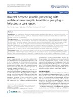
nous saline and blood transfusions. Computed tomogra-
phy showed extravasatio n of contrast medium into the
cecum, and active bleeding was confirmed by angiogra-
phy (Figure 1). After angiographic occlusion of a branch
of the ileocaecal artery, bleeding stopped and did not
recur. Colonoscopy revealed a large caecal ulcer with
irregular margins and a fibrinous base (Figure 1), located
at the site of the hemorrhage.
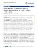
A biopsy of the ulcer showed acute inflammation in
the lamina propria, consisting of an increase in poly-
morphonuclear cells without a concomitant increase of
lymphocytes and plasma cells, associated with crypt
abscesses and flattened epit helial cells (Figure 2), com-
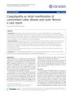
patible with an edge of the ulcer. The adjacent colonic
mucosa showed a mildly inflamed lamina propria with
regenerative glands (Figure 3). Although lymphocytes
and p lasma cells were not increased in the lamina pro-
pria, immunohistochemistr y with anti-CD3, - CD4 and
-CD8 demonstrated fewer CD4
+
T cells than CD8
+
Figure 1 Angiography and colonoscopy. Caecal bleeding on angiography (arrow) and caecal ulcer on colonoscopy (arrow).
Emonet et al. Journal of Medical Case Reports 2010, 4:279
/>Page 2 of 5
T cells (Figure 3). Grocott, periodic acid Schiff (PAS),
Giemsa, and Ziehl-Neelsen stains, as well as immuno-
histochemical methods using antibodies to CMV, HSV-
1, and HSV-2, did not reveal any pathogens. P24 antigen
was undetectable. Tissue c ulture was not performed.
The CMV viral l oad in the blood was very low (29
copies/mL).
Serologic investiga tions to identify a c ause for th e
patient’s m ononucleosis syndrome indicated only past
infections with EBV, CMV, and T. gondii (the presence
of IgG, but not IgM antibodies). However, the patient
was f ound to have a low CD4 count (180/mm
3
,10%of
total lymphocytes), and a fourth-generation HIV test
was positive. The patient’s history suggested that he had
acquired his HIV infection recently. He reported having
had a negative HIV rapid test ( Determine) five months
earlier and had had unprotecte d sex with a man one
month previously. Primary HIV infection was conf irmed
by a very high viremia (3.6 × 10
7
copies/mL HIV-1
RNA tested by CAP/CTM, Roche) and an evolving
immunoblot (INNOLIA, Innogenetics) on the blood
sample obtained one day before his gastrointestinal
hemorrhage.
In view of a persisting mononucleosis syndrome and a
further decrease in his CD4 count (101/mm
3
,7%),anti-
retroviral therapy with emtricitabine, tenofovir, and lopi-
navir/r was started. The pharyngitis, fever, and
lymphadenopathy improved rapidly. On follow-up five
monthslater,hisviremiawaslessthan40copies/mL,
and his CD4 count was 310/mm
3
.
Discussion
Oral and esophagea l ulcers have been describe d in PHI
[2,3], whereas colonic ulcerations are usually associat ed
with AIDS [4]. However, a report exists of a lorry driver
in Rwanda with melena in the context of acute HIV and
oropharyngeal and rectal ulcers [5]. Esophageal ulcera-
tion and rectal fissures also were identified in a patient
with HIV-1-seronegative AIDS [6]. Pancreatitis [7] and
hepatitis [8] have been reported occasionally.
We believe that PHI could be the cause of this colonic
ulcer for a number of reasons. First, despite extensive
investigations, we could not identify any alternative
infectious or tumoral etiology. Even if “special histologic
stains are rarely beneficial for the evaluation of HIV-
related g astrointestinal infections” [9], the failure to
detect pathogens on Grocott, PAS, Giemsa, and Ziehl-
Neelsen stains, as well as immunohistochemically by
using antibodies to CMV, HSV-1, and HSV-2, indirectly
supports the role of HIV as the causative pathogen. Sec-
ond, the existing literature suggests an extensive loss of
intestinal mucosal CD4
+
T cel ls associated with an
increase of cytotoxic CD8
+
T cel ls during PHI [10].
Third, the exceptionally high level of HIV viremia, the
Figure 2 Biopsy of the caecal ulcer. The col ic biopsy shows an acute inflammation in the lamina propria, consisting of an increase in
polymorphonuclear cells. The neutrophils infiltrate the mucosa, leading to crypt abscess (arrowhead), flattened epithelial cells, and erosion
(arrow).
Emonet et al. Journal of Medical Case Reports 2010, 4:279
/>Page 3 of 5
temporal correlation between ulcer and pri mary HIV
infection, and the rapid improvement of the ulcer after
the initiation of highly active antiretroviral therapy,
strongly suggest that HIV itself was the cause of this
ulcer.
Conclusion
This case is a reminder to always consider HIV in the
differential diagnosis, especially when confronted with
non-resolving symptoms or an unusual presentation.
The inte stinal complication described is unusual but
relevant, because it links pathophysiolo gy and clinical
events.
This report also adds to the debate o n treatment of
acute HIV infection. Even if no docume nted proven
advantage exists after six months of treatment [11], anti-
retroviral therapy (ART) is effective in alleviating the
symptoms of PHI, as shown in this case. In the future,
the use of CCR5 inhibitors in the context of a very
recent HIV infection could become an area of special
interest, because CCR5 + CD4
+
T cells of the gut are
the primary target of HIV [12].
Consent
Written informed consent was obtained from the patient
for publication of this case report and accompanying
images. A copy of the written consent is available for
review by the Editor-in-Chief of this journal.
Author details
1
Department of Internal Medicine, University Hospitals Geneva, Rue
Gabrielle-Perret-Gentil 4, Geneva, 1211, Switzerland.
2
Department of Genetic
Medicine and Laboratories, University Hospitals Geneva, Rue Gabrielle-Perret-
Gentil 4, Geneva, 1211, Switzerland.
3
Department of Community Medicine,
University Hospitals Geneva, Rue Gabrielle-Perret-Gentil 4, Geneva, 1211,
Switzerland.
4
Infection Control, Medical Directorate, University Hospitals
Geneva, Rue Gabrielle-Perret-Gentil 4, Geneva, 1211, Switzerland.
5
Department of Imaging, University Hospitals Geneva, Rue Gabrielle-Perret-
Gentil 4, Geneva, 1211, Switzerland.
Authors’ contributions
SE and BH cared for the patient during and after his hospitalization and
wrote the manuscript.
TH contributed to writing and editing of the manuscript. SD did the
histologic workup of the biopsy. IDH treated the patient in the emergency
center. SY determined the viral load and the immunoblot. SS performed the
arterial occlusion (angiography). All authors read and approved the final
manuscript.
Competing interests
The authors declare that they have no competing interests.
Received: 18 September 2009 Accepted: 20 August 2010
Published: 20 August 2010
References
1. Rosenberg ES, Caliendo AM, Walker BD: Acute HIV infection among
patients tested for mononucleosis. N Engl J Med 1999, 340:969.
2. Rabeneck L, Popovic M, Gartner S, McLean DM, McLeod WA, Read E,
Wong KK, Boyko WJ: Acute HIV infection presenting with painful
swallowing and esophageal ulcers. JAMA 1990, 263:2318-2322.
Figure 3 Biopsy of colonic mucosa adjacent to the ulcer;
immunohistochemistry with anti-CD3, -CD4, and -CD8. The colic
mucosa is regenerative, with less mucus in the cytoplasm of the
epithelial cells. The lamina propria contains some neutrophils, which
infiltrate the epithelial cells (arrow), without an increase of
mononuclear cells. In this inflamed mucosa, fewer CD4
+
cells
(arrow) than CD8
+
T cells (arrow) are found.
Emonet et al. Journal of Medical Case Reports 2010, 4:279
/>Page 4 of 5
3. Ehrenpreis ED, Bober DI: Idiopathic ulcerations of the oesophagus in HIV-
infected patients: a review. Int J STD AIDS 1996, 7:77-81.
4. Monkemuller KE, Wilcox CM: Diagnosis and treatment of colonic disease
in AIDS. Gastrointest Endosc Clin North Am 1998, 8:889-911.
5. Melzer M, Ong EL: A spotted fever and melaena: acute HIV
seroconversion. Int J STD AIDS 1997, 8:535-536.
6. Monkemuller K, Fry LC, Decker JM, Rickes S, Smith PD: Severe
gastrointestinal disease due to HIV-1-seronegative AIDS. Z Gastroenterol
2007, 45:706-709.
7. Rizzardi GP, Tambussi G, Lazzarin A: Acute pancreatitis during primary
HIV-1 infection. N Engl J Med 1997, 336:1836-1837.
8. Molina JM, Welker Y, Ferchal F, Decazes JM, Shenmetzler C, Modai J:
Hepatitis associated with primary HIV infection. Gastroenterology 1992,
102:739.
9. Monkemuller KE, Bussian AH, Lazenby AJ, Wilcox CM: Special histologic
stains are rarely beneficial for the evaluation of HIV-related
gastrointestinal infections. Am J Clin Pathol 2000, 114:387-394.
10. Mattapallil JJ, Douek DC, Hill B, Nishimura Y, Martin M, Roederer M: Massive
infection and loss of memory CD4+ T cells in multiple tissues during
acute SIV infection. Nature 2005, 434:1093-1097.
11. Streeck H, Jessen H, Alter G, Teigen N, Waring MT, Jessen A, Stahmer I, van
Lunzen J, Lichterfeld M, Gao X, et al: Immunological and virological
impact of highly active antiretroviral therapy initiated during acute HIV-
1 infection. J Infect Dis 2006, 194:734-739.
12. Johnson RP: How HIV guts the immune system. N Engl J Med 2008,
358:2287-2289.
doi:10.1186/1752-1947-4-279
Cite this article as: Emonet et al.: Unusual primary HIV infection with
colonic ulcer complicated by hemorrhagic shock: a case report. Journal
of Medical Case Reports 2010 4:279.
Submit your next manuscript to BioMed Central
and take full advantage of:
• Convenient online submission
• Thorough peer review
• No space constraints or color figure charges
• Immediate publication on acceptance
• Inclusion in PubMed, CAS, Scopus and Google Scholar
• Research which is freely available for redistribution
Submit your manuscript at
www.biomedcentral.com/submit
Emonet et al. Journal of Medical Case Reports 2010, 4:279
/>Page 5 of 5