BASIC AND CLINICAL DERMATOLOGY - PART 2 docx
Bạn đang xem bản rút gọn của tài liệu. Xem và tải ngay bản đầy đủ của tài liệu tại đây (920.83 KB, 35 trang )
The lesions are essentially asymptomatic. Howe ver, in an advanced degree of cellu-
lite, symptoms such as a sensation of weight and pain may occur in the affected areas
(10,20,29). These probably occur as a result of compression of the nervous terminals or
the presence of inflammatory reactions (16,19).
The main manifestations of clinical cellulite are:
1. flaccid ‘‘mattress-like’’ skin, with multiple depressions and some elevations, caused by
irregular retraction of the skin, form ing a surface where protuberances and depressed
areas alternate (Fig. 1) (6,16,34);
2. ‘‘orange peel’’ skin due to the tumefact ion of the epidermis and dilation of follicular
pores (6,16,34).
The cutaneous surface alte rations that ch aracterize cellulite are predominantly
depressed, when compared to cutaneous surface of the affected area (29). These depres-
sions have the same color and consistency as normal skin, and the number of lesions
mayvaryfromonetomany(29).Theshape of these lesions is varied (29): rounded,
oval, or linear (Fig. 3). Most lesions are oval, as the longest axis of the lesions lies par-
allel to the relaxed skin tension lines (Figs. 4A–E). It is interesting to note that those
lesions that do not have the same disposit ion in relation to the relaxed skin tension lines
in general originate from secondary fibrosis of the subcutaneous tissue, such as injec-
tions, trauma, etc. They are usually found in the lower portion of the buttocks and
the upper thigh (Fig. 4). In both the buttocks and the upper thigh, just below the gluteal
fold, the longest axis is in the horizontal direction, with the lateral extremities slightly
elevated. I n these locations, cellulite may be more evident due to flaccidity of the epider-
mis, which tends to become a ggrava ted with age. This can be demonstrated by the
diminishing or even the disappearance of the lesions when the buttocks are lifted to their
original position.
CLASSIFICATION
Several authors have classified cellulite into four clinical stages or degrees (Table 2), based
on the clinical alterations observed with the patient at rest and after the application of the
pinch test or muscular contraction (6,29,34).
Because cellulite is diagnosed by clinical alterations, without histopathological find-
ings or anatomical or pathognomonic charact eristics, it can also be classified into primary
and secondary cellulite. In primary cellulite, there are no aggravating factors involved. In
secondary cellulite, the alterations are provoked by secondary factors such as localized fat,
flaccidity, surgical or accident trauma mainly from lipos uction, after injections that cause
lipoatrophy, or after subcutaneous fibrosis from any inflammatory or infectious process.
These circumstances may aggravate or even bring about primary cellulite and should be
detected through the medical history and physical examination. Treatment, in this case,
implies the correction of the prim ary factor.
CLINICAL APPROACH
As with other pathologies, the medical history should be detailed in the evaluation of cel-
lulite. The patient should be questioned regarding the age at which cellulite appeared,
prior occurrence of trauma, liposuction or injections in the affected area, history of prior
DEFINITION, CLINICAL ASPECTS, ASSOCIATED CONDITIONS, AND DIFFERENTIAL DIAGNOSIS
&
11
disease or surgery, family history, presence of chronic vascular or associated hormonal
diseases, the occasional or regular use of medications, and previous or current history
of hormonal treatment or the use of any medicine that may contribute to the increase
in the deposit of fat in the affected areas, such as corticosteroids and estrogens. Other
aspects that should be researched are sedentarism, diet, psychosomatic factors, smoking,
prior pr egnancy, and the behavior of cellulite during pregnancy.
Although smoking and circulatory problems are frequently cited as causative agents
of cellulite, in the experience of the present authors, in a sample of 1200 patients with
advanced cellulite, the vast majority were neither smokers (more than 80%) nor those hav-
ing varicose veins or other circulatory problems.
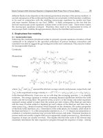
Figure 3
Oval and linear lesions of cellulite.
12
&
HEXSEL ET AL.
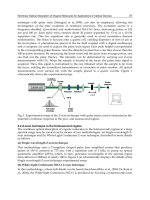
Figure 4
(A) Relaxed skin tension lines mapped on a body scheme. The left half shows the frontal view and
the right half, the back view. (B–E) Cellulite lesions follow the relaxed skin tension lines.
DEFINITION, CLINICAL ASPECTS, ASSOCIATED CONDITIONS, AND DIFFERENTIAL DIAGNOSIS
&
13
Physical Examination
The physical examination should be performed with the patient in a standing position, with
muscles relaxed (9,10,29). Cellulite can be better observed with the application of the pinch
test, in which the skin in the area to be examined is pinched between the thumb and index
finger to form a fold by skinfold plicometry or through the contraction of the muscles in the
Table 2
Classification of Cellulite
Classification Evaluation results
Degree or stage 0 There is no alteration to the skin surface
Degree or stage I The skin of the affected area is smooth while a subject is standing
or lying down, but undulations on the skin surface can be seen
on pinching the skin or during muscle contraction (Fig. 5)
Degree or stage II The ‘‘orange peel’’ or ‘‘mattress’’ appearance is evident when
standing, without the use of any manipulation (skin pinching or
gluteus muscle contraction) (Fig. 6)
Degree or stage III Presence of alterations described in second degree or stage II, plus
presence of raised and depressed areas and nodules (Fig. 7)
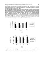
Figure 5
First degree cellulite, in which there are no alterations to the skin surface in a standing position and
with relaxed gluteous muscles. Alterations are found under the pinch test applied to the skin of the
affected area.
14
&
HEXSEL ET AL.
area (Figs. 8 and 9) (9). Overhe ad or tangential illumination of the patient facilit ates the
visualization of cellulite (29). There are significant differences in the appearance of cellulite,
depending on the position and the method used for its classification. For this reason, the
standing position is recommended for the examination of a patient with cellulite.
Palpation should always be performed to check the elasticity of the skin (6) and sub-
cutaneous tissues. However, at present there are no exact parameters for the classification
of skin elasticity. Venous or lymphatic insufficiency may, in theory, aggravate cellulite and
should also be checked during the physical examination (35). One should make note of the
presence of varicose and telangiectatic leg veins as well as any pitting edema or induration
of the skin. A Doppler or duplex ultrasound examination of the superficial venous system
will also he lp to classify the significance of venous insufficiency. Even if venous insuffi-
ciency is not found to be an etiologic factor in the pathogenesis of cellulite, its presence
or absence will help direct appropriate treatment regarding graduated compression.
Figure 6
‘‘Orange peel’’ or ‘‘mattress’’ appearance of second degree cellulite.
DEFINITION, CLINICAL ASPECTS, ASSOCIATED CONDITIONS, AND DIFFERENTIAL DIAGNOSIS
&
15
AGGRAVATING FACTORS
A number of clinical conditions or circumstances frequently accompany or aggravate cel-
lulite, especially obesity, localized fatty accumulations, and skin flaccidity.
Obesity promotes a generalized increase in body weight (skeletal, muscular, intersti-
tial fluid, organ hypertrophy, etc.). After a return to the original baseline weight is
achieved, an increased accumulation of fat is observable (36). The clinical manifestation
of localized adiposity is an increase in the ill-defined symmetrical and bilateral diffuse
volume, owing to an increase in the adipose tissue (29). The localized increase in adipose
tissue in the subcutaneous tissue leads to the aggravation of cellulite lesions by contribut-
ing to a worsening of the irregular undulations of the skin. The increase in fat volume
leads to an augmentation of tension forces within the fat lobules. This tension is projected
to the skin surface and aggravates the depressions, causing an effect similar to that of a
stuffed quilt (29). These alterations contribute to the appearance of the mechanical and
circulatory alterations that occur in cellulite. Greater thickness of the subcutaneous fat
in the affected areas may be seen by histopathological ex amination and can be measured
by specia l instruments or by the pinch test (Fig. 9) (36).
Rosenbaum et al. described the exacerbation of cellulite with weight gain and its cor-
relation with the body mass index (BMI). This study demonstrates the protrusion of adi-
pose tissue into the dermis when the volume of subcutaneous fat is augmented, which
explains the mattress-like appearance (31).
Flaccidity is caused by physiological ptosi s of subcutaneous structures, making the
skin permanently distended and loose. This condition frequently occurs in the buttocks,
Figure 7
Third degree cellulite, showing raised and depressed areas and modules plus orange peel
or mattress appearance.
16
&
HEXSEL ET AL.
thighs, the region above the knee, and the inner surface of the arms, regions where the skin
probably has less retent ive capacity and suffers the mechanical action of weight exerted by
the adipose tissue and by the other subcutaneous structures (29). The weight of these struc-
tures increases the effect of gravity, causing alterations to the skin surface in these areas,
which is seen as laxity and looseness (29). The reduced elasticity of the skin and sudden
loss of weight (29) or subcutaneous fat due to liposuction (37) are conditions that can
bring about or aggravate skin flaccidi ty.
Although it is of great importance, the presence of flaccidity or other aggravating
conditions is usually not mentioned in present day classifications of cellulite. In the
absence of flaccidity, a distension test in the antigravity direction tends not to diminish
the lesions. In the presence of flaccidity, however, such a test can lead to a reduction or
even disappearance of cellulite lesions (Fig. 10). The pinch test causes an increase in
Figure 8
Pinch test using a special device, the skinfold plicometry.
DEFINITION, CLINICAL ASPECTS, ASSOCIATED CONDITIONS, AND DIFFERENTIAL DIAGNOSIS
&
17
Figure 10
The patient shown in Figure 9 showing improvement to the skin surface when stretching the skin in
the direction opposite to forces of gravity.
Figure 9
Patient with cellulite secondary to flaccidity or loose skin. Alterations to the skin surface became
more evident on pinching the skin.
18
&
HEXSEL ET AL.
tension inside the lobes, and the cellulite becomes apparent as the lobes bulge and aggra-
vate the traction of the septa in the pinched area (Fig. 11). Moreover, flaccidity has an
effect similar to that of pinching by compressing the lobes and, thus, augmenting the ten-
sion within them. This situation is respon sible for the emergence or worsening of cellulite
lesions, especially after the fourth or fifth decade of life when the elastic properties of the
skin diminish (38). This, together with the weight of the subcutaneous fat, determines the
worsening of distension of the skin.
Other notable conditions that cause secondary cellu lite or that aggravate cellulite are
subcutaneous fibrosis caused by previous surgery, mainly liposuction, and the subcuta-
neous fibrosis and lipoatrophy originating from the trauma caused by injections in the
affected areas. Alterations to the cutaneous surface resulting from liposuction usually
appear late, from three months to one year after surgery. They may be slight, moderate,
or severe, and always emerge in previously treated areas, such as the lateral and posterior
thighs, buttocks , abdomen (Fig. 12), flanks, and the region above the knees. Like cellulite,
the cutaneous sequelae from liposuction are predominantly depressed subcutaneous tissue,
but raised and depressed areas may intercalate and vary in number and shape as a reflec-
tion of the number and variety of liposculpture cannula insertions, as well as the size and
type of cannulas. Generally, they form larger depressions with bizarre shapes and do not
necessarily follow the direction of the relaxed skin tension lines. Instead, they follow the
direction of cannula insertion (Fig. 12).
The cutaneous surface alterations caused by previous injections (such as insulin
injections in diabetics) occur in places where the injections are normally applied, that is,
in the upper, outer quarter of the buttocks. They also vary in number and shape, and
do not follow the force lines of the skin.
The presence of atrophic scars in the areas frequently affected by cellulite can also
simulate or aggravate cellulite.
Many factors can cause cellulite, and other factors can make it worse. The classifica-
tion in Table 2 is useful for generic diagnostic purposes, but is not appropriate for an accu-
rate measure of the results of treatments, other than surgical treatment. To evaluate the
results of other treatments, such as topical or systemic treatments, alternative objective
and subjective measures are needed; these are presented in the appendix to this chapter
in the form of a protocol used in our clinics.
COMPLEMENTARY EXAMINATIONS
The BMI is widely used and cited by some authors as a simple, low-cost examination
considered fundamental for the evaluation of the clinical cellulite (6,39). This is a quan-
titative method that uses measures of weight and height to assess the degree of obesity
(39). By using this index, it is not possible to distinguish the percentage of body fat in
the muscular mass. BMI is an uncertain diagnostic index of obesity (40). Studies reveal
that the estimated standard error of the percentage of body fat of BMI is approximately
5% to 6% (39).
A clinical evaluation of a sample of 32 patients ranging from 18 to 45 years of age,
performed by the present authors by means of physical examination, BMI calculation, and
assessment of body fat percentage by skinfold plicometry (39), revealed that cellulite man-
ifested even in patients with a low percentage of body fat and a normal BMI.
DEFINITION, CLINICAL ASPECTS, ASSOCIATED CONDITIONS, AND DIFFERENTIAL DIAGNOSIS
&
19
Two-dimensional ultrasound is a noninvasive method of evaluating variations
(41,42) and alterations of the subcutaneous fatty tissue, and with the assistance of Doppler,
it evaluates the local circulation (6). This examination has been used in some studies for the
evaluation of cellulite, and has demonstrated a diffuse pattern of extrusion of underlying
adipose tissue into the reticular dermis in affected individuals, but not in unaffected
individuals (2,31).
Computed tomography (43) and magnetic resonance imaging (44,45) are exami-
nations used for measuring the thickness of adipose tissue, which do not allow evaluation
of the dermis or microcirculation (6). In one study, the magnetic resonance imaging quan-
tified deeper indentations of adipose tissue into the dermis and evidenced for the first time
a great increase in the thickness of the inner fat layer in women with cellulite (46) .
Although invasive, histological examination may be useful as a method for evaluat-
ing cellulite (3,6,13). The stains used in this examination include hematoxylin–eosin for
routine histological examination; Alcian blue for polysaccharides; periodic acid–Schiff
for basement membranes; Weigert–Van Gieson (fuchsin–resorcin and acid fuchsin) for
highlighting elastic, collagen, and flat muscle fibers; and Masson trichromic, which
demonstrates contrast between collagen and muscle fibers (6). With this exami nation, it
Figure 11
Pinch test, which makes the septa pulling the
skin surface more evident.
20
&
HEXSEL ET AL.
is also possible to observe the diffuse extrusion pattern of underlying adipose tissue
distending the reticular dermis in people with cellulite (31). The macroscopic aspect of sub-
cutaneous fat from corpses is shown in Figure 13.
&
DIFFERENTIAL DIAGNOSIS
Of particular importance in the differential diagnosi s of cell ulite are the localized deposits
of fat (13) , flaccidity, surgical sequelae (from liposuction) (Fig. 12) or other trauma (47),
Figure 12
‘‘Cellulite-like’’ liposuction sequelae on the abdomen, one year after the surgery.
Figure 13
Macroscopic aspect of subcutaneous fat from a corpse.
DEFINITION, CLINICAL ASPECTS, ASSOCIATED CONDITIONS, AND DIFFERENTIAL DIAGNOSIS
&
21
Figure 15
Lipomatosis from cellulite.
Figure 14
Lipomatosis from cellulite.
22
&
HEXSEL ET AL.
the presence of lipomas or lipomatosis (Figs. 14 and 15), and depressions that occur in
multiple atrophic scars, after furunculosis or other pathologies in the affected areas. It
is also ne cessary to differentiate from cellulite, those cutaneous depressions that occur
as a result of injections of medicines that cause fibrosis or atrophy of the subcutaneous
tissue; for example, corticosteroid injections (48). When unilateral, localized scleroderma
or morphea should be part of the differential diagnosis (29). In these cases, the treatment
of the primary condition is fundamental and mandatory.
DEFINITION, CLINICAL ASPECTS, ASSOCIATED CONDITIONS, AND DIFFERENTIAL DIAGNOSIS
&
23
&
REFERENCES
1. Draelos ZD. Cellulite. Etiology and purported treatment. Dermatol Surg 1997; 23:1177–1181.
2. Lucassen GW, Van-Der-Sluys WLN, et al. The effectiveness of massage treatment on cellulite
as monitored by ultrasound imaging. Skin Res Technol 1997; 3:154–160.
3. Segers AM, Abulafia J, Kriner J, Cortondo O. Celulitis. Estudo histopatolo
´
gico e histoquı
´
mico
de 100 casos. Med Cutan Ibero Lat Am 1984; 12:167–172.
4. Scherwitz C, Braun-Falco O. So-called cellulite. J Dermatol Surg Oncol 1978; 4(3):230–234.
5. Ronald M, Di Salvo. Controlling the appearance of cellulite. Cosmet Toilet 1995; 110:
50–58.
6. Rossi ABR, Vergnanini AL. Cellulite: a review. J Eur Acad Dermatol Vener 2000; 14:251–262.
7. Hexsel DM, Gobbato D, Mazzuco R, Hexsel CL. Lipodistrofia gino
´
ide. In: Kede MPV, Saba-
tovich, eds. Dermatologia Este
´
tica. 1st ed. Sa˜o Paulo: Atheneu, 2003:350–359.
8. Murphy GF. Histopathology of the skin. In: Elder DE, Elenitsas R, Jaworsky C, Johnson BL
Jr, eds. Lever’s Histopathology of the Skin. Philadelphia: Lippincott-Raven, 1997:5–50.
9. Hexsel D, Mazzuco R. Subcision: uma alternativa ciru
´
rgica para a lipodistrofia gino
´
ide
(‘‘celulite’’) e outras alterac¸o˜es do relevo corporal. Ann Bras Dermatol 1997; 72(1):27–32.
10. Hexsel DM, De Oliveira NIM. Tratamento da celulite pela subcisa˜o. In: Horibe EK, ed. Este
´
-
tica Clı
´
nica e Ciru
´
rgica. Rio de Janeiro: Revinter, 2000:261–264.
11. Nurnberger F, Mu
¨
ller G. So-called cellulite: an invented disease. J Dermatol Surg Oncol 1978;
4:221–229.
12. Burton JL, Cunliffe WJ. Subcutaneous fat. In: Champion RH, Burton JL, Ebling FJG, eds.
Textbook of Dermatology. 6th ed. Oxford: Blackwell Science, 1992:2140.
13. Braun-Falco O, Buddecke E, et al. Zellulitis. Round-Table Gesprach. Med Klin 1971; 66:
827–832.
14. Salache SJ, Bernstein G, Senkarik M. Superficial musculoaponeurotic system. In: Salache SJ,
Bernstein G, Senkarik M, eds. Surgical Anatomy of the Skin. Norwalk: Appleto e Lange,
1988:89–97.
15. Franchi J, Pellicur F, Andre P, Schnebert S. The adipocyte in the history of slimming agents.
Pathol Biol 2003; 51(5):244–247.
16. Bacci PA, Leibaschoff G. La Cellulite. Gasgo
´
n: Medical Books, 2000; 19:196.
17. Laguese P. Sciatique et infiltration cellulalgique. These Me
´
d Lyon, 1929.
18. Curri SB. Las paniculopatias de estasis venosa: diagno
´
stico clı
´
nico e instrumental. Barcelona:
Hausmann, 1991.
19. Curri SB. Aspects morphohistochimiques e bioquimiques du tissue adipeaux dans la dermo
hypodermose cellulitique. J Med Esth 1976; 5:183.
20. Medeiros LB. Lipodistrofia gino
´
ide. Abordagem terape
ˆ
utica. In: Kede MP, Sabatovich, eds.
Dermatologia Este
´
tica. 1st ed. Rio de Janeiro: Atheneu, 2003:337–342.
21. Di Salvo RM. Controlling the appearance of cellulite: surveying the cellulite reduction effective-
ness of xantines, silanes, Coa, 1-carnitina and herbal extracts. Cosmet Toilet 1995; 110:50–59.
22. Binazzi M, Grilli-Cicioloni E. A propo
´
sito della cosidetta cellulite e della dermato-paniculopa-
tia edemato fibrosclero
´
tica. Ann It Derm Clin Sper 1977; 31:121–125.
23. Binazzi M. Cellulite. Aspects cliniques et morpho-histologiques. J Med Esth Et Chir Derm
1983; 10(40):223–229.
24
&
HEXSEL ET AL.
24. Ciporkin H, Paschoal LHC. Clı
´
nica da L.D.G. In: Atualizac¸a˜o Terape
ˆ
utica e Fisiopatoge
ˆ
nica
da Lipodistrofia Gino
´
ide (LDG) ‘‘celulite’’. Sa˜o Paulo: Santos, 1992:141–154.
25. Francischelli RT, Francischelli MN. Hidrolipodistrifia. Avaliac¸a˜o epidemiolo
´
gica e uma pro-
posta de classificac¸a˜o. SBME 2001; 12:27–36.
26. Pierard GE, Nizet JL, Pierard-Franchimont C. Cellulite: from standing fat herniation to hypo-
dermal stretch marks. Am J Dermatopathol 2000; 22(1):34–37.
27. Hay RJ, Adriaans BM. Bacterial infections. In: Champion RH, Burton JL, Ebling FJG, eds.
Rook/Wilkinson/Ebling, Textbook of Dermatology. Vol 2. 6th ed. Oxford: Blackwell Science,
1998:1112–1116.
28. Sanches CF. Celulitis. 3rd ed. Buenos Aires: Celsius, 1992:3–225.
29. Hexsel DM. Body repair. In: Parish LC et al., eds. Women’s Dermatology. Nova Iorque:
Parthenon Publishing, 2001:586–595.
30. Garder AS. New insight on the etiology and treatment of cellulite according to Chinese medi-
cine: more than skin deep. Am J Acupunct 1995; 23(4):339–346.
31. Rosenbaum M, Prieto V, Hellmer J, et al. An exploratory investigation of the morphology and
biochemistry of cellulite. Plast Reconstr Surg 1998; 07(101):1934–1939.
32. Hexsel DM, Mazzuco R. Subcision: a treatment for cellulite. Int J Dermatol 2000; 39:539–544.
33. Gruber DM, Huber JC. Gender-specific medicine: the new profile of gynecology. Gynecol
Endocrinol 1999; 13(1):1–16.
34. Paschoal LHC. Tratamento da ‘‘celulite’’-lipodistrofia gino
´
ide (LDG). In: Horibe EK, ed. Este
´
-
tica Clı
´
nica e Ciru
´
rgica. Rio de Janeiro: Revinter, 2000:257–260.
35. Bertin C, Zunino H, Pittet JC, et al. A double-blind evaluation of the activity of an anti-cellulite
product containing retinol, caffeine, and ruscogenine by a combination of several non-invasive
methods. J Cosmet Sci 2001; 52:199–210.
36. Coleman WP. Liposuction. In: Wheeland RG, ed. Cutaneous Surgery. Philadelphia: WB Saun-
ders Company, 1994:549–567.
37. Matarasso A, Matarasso SL. When does your liposuction patient require an abdominoplasty?
Dermatol Surg 1997; 23:1151–1160.
38. Benaiges A, Marcet P, Armengol R, Betes C, Girone
´
s E. Study of the refirming effect of a plant
complex. Int J Cosmet Sci 1998; 20(4):223–233.
39. Fernades JF. Avaliac¸a˜o antropome
´
trica. In: Fernades JF, ed. A Pra
´
tica da Avaliac¸a˜o Fı
´
sica.
Sa˜o Paulo: Shape, 2003:99–100.
40. Wellens RI, Roche AF, Khamis HJ, Jackson AS, Pollock ML, Siervogel RM. Relationships
between the body mass index and body composition. Obes Res 1996; 4(1):35–44.
41. Radie R, Nikolic V, Karner I, Kurbel S, Selthofer R. Ultrasound measurement in defining the
regional distribution of subcutaneous fat tissue. Coll Antropol 2002; 26:59–68.
42. Perin F, Pittet JC, Schnebert S, Perrier P, Tranquart F, Beau P. Ultrasonic assessment of varia-
tions in thickness of subcutaneous fat during the normal menstrual cycle. Eur J Ultrasound
2000; 11(1):7–14.
43. Ferland M, Depres JP. Assessment of adipose tissue by computed axial tomography in obese
women: association with body density and anthropometric measurements. Br J Nutr 1989;
61(2):139–148.
44. Ross R, Shaw KD, Rissanen J, Martel Y, de Guise J, Avruch L. Sex differences in lean and
adipose tissue distribution by magnetic resonance imaging: anthropometric relationships. Am
J Clin Nutr 1994; 59:1277–1285.
DEFINITION, CLINICAL ASPECTS, ASSOCIATED CONDITIONS, AND DIFFERENTIAL DIAGNOSIS
&
25
45. Thomas EL, Saeed N, Hajnal JV, et al. Magnetic resonance imaging of total body fat. J Appl
Physiol 1998; 85:1778–1785.
46. Querleux M, Cornillon C, Jolivet O, Bittoun J. Anatomy and physiology of subcutaneous adi-
pose tissue by in vivo magnetic resonance imaging and spectroscopy: relationships with sex and
presence of cellulite. Skin Res Technol 2002; 8(2):118–124.
47. Gruber PC, Fuller LC. Lipoatrophy semicircularis induced by trauma. Clin Exp Dermatol
2001; 26(3):269–271.
48. Perrot H. Localized lipo-atrophies. Ann Dermatol Venerol 1988; 115(4):523–527.
26
&
HEXSEL ET AL.
& APPENDIX
CELLULITE ASSESSMENT PROTOCOL
Name: ________________________________________________________________________
Age: __________________________________________________________________________
Skin color: ____________________________________________________________________
Phototype: ____________________________________________________________________
Ethnic descent: ________________________________________________________________
Height : ______________________________________________________________________
Weight: _______________________________________________________________________
BMI: _________________________________________________________________________
Cellulite family history: & Yes & No
Age of onset: __________________________________________________________________
Compromised areas: ____________________________________________________________
Previous treatments: ____________________________________________________________
Concomitant diseases: __________________________________________________________
Drug utilization: _______________________________________________________________
_______________________________________________________________________
Assessed region: _______________________________________________________________
Date: _________________________________________________________________________
1. Predominant lesions and shapes (over 75%):
& depressions & round
& elevations & linear
& mixed & orange peel appearance
2. Number of lesions:
& less than 5
& over 5 and less than 10
& over 10 and less than 20
& over 20
3. Relief in relation to normal skin:
a. Depressed:
& superficial (up to 1 mm underneath the cutaneous surface)
& medium (1 to 3 mm underneath the cutaneous surface)
& profound (over 3 mm underneath the cutaneous surface)
b. Elevated:
& discrete elevation (up to 1 mm over the cutaneous surface)
& moderate elevation (1 to 3 mm over the cutaneous surface)
& severe elevation (over 3 mm over the cutaneous surface)
DEFINITION, CLINICAL ASPECTS, ASSOCIATED CONDITIONS, AND DIFFERENTIAL DIAGNOSIS
&
27
4. Associated factors:
a. Localized fat: & Yes & No
Localization: ______________________________________________________________
Thickness by skinfold plicometry: ____________________________________________
b. Flaccidity: & Yes & No
& unapparent (only evidenced by the distension test)
& apparent (noticeable without the distension test)
& slight (does not determine relief alterations)
& moderate (determines relief alterations classified as cellulite degree II)
& severe (determines relief alterations classified as cellulite degree III)
5. Other lesions:
a. Surgical sequelae: & Absent & Present
Localization: ______________________________________________________________
b. Scars: & Absent & Present
Localization: ______________________________________________________________
c. Other: ___________________________________________________________________
28
&
HEXSEL ET AL.
3
Anatomy of Cellulite and the Interstitial Matrix
Pier Antonio Bacci
University of Siena, Siena, Italy and Cosmetic Pathologies Center, Arezzo, Italy
&
INTRODUCTION
The understanding of the structure and function of the interstitial (or extracell ular) matrix
constitutes a relative ly recent conceptual revolution. Prof. Francesco Albergati of Milan,
student of Prof. Sergio Curri, was the first to study and describe the clinical relev ance of
this microvascular-tissue unit (1). A brief overview is given here.
&
CELLULITE
The body’s silhouette is characterized by a particular localization of the subcutaneous
adipose tissue over the osteomuscular structure. The human body is characterized by the
presence of rigid fasciae and especially deep muscular fasciae that start from the base of
the cranium and continue to the ankles and metatarsus promoting various physiological
functions: vascular, neurophysiologic, and orthopedic. Cellulite is a degenerative and evo-
lutional affect on subcutaneous tissue. The authors describe cellulite from a histomorpho-
logically point of view, defining it as a PEFS: ‘‘panniculopatia edematofibrosclerotica
(edematofibrosclerotic dermo-lipodermic pathology)’’ (2).
Cellulite is considered as a series of events characterized by interstitial edema,
secondary connective tissue fibro sis, and consequent sclerotic evolution. Recent clinical
observations demonstrated that if PEFS is a true part of cellulite, it does not represent
all the various clinical aspects of cellulite. In fact there are often particular forms of
connective and interstitial damage or diffuse syndromes characterized by a lipedema asso-
ciated with a lymphedema and/or lipodystrophy. Such pathologies are mainly observed
on the gluteal muscle and on the lower limbs of women.
Fundamental here is acceptance that cellulite is not a female whim or something con-
sidered unsightly, but a real disorder, or rather, different disorders that represent aesthetic
pathologies that must be cared for from a medical and cosmetic point of view.
&
The cellulite disorder normally is an expression of lipolymphedema, or more precisely
a typical expression of mesenchimopathy with microvessel alterations.
29
&
It is, above all, an endocrine-metabolic disorder that may or may not be associated with
lipolymphedema, localized adiposity, and lipodystrophy with an alteration of the inter-
stitial matrix and connective tissue. It, therefore, presents various aspects that call for
different therapies.
&
First and foremost, it displays alterations of the purifying organs that must be con-
trolled and brought back into balance. There are also alterations of the basic regulation
of temperature, pH, and the oxidation–reduction systems.
&
Such alterations can be discovered with tests to assay free radicals and heavy metals,
and by video capillaroscopy. These dismetabolic situations can be corrected through
diet (especially protein therapy in two-week cycles), physical activity, and polyvitami-
nic, alkal inizing, and orthomolecular therapy (3–10).
&
Cellulite is often also associated with venous lymphatic insufficiency; however,
cellulite formation occurs before, not after, the venous disease. It is the cause, not
the effect.
&
Lipolymphedema and cellulite are the greatest expressions of an alteration of the func-
tionality of the cleansing organs. We also know that unnecessary nongraduated elastic
stockings are one of the causes of superficial cellulite due to compression and the slow-
ing of microcirculation (11).
We know that three forms of edema can be associated with cellulite disorder: venous
edema, lymphatic edema, and lipedema.
1. What is venous edema?
Venous edema is basically characterized by a release of kinins, toxic substances, and
iron that carries calcium with it. It is an edema associated with phlogosis of the tissues
and deposition of hemosiderin.
2. What is lymphedema?
Lymphedema is a pathological condition characterized by a state of tumescence of the
soft tissues, usually superficial, due to accumulation by stasis of high protein-content
lymph caused by primary and/or secondary alterations of the lymphatic vessels. Lym-
phatic edema is linked to alterations of the lymphatic vessels, and is characterized by
free water in the interstices that has bonded with proteins and solutes, forming an
edema of lymph with interstitial hyperpressure (12).
3. What is lipedema?
Lipedema is a particular syndrome characterized by subcutaneous deposition of fatty
tissue and water, especially in the buttocks and lower limbs, which may or may not
be associated with lymphedema and/or lipodystrophy (13,14). It is an edema
characterized by an increase of free water in the interstices; it is not lymph—it is free
water and fatty tissue.
LYMPHEDEMA
Lymphedema is a chronic and progressive affliction that is very difficult to cure. The aim
of treatment is to keep the disease stable in order for the patient to live normally. In this
type of pathology, the first component is edema and the second is fibrosis. The increase of
protein levels in the tissues contributes to the development of edema and probably causes
chronic inflammation and subsequently the fibrosis.
30
&
BACCI
The basic clinical sign of lymphatic problems, either mechanical or dynamic, is a cold
and pale swelling, which is initially viscous and later hardens but is not painful in most
cases. With the increase in severity of edema, there is an increase in limb volume. At this
point, it is not sufficient to hold the limb in an elevated position in order to reduce edema;
fibrosis is already present.
LIPEDEMA AND LIPOLYMPHEDEMA
Lymphedema is described as a pathology characterized by a tumescent state of soft tissues,
usually superficial (15), and is related to an accumulation of lymph with high protein con-
tent due to stasis in the interstitial space. It is determined by primary and/or secondary
damage of the transport vessels. In contrast, lipedema is a particular syndrome with a
poorly understood etiology characterized by fat and water deposits in the subcutaneous
tissue (particularly in lower limbs and gluteal muscle), and associated with lymphedema
and/or lipodystrophy.
Lipedema was described for the first time as an accumulation of subcutaneous fat
with hard leg edema excepting the feet. In various descriptions (16), the following observa-
tion has always been underlined: foot hypothermia with a localized gradient of tempera-
ture. Such pathology, often superficially defined as a lymphedema or venous insufficiency
or cellulite, is observed in more than 65% of women between the ages of 14 and 35 years,
becoming lipodystrophic lipolymphedema after the age of 40. The common characteristics
of a lipolymphedema are the absence of ven ous insufficiency (eventually secondary) and
the close relation with the fat tissue metabolism.
Lipolymphedema is a syndrome of unknown etiology, characterized with fat deposi-
tion in the subcutaneous tissue and associated with orthostatic and recurrent edema in the
legs and gluteal muscle that induces the impression of an increased volume in the limbs.
Lipedema always begins in the legs, exclud ing the ankle and foot, which makes it different
from lipolymphedema. It can be related to weight increase but is often independent of it. It
is often related to familial factors. The characteristic of this extremely frequent disease is
that edema always succeeds fat deposition. The latter is subsequent to endocrinometabolic
disorder of the interstitial matrix and is not accompanied with obesity.
The edema here is not caused by structural changes of veno-lymphatic vessels, but
by the modified ratio of the distance from the adiposity and connective structure with a loss
of support. It is an edema that worsens with walking and standing, in contrast to phlebo-
lymphedema. Another difference from lymphedema is its softness and the possibility of
its making a skin fold that is not obstructed by the viscosity. Thus it is different from lipo-
lymphedema, phlebolymphedema, Barraquer–Simmond disease (characterized by upper
body thinness), and Dercum syndrome; the latter, which is clinically similar, has an etiology
related to toxicities of the autonomous nervous system linked to an intestinal dysbiosis.
DERCUM SYNDROME
The word ‘‘lipodystrophy’’ means a pathology characterized by structural and functional
damage of adipose tissue. Lipodystrophy can be associated with some form of lipolymph-
edema, the more typical being Dercum lipodystrophy or painful lipodystrophy. Women
are affected early with recurrent lipedema. Typically painful fat nodules are often preceded
by the appearance of lipedema and are often associated with asthenia, neuropsychic and
ANATOMY OF CELLULITE AND THE INTERSTITIAL MATRIX
&
31
adynamical troubles (depression or anxiety), and intestinal dysbiosis. Limb pain is differ-
ent from the pain of lipolymphedema or from superficial hypoxia, where pain is induced
by pinching the subcutaneous tissue and is associated with tissue viscosity due to intersti-
tial hyperpressure of toxic lymph.
Pathogenesis of the Dercum syndrome is not endocrine or metabolic (as in recurrent
lipedema), but from nerve damage of the neurovegetative—either the hypothalamic or the
peripheral—system. Interstitial inflammation phenomena have been demonstrated to be
related to the nervous network linked to the adipose tissue in the environment of the extra-
cellular matrix. In this context, bacteria from intestinal origin have also been found. This
disease is certainly attributable to a suffering of interstitial mesenchyma with exaltation of
the lipogenesis (slowdown of the microcirculatory flux and damage of the a-2-fibers) due
to the damage to the peripheral neurovegetative regulatory system.
‘‘BIG LEG’’
For Robert Stemmer (16), famous French phlebologist and memorable president of Inter-
national Union of Phlebo logy, ‘‘big leg’’ means a lower limb in which volume increa se is
measurable and palpable. A total or partial big leg can be observed, but there are also
different kinds of big leg such as venous, post-phlebitis syndrome, posttraumatic, angiodis-
plasic, lymphatic, adipose, or cellulitic big leg. The main characteristic of big leg is
edema—systemic, lymphatic, venous, or interstitial edema. Considering that lymphatics
run in the interstitial subcutaneous tissue, it is easy to assume that the increase of lym-
phatic edema or of adipose tissue could induce a lymph slowdown. We know that there
is a neoangiogenesis, stimulated by collagen production, obtained after adipocyte rup-
ture. Such collagen production also stimulates fibrinogenesis and vascular formation.
The difference between localized adiposity and lipodystrophy or angiolipodystrophy is
this: Localized adiposity means physiological or pathological accumulation of fat tissue
in determined body areas, without a dystrophic process. Lipodystrophy means a patho-
logic affection of both supporting tissue and subcutaneous adipose tissue, characterized
by various circulatory and metabolic damages. For this type of pathology, we now
essentially use liposculpture.
&
INTERSTITIAL MATRIX
These cells represent the functional units of all living organisms by virtue of their specific
structural organization. They possess complicated biochemical and molecular systems,
complexly organized and highly sophisticated. Such systems not only guarantee the survi-
val of the cell, but they also (above all) allow numerous fundamental activities to take
place for the biological life of the cell. This affirmation could appear banal at first: In rea-
lity the cell and its functional organization represent an extraordinary example of ‘‘natural
functionality,’’ as the natures of both are able not only to organize the constitutive ele-
ments of the tissues but also to predispose them, in the functional sense, to their precise
and mutable adaptation in answer to the different biological changes that happen every
second in the living organism.
An example of the importance of such sophisticated mechanisms is that some cellular
passages are open only to sodium but not to potassium ions, whi le others are open only to
32
&
BACCI
glucose but not to amino acids. The protein in the transport membrane functions as a real
‘‘organ’’ to a degree that allows, through specific sites of recognition, the selective entry of
substances into cells, determined by some precise passages. The ionic transport has
extreme importance in biology. Perfect operation of the ionic pumps is vital for cellular
life. The ionic movement through the membrane is also at the base of the production of
adenosine triphosphate (ATP) in all cells, and particularly for the nervous system.
The ionic concentrations in the intra- and extracellular environments are shown in
Figure 1 (18).
As is known, the large concentration of Na
þ
outside of the cell is compensated by the
concentration of Cl
À
, while the strong concentration of K
þ
is counterbalanced by a series of
negative intracellular ions. For example, this narrow joining ensures the activity of the pump
only when there are proper ions to transport, so that there is no wastage of ATP (Fig. 2).
Every cell, as a separate living cellular mechanism, has the vital necessity ‘‘to feel’’ its
environment and ‘‘to interact’’ with it, to be able to survive dispatching its vital functions.
We could say that every cell necessarily has to have a ‘‘social life,’’ and it therefore
must develop ‘‘senses’’ that allow it to communicate with other cells and with the whole
extracellular environment, or rather with the ‘‘extracellular matrix.’’
In a multicel lular organism, cells have to coordinat e their behavior in many differ-
ent ways, exactly as happens in a community of human beings; here, in fact, communica-
tion is constant and fervent: Nearby individuals are spoken to and discussions are held
with them; public announcements are transferred to whole populations; urgent messages
are delivered from near or far to precise individuals; and precise alarms are sounded
when dangers or threats draw near. What would seem difficult to humans is in reality
Figure 1
The ionic concentrations in the intra- and extracellular environments.
ANATOMY OF CELLULITE AND THE INTERSTITIAL MATRIX
&
33
even more difficult (but not impossible) for the individual constituents of our body ,
firmly created to be gathered in ‘‘organs’’ and ‘‘apparatuses’’ developing precise and
defined functions.
To transmit a message ‘‘person to person,’’ we can write it on paper, then repeat it by
voice, sending it in the form of ‘‘sensorial’’ impulses, for example by telephone. This sen-
sorial impulse will come to another individual that will turn it into nervous impulses.
In the various phases of this simple communicative run, the same message is repre-
sented with different forms of signals: The real critical points of the transmission meet
when information is converted from one form into another. This process of conversion
is known as ‘‘translation of the signal.’’
Figure 2
The ionic pumps.
34
&
BACCI
Cells come into contact with the complicated extracellular world through their
surface, constituted by lipid and protein molecules composing the plasma membrane.
Additionally, they come into contact with the specific areas of these molecules that are
found, because of their steric and biochemical encumbrance and their conformation, in
the extracellular environment, forming intimate and complex biochemical–functional rela-
tionships with the extracellular matrix. Thanks to the continuous activity of this real inter-
face of cellular contact, the cells are able to recognize other cells, near or distant, as real
functional entities of a similar subject, or as structures extraneous to them; to send and
to continually receive chemical and physical signals; and to stick to other cells or other
substances present in the interstitial spaces of the extracellular matr ix. For example, cel-
lular receptors have great importance, especially the receptors that tie the molecular
protein conducting the signal to the extracellular matrix, where the union happens with
the membrane (Fig. 3).
The cellular membranes are responsible for the internal organization of cells as
well as for interaction with external stimuli and for ‘‘structural integrity.’’ The plasma
membrane prevents a mixing of cellular contents with extracellular molecules and acts
as the first element of ‘‘contact’’ between the cells and the extracellular environment.
Figure 3
Schematic representation of a G protein coupled receptor. The receptors that tie the protein molecules
(signal protein) a sk for a site at the e xtracellular matrix level, formed by the polypeptidical substance
identified on the fi gure. Smaller molecules (signal protein), such as adrena line, ask for a sm all
extracellular site.
ANATOMY OF CELLULITE AND THE INTERSTITIAL MATRIX
&
35