BASIC AND CLINICAL DERMATOLOGY - PART 6 pptx
Bạn đang xem bản rút gọn của tài liệu. Xem và tải ngay bản đầy đủ của tài liệu tại đây (756.51 KB, 35 trang )
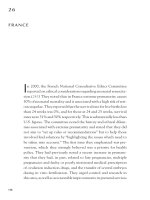
Figure 5
Before and after liposurgery, Cellulase Gold
1
, and Liposhape
TM
. Source: Photo courtesy of
M. Gasparotti.
Figure 6
The effects of Cellulase Gold
1
and Lipopanthy
TM
after four months. Source: Photo courtesy of
M. Gasparotti.
MEDICAL TREATMENT OF CELLULITE
&
151
a period of 60 days prior to superficial liposculpture, and continuing for two month after
the surgery.
Cellulase Gold
1
is a membrane flow activator and a dietary supplement based on
C. asiatica, Bladderwrack, M. officinalis, G. biloba, R. aculeatus, bioflavonoids, and Recapta-
cell
TM
. It increases the cell membrane fluidity for a better intracellular–extracellular exchange,
stimulates microcirculation, activates the anti–free-radical defences, contrasts vessal perme-
ability and enhances drainage of the excess of fluids in the tissue.
As a result, the use of Cellulase Gold
1
helps the transformation of fatty deposits
into metabolic energy, prevents the fibrous and sclerotic conditions of the connective tis-
sue, and helps reduce volumes and circumferences. In our opinion, the use of Cellulase
Gold
1
appears to optimize the outcome of three-dimensional liposuction and increases
overall patient compliance (Fig. 4–6) (17).
152
&
LEIBASCHOFF
&
REFERENCES
1. Vassallo C, Berardesca E. Efficacy of a multifunctional plant complex in the treatment of a
localised fat-lobular hypertrophy. Am J Cosmet Surg 2001; 18(4):203–208.
2. Bacci PA, Izzo M, Botta G, Mancini S. Valutazione dell’azione antiossidante di un prodotto
fitofarmacologico nelle sindromi cellulitiche, Podologia, Napoli, 2002.
3. Barracchini A, Franceschini N, Filippello M, et al. Leukocyanidines and collagenases: in vitro
enzyme inhibition activity. Clin Ther 1999; 150:275–278.
4. Costantini A, De Bernardi T, Gotti A. Clinical and capillaroscopic evaluation of chronic
uncomplicated venous insufficiency with procyanidins extracted from Vitis vinifera. Minerva
Cardioangiol 1999; 47(1–2):39–46.
5. Maffei Facino R, Carini M, Aldini G, Bombardelli E, Morazzoni P, Morelli R. Free radicals
scavenging action and anti-enzyme activities of procyanidines from Vitis vinifera. A mechanism
for their capillary protective action. Arzneimittelforschung 1994; 44(5):592–601.
6. Kleijnen J, Knipschild P. Ginko biloba. Lancet 1992; 340:1136–1139.
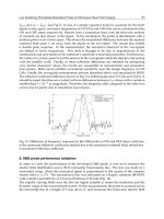
7. Pepe C, Rozza A, Veronesi G. The evaluation by video capillaroscopy of the efficacy of a
Ginkgo biloba extract with l-arginine and magnesium in the treatment of trophic lesions in
patients with stage-IV chronic obliterating arteriopathy. Minerva Cardioangiol 1999;
47(6):223–230.
8. Loiseau A, Mericer M. Centella asiatica and skin care. Cosmet Toilet 2000; 115:63–66.
9. Bonte F, Dumas M, Chaudagne C, Meybeck A. Influence of asiatic acid. Madecassic acid and
asiaticoside on human collagen I synthesis. Plant Med 1994; 60:133–135.
10. Vettorello G, Cerreta G, Derwish A, et al. Contribution of a combination of alpha and beta
benzopyrones, flavonoids and natural terpenes in the treatment of lymphedema of the lower
limbs at the 2nd stage of surgical classification. Minerva Cardioangiol 1996; 44:447–455.
11. Bolton T, Casley Smith J. The in vitro demonstration of proteolysis by macrophages and its
increase with Melilotus and Coumarine. Experentia 1975; 31:271–273.
12. Martignani A, Scondotto G. Terapia farmacologica del linfedema con estratto narurale del
meliloto. Gazzetta Medica Italiana 1997; 156(2):85–58.
13. Morris CA, Nicolaus B, Sampson V, Harwood JL, Kille P. Identification and characterization
of a recombinant metallothionein protein from a marine alga, Fucus vesiculosus. Biochem J
1999; 338:553–560.
14. Durig J, Bruhn T, Zurborn KH, Gutensohn K, Bruhn HD, Beress L. Anticoagulant fucoidan
fractions from Fucus vesiculosus induce platelet activation in vivo. Thromb Res 1997; 85:479–491.
15. Bacci PA, Izzo M, Botta G, Mancini S. Evaluacion de la accion antioxidante de productos fito-
farmacologicos empleados en los sindromes de celulitis con respecto al cigarro y a las hormo-
nas. Int J Aesthetic Surg 2003; 5:1.
16. Leibaschoff GH, Coll L, Desimone JG. Non-invasive assessment of the effectiveness of Cella-
sene in patients with oedematous fibrosclerotic panniculopathy (cellulitis): a double-blind pro-
spective study. Int J Cosmet Surg Aesthetic Dermatol 2001; 3(4):265–273.
17. Gasparotti M. Perspectives in plastic surgery. In: Three Dimensional Superficial Liposculpture
Reconstructive Plastic Surgery. Baltimore: Williams & Wilkins. Accepted for publication.
MEDICAL TREATMENT OF CELLULITE
&
153
9
Theory and Working Principles of Beautytek
1
in Cosmetic Medicine
Valerio Genitoni
Universita
`
di Urbino, Urbino, Italy
All of the conventional physical stimulation systems used in cosmetic medicine such as
laser, ultrasound (US), transcutaneous electrical nerve stimulation (TENS), and magnetic
fields share one common characteristic, i.e., they are unfocused. This means that they all
emit large amounts of energy in different ways in a repetitive fashion, following logical but
preestablished patterns.
With lasers, this energy takes the form of consistent light, while magnetotherapy uses
electromagnetic waves. US relies on sound waves, while TENS uses electrical stimulation.
These types of emissions share one characteristic. They are not suited to the requirements
of correction. They are therefore quantitatively and qualitatively unfocused. They are used
because they are backed by medical tradition, but unfortunately they produce very few
truly satisfying results in the correction of blemishes. Beautytek
1
encompasses the bio-
logical requirements that are unrecognized by conventional therapies. Time after time,
day after day our bodies require a whole range of different corrections. The instruments
and methods used in conventional physical therapies emit energies of different types
and characteristics in an imprecise way. This means that they have an unpredictable effect
on biological structures.
The most important information syst em in living biological systems is the neuronal
network. Biological systems have many ways of transferring information, but the most
important is probably via the neuronal network. Advances made in neurophysiological
research mean that we can now measure the chemical activity that occurs in individual
cells or in groups of cells. Many of the functions of the neuronal and muscle cells are
chemical in nature. Nonetheless, these functions produce changes in the electrical field,
which can be monitored using electrodes. The so-called electrical potentials help neuro-
physiologists to study cell function by directly measuring the chemical potential relating
to ion concentrations. These phenomena can be detected using special transducers such
as selective electrodes.
The source of the electrical signal is the individual neuronal or muscular cell.
However, such cells do not function alone; they function in large groups. The cumulative
effects of such cellular activity result in the generation of an electrical field that propagates
155
in the conduction volume, which consists of various types of tissues. Thus, the activity of
the muscle or certain neuronal networks can be indirectly improved by applying electrodes
to the skin. This type of information is not simple to collect, and the electrodes must be
properly positioned on the skin. Even then, it is very difficult to analyze the information
process involved. The results of all of the neuronal and muscular activity in unknown
anatomical sites are transmitted using a homogenous medium. The electrical signals moni-
tored on the surface of the skin are of enormous clinical and physiological importance.
Electroencephalograms, electrocardiograms, electromyograms, and other signals are
already being used in clinical medicine to measure the acti vity of muscular and neuronal
systems. The way in which the information supplied by these systems is interpreted is
based principally on statistical experience built up over the years. The plasma cell mem-
brane is a medium that separates the intercellular fluids from the extracellular ones. These
two types of fluids have different ions concentrations, and the membrane has different
levels of permeability for the different ions dissolved in the solution. A membrane poten-
tial is generated by the ion transfer, principally as a function of diffusion mechanisms. If
we take into consideration the effects of the three main ions alone, potassium, sodium, and
chlorine, we obtain the membrane potential via the following equation:
E ¼ ln RTP
X
½K
þ
þP
Na
½Na
þ
þP
C
½Cl
À
FP
X
½K
þ
þP
Na
½Na
þ
þP
C
½Cl
À
where R, T and F are the universal gas constant, the absolute temperature, and Faraday’s
constant, respectively; P
X
is the permeability of the remaining membrane to X ions and X
o
and X
i
are the concentrations of X ions in the extracellular and intracellular fluids. The
remaining membrane potential calculated in this way is approxim ately 80 mV; the interior
of the cell becomes negative in relation to the exterior.
Some membranes have different levels of excitability. When the membrane is excited
by an electrical or mechanical signal or by a chemical stimulus, its permeability changes in
relation to the ion transfer. These changes in turn cause an increase in the remaining
potentials of the membrane, which become positive for a short period of time and then,
when the membrane changes its sign, return to the resting potential.
The type and duration of the action potential differs from one cell type to another.
The membrane only becomes excited when the stimulus exceeds a threshold level of
around 20 mV. Once this threshold has been exceeded and the action potential appears,
there is also a change in the sensitivity of the threshold. After the potential has been acti-
vated, there is a period of time (approximately 1 or 2 msec) during which the threshold
becomes infinite. This period is called the period of total refractoriness during which no
new action potential can be activated. The threshold thus returns to its nominal value
in accordance with the computation of the decay function. The period during which the
threshold falls to its normal level is known as the relative refractoriness period. In that per-
iod, a new action potential can be activated by a stimulus that is sufficiently large to cross
the relatively high threshold.
The source of electrical signals is the action potential generated by individual
neurons and muscle fibers.
The current density generated by the membrane activity can give rise to a change in
the surrounding medium. The surrounding tissue in which the current chan ge took place is
called the conduction volume. In many clinical and neurophysiological applications, we
can monitor the conduction volume field but not the bioelectrical sources that generate it.
156
&
GENITONI
This is definitely the case when electrodes are attached to the skin to monitor the electrical
activity of the heart and brain. It is therefore extremely important to be able to precisely
deduce the underlying bioelectric source producing the conduction volume activity.
This operation involves a very complex computation, especially if the characteristics
of the biological medium are taken into consideration.
Mathematical models of flow fields of currents in the conduction volumes have been
developed with varying degrees of success.
Beautytek
1
creates a loop—a closed circuit—with the area to be stimulated. If, for
example, the two electrodes are situated in a position that will permit a reading of the sys-
tem in an inflamed area, the machine performs a very fast physiochemical analysis of the
tissue once the circuit is closed. Using a series of algorithms, Beautytek
1
reads and inter-
prets the physiochemical situation and then makes the necessary correction. Even as the
correction is being made, the syst em is already moving to the next reading so that the
closed system ensures that the machine can take hold of the tissue and bring it to a differ-
ent physiochemical state of equilibrium. Because the system’s algorithms are aimed at
bringing about tissue equilibrium, the electronic system cannot cause any damage even
though the goal is to br ing about a biological change. Once a state of equilibrium has been
reached in the area of the tissue under examination, the machine stops the treatment, so it
cannot overstimulate or understimulate it. The stimulation is always by definition the level
required to reach equilibrium.
Instant by instant, several hundred times a second, the machine takes readings, inter-
prets the data, and makes a correction. Then it starts from the beginning again with a
reading of the tissue modifications obtained, calculates, and corrects once again. It inter-
venes in a cyclical and interactive fashion so that the tissue is forced to modify itself and all
of its physiochemical compensation systems and to establish a new equilibrium.
Thus, the polarization of the chemical–physical constituents of the tissue is modified;
this is an expression of the chain of overlaps of substances commonly involved in
biological and bioelectric processes.
BEAUTYTEK
1
IN COSMETIC MEDICINE
&
157
10
Topical Management of Cellulite
Doris Hexsel
School of Medicine, University of Passo Fundo, Passo Fundo,
Rio Grande do Sul, Brazil
Debora Zechmeister do Prado
Doris Hexsel Dermatologic Clinic, Porto Alegre, Rio Grande do Sul, Brazil
Jaggi Rao
American Academy of Cosmetic Surgery Fellow Trainee and La Jolla Spa MD, La Jolla,
California, U.S.A.
Mitchel P. Goldman
University of California, San Diego, California and La Jolla Spa MD, La Jolla,
California, U.S.A.
&
INTRODUCTION
Cellulite is the unsightly skin dimpling that is frequently found on the thighs and buttocks
of women. Approximately 85% of post-adolescent women have some degree of cellulite
(1–3). Many allegedly successful cosmetic and medical treatments show little effect in
improving cellulite, and none of them has been shown to cause its complet e disappearance.
The anatomy and pathophysiology of cellulite are poorly understood. A review of the lit-
erature demonstrates a paucity of studies to validate currently popular theories and treat-
ments. However, a thorough understanding of cellulite pathophysiology is necessary for
successful treatment modalities to be developed. Until this is clearly delineated, accepting
a less-than-ideal outcome from treatment of this unwanted skin condition will continue to
be necessary.
This chapter describes the role of topical agents in reducing the appearance of cellu-
lite. The effect of supplementary aids, such as occlusive garme nts, will be addressed as
well. The various therapies are presented with a focus on how the therapy addresses
current concepts of the origin and nature of cellulite.
159
&
DEFINITION AND NATURE OF CELLULITE
The term ‘‘cellulite’’ is used in modern times to describe the dimpled or puckered skin
of the posterior and lateral thighs and buttocks seen in both trim and overweight women.
The appearance is often described as resembling the surface of an orange peel or that of
cottage cheese. The condition is be st described by Goldman as a normal physiologic state
in post-adolescent women, which maxi mizes adipose retention to ensure adequate caloric
availability for pregnancy an d lactation (4). Adipose tissue is also essential for nutrition,
energy, support, protection, and thermal insulation (5).
At the histological level, cellulite is the result of localized adipose deposits and edema
within the subcutaneous tissue. In women, fascial bands of connective tissue are orient ed
longitudinally and extend from the dermis to the deep fascia. These bands form fibrous
septa, which segregate fat into channels resembling a ‘‘down quilt’’ or mattress, and the
subcutaneous fat is projected superficially into the reticular and papillary dermis. As
the fat layer expands, the perpendicular connective tissue remains fixed and anchored
to the underlying tissue, creating a superficial puckered appearance of the skin (5–8). Fatty
acids are then believed to be modified through peroxidation by free radicals. These events
are thought to contribute to the worsening of local microcirculation by disrupting venous
and lymphatic drainage. This skin phenomenon is rarely found in men because the connec-
tive tissue in male s is not normally arranged vertically, but rather in a crisscrossing pattern
that is gender-typical for the skin of the thighs and buttocks (5,7).
&
PATHOPHYSIOLOGIC MECHANISMS OF
CELLULITE FORMATION
Hormones, specifically estrogens and androgens, are thought toinfluence the formation of cel-
lulite. Estrogen is known to stimulate lipogenesis and inhibit lipolysis, resulting in adipocyte
hypertrophy (9). This may explain the onset of cellulite at puberty, the condition being more
prevalent in females, and the exacerbation of cellulite with pregnancy, nursing, menstruation,
and estrogen therapy (oral contraceptive use and hormone replacement) (9). The opposite
seems true for men. From the limited number of studies involving men, it is hypothesized that
the combination of gender-specific soft tissue histology at the cellulite-prone anatomic sites,
with a relatively lower circulating estrogen level, may be responsible for the lower incidence
of cellulite in males (10,11). Although not proven, it is possible that circulating androgens
may have an inhibitory effect on cellulite development by contributing to a different pattern
of adipose tissue storage (that is, more on the trunk than on the buttocks and thighs).
Adipose tissue is very vascular, leading to the theory that cellulite may worsen in pre-
disposed areas where circulation and lymphatic drainage have be en decreased, possibly
due to local injury or inflammation. In response to impairment of microvascular circula-
tion, there is increased microedema within the subcutaneous fat layer, causing further
stress on surrounding connective tissue fibers and on the accentuation of skin irregularities
(2,4). Many of the currently accepted cellulite therapies target deficiencies in lymphatic
drainage and microvascular circulation. The lipids within adipocytes are derived from
plasma-circulating lipoproteins. In a dynamic process, the stored fat is hydrolyzed and
eliminated again to the plasma as free fatty acids and glycerol. Various enzymes including
160
&
HEXSEL ET AL.
insulin and cyclic adenosine monophosphate (cAMP) participate in this process. In parti-
cular, triglyceride lipase is very important in the promotion of lipolysis. This enzyme is
activated by adenylyl cyclase stimulation by means of an antagonist effect. This inhibitory
process causes triacylglycerol hydrolysis and releases free fatty acids and glycerol into the
interstitial space and plasma.
On the surface of adipocytes, there are receptors that promote the storage of fat and lipo-
genesis, such as neuropeptide Y and peptide YY. Conversely, other surface receptors promote
the elimination of fat and lipolysis, such as b1andb2. Manipulation of these surface enzymes
by topical medications is a new mechanism by which cellulite development can be controlled.
&
TOPICAL MANAGEMENT
When using topical treatments to reduce the appearance of cellulite, the concentration and
pharmacokinetics of the active drugs as well as the nature of the vehicle must be consid-
ered. Vehicles can be in the form of gels, ointments, foams , creams, and lotions, all of
which aim to efficiently deliver active product to the skin. Factors that affect the clinical
response to treatment are: (i) the interaction of the drug with the vehicle and the skin, (ii)
the method by which the drug is applied, and (iii) other biological and environmental fac-
tors (12–14). The main barrier to drug penetration is the stratum corneum, the cornified
outermost layer of the epidermis. Formulations for topical use may include ‘‘skin enhan-
cers,’’ which significantly increase cutaneous penetration when included in the formula-
tion. Skin enhancers can be common solvents (water, alcohol, and methyl alkyl
sulphoxide) or surfactants. They may also be phospholipid molecules called phytosomes,
which, when attached to the active drug, increase their lipid solubility. A novel percuta-
neous delivery system utilizes liposomes, which are specially designed lipid vesicles that
are filled with active medication (15,16). Topical anticellulite preparations can be divided
into four major groups according to their proposed mechani sm of action (Table 1).
1. Agents that increase microvascular flow.
This includes most of the active ingredients in cellulite treatments. They are included
to increase microvascular flow and lymphatic drainage, which is thought to play a
role in cellulite pathogenesis.
2. Agents that reduce lipogenesis and promote lipolysis.
With the goal of reducing the size and volume of adipocytes, decreased tension
on surrounding connective tissue is thought to decrease the clinical appearance of
puckering.
3. Agents that restore the normal structure of the dermal and subcutaneous tissue.
By thickening the dermis or preventing fat herniation into superficial tissue, the
appearance of cellulite may be reduced.
4. Agents that prevent or destroy free-radi cal formation.
It is belie ved that free radicals modify free fatty acids by peroxidation, contributing
to the availability of lipids for cellulite formation. Free radicals may also damage
elements of the microcirculation, further assisting cellulite development.
The following discussion summarizes the current knowledge of individual and com-
bination topical therapies used to reduce cellulite.
TOPICAL MANAGEMENT OF CELLULITE
&
161
AGENTS THAT INCREASE MICROVASCULAR FLOW
Drugs that act on the microcirculation of the skin, include the ivy and Indian chestnut
vegetable extracts, which are rich in saponins, Gingko biloba , and rutin, whi ch contain bio-
flavonoids. These compounds decrease capillary hyperpermeability and increase venous
tone by stimulation of proline hydroxylase and inhibition of prostaglandin E
2
. These
agents also decrease platelet aggregation, thereby inhibiting microthrombus formation.
Studies using oscillometry, Duplex ultrasound, hemodynamic methods, and capillaro-
scopy have demonstrated that G. biloba extract is anti-edematous and improves venous
return and arterial circulation (17,18). This is accomplished by decreasing capillary hyper-
permeability and is employed as an active agent in many topical anticellulite formulations.
G. biloba is a member of the Ginkgoaceae family. The leaf extracts contain sub-
stances such as flavonoids (quercetin, campherol epicathecol derivates, etc.), biflavones
(ginkgetin), and terpenes (ginkgolide B) among others (19) . G. biloba is used in the treat-
ment of cellulite due to its several effects on peripheral circulation, such as reducing blood
Table 1
Topical Therapies for Cellulite, Based on Proposed Mechanism of Action
Agents that increase microvascular flow
Ivy
Indian or horse chestnut (Aesculus hippocastanum)
Ginkgo biloba
Rutin
Pentoxyfylline
Butcher’s broom (Ruscus aculeatus)
Asiatic centella
Silicium
Chofitol or artichoke (Cynara scolymus)
Common ivy (Hedera helix)
Ground ivy (Glechoma hederaceae)
Sweet clover (Melilotus officinalis)
Red grapes (Vitis vinifera)
Papaya (Carica papaya)
Pineapple (Ananas sativus, Ananas comosus)
Agents that reduce lipogenesis and promote lipolysis
Methylxanthines (theobromine, caffeine, aminophylline, theophylline)
Beta-adrenergic agonists (isoproterenol, adrenaline)
Alpha-adrenergic antagonists (yohimbine, piperoxan, phentolamine, dihydroergotamine)
Agents that restore the normal structure of the dermal and subcutaneous tissue
Retinol (vitamin A)
Ascorbic acid (vitamin C)
Bladderwrack (Fucus vesiculosus)
Agents that prevent or destroy free-radical formation
Alpha-tocopherol (vitamin E)
Ascorbic acid (vitamin C)
Gingko biloba
Red grapes (Vitis vinifera)
162
&
HEXSEL ET AL.
viscosity. The terpene s, especially ginkgolide B, inhibit the platelet-activating factor. They
increment red blood cell deformability, diminish vascular permeability, and improve vas-
cular wall tonus. All these actions improve the microcirculation. The methylxanthine
‘‘pentoxyfylline’’ improves microcirculatory perfusion through its effect on hemato logical
factors such as erythrocyte shape, plate let aggregation, and plasma fibrinogen concentra-
tion. It also has immunomodulatory activity. It has been utilized for peripheral vascular
disease treatment with significant benefit. For the treatment of cellulite (20), it has been
used transdermally with other drugs, making its evaluation difficult.
Butcher’s broom (R. aculeatus) is a potent venous vasoconstrictor and has the ability
to decrease edema. It acts as an alpha-adrenergic receptor agonist of the smooth muscle
of veins and therefore reduces vascular permeability. The main active ingredients are
saponins, ruscogenin, and neororuscogenina (21).
Asiatic centella extract, both topically and systemically, has been used for treating cel-
lulite and has been demonstrated through capillaroscopy to have an effect on the microcir-
culation in patients with chronic venous insufficiency, who were treated for venous ulcers
(22). Chemically consisting of 40% asiaticosideo, 30% madecassic acid, and 30% Asiatic
acid, topical and systemic Asiatic centella have been shown to be harmless by toxicity tests.
Asiatic centella also acts in vitro on fibroblasts, stimulating colla gen and mucopolysacchar-
ide synthesis. This compound also acts as an anti-inflammatory agent, which may be ben-
eficial in protecting dermal and subcutaneous structures from inflammatory cell injury (19).
Silicium is a structural element of connective tissue, which regulates and normalizes
cellular metabolism and cellular division. In the microcirculation, it modifies venous capil-
lary and lymphatic permeability and, in the fatty tissue, it stimulates cAMP synthesis as
well as triglyceride hydrolysis, likely activating adenylcyclase in the cellular membrane
(23). For this reason, it has been used in topical cellulite treatment products.
Chofitol or artichoke (Cynara scolymus) is a member of Arteraceae family, and it is
found in northern Mediterranean soil. Its principal active chemical constituents are numer-
ous enzymes, cynarin, ascorbic acid, caffeoylquinic acid derivates, and flavonoids. It has an
antiedematous and diuretic effect, as well as a stimulating effect on the circulation (19).
Common ivy (Hedera helix) is a phytomedicine that grows in places with rich soil, sun,
or shade. The parts of the plant used are dried leaves and stems. The leaves have flavonoids
such as rutosid and rutinosid, and saponins such as hederin, hederacosid, and hederagenin
(19,24). The fruits have saponins, especially hederin, and the trunk has gomoresins and sapo-
nins. All saponins improve venous and lymphatic drainage and reduce edema. One of these
compounds, hederin, also has an analgesic and anti-inflammatory effect. It has vasoconstric-
tive and antiexudative properties and can also reduce capillary permeability. It increases cir-
culation and therefore assists drainage of the infiltrated tissue and reduces inflammation.
Ground ivy (Glechoma hederaceae) is from the Lamiaceae family and is also used in
anticellulite treatment. The main constituents are flavonoids, triterpenoids, and phenolic
acids. It grows in moist soil in Europe, especially the Caucasus, and in North America
(19). Both types are used in concentrations of 2%.
Indian or horse chestnut (Aesculus hippocastanum) belongs to the Hippocastanaceae
family. The seeds and the shells are used in the elaboration of the standard extract (25).
The active ingredients contained in the seeds are triterpenoid saponins, such as escin
and aesculin, and flavones, coumarins, and tannins (25), with anti-inflammatory and anti-
edematous properties (26). Escin is the principal component of horse chestnut, and it has
the capacity to reduce lysosomatic enzyme activity by up to 30%, probably by stabilizing
TOPICAL MANAGEMENT OF CELLULITE
&
163
the cholesterol content of the lysosome membranes, thus reducing enzyme release and
capillary permeability. The recommended concentration is 1% to 3%.
Sweet clover (Melilotus officinalis) is a plant from the Fabaceae family. The active
ingredient is contained in the flowers and leaves . One of the components of this botanical
extract is coumarin, which reduc es lymphatic edema and diminishes capillary permeability
(27). It is usually recommended to patients with chronic venous insufficiency and lympha-
tic congestion—conditions that are believed to be associated with cellulite. The recom-
mended concentration is 2% to 5% (27).
Red grapes (Vitis vinifera) have procianidins that increase the permeability of
lymphatic and microarterial vessels (27). In topica l products, the essential oil is used at
a concentration of 2% to 7% (27).
The fruits and leaves of papaya (Carica papaya) and pineapple (Ananas sativus, Ana-
nas comosus) have anti-inflammatory and anti-edematous effects (28). They contain the
proteolytic enzymes papain and bromelain, respectivel y. These plants are originally from
tropical America and were introduced to southern Florida. The recommended concentra-
tion is 2% to 5%. Extracts from the fruits and leaves of pineapple (A. sativus, A. comosus)
may be associated with the so-called ‘‘pineapple itch,’’ a contact dermatitis due to a mite
that infests pineapple plantations (29).
AGENTS THAT REDUCE LIPOGENESIS AND
PROMOTE LIPOLYSIS
Drugs that have a lipolytic effect on adipose tissue include the methylxanthines (theobromine,
caffeine, aminophylline, and theophylline). These act through phosphodiesterase inhibition
and are the most common active ingredients in commercial anticellulite formulations (30).
The most useful and safest methylxanthine is caffeine, normally used at a concentration of
1% to 2%. It offers good skin penetration and is therefore rapidly absorbed, leading to rapid
action. Caffeine acts directly on adipocytes, promoting lipolysis through the inhibition of
phosphodiesterase by augmentation of cAMP (31). All methylxanthines activate the enzyme
triglyceride lipase and transform triglycerides into free acids and glycerol. Caffeine also has a
stimulating effect on the cutaneous microcirculation. Table 1 lists botanical sources of methyl-
xanthines, extracts of which are very common in anticellulite agents.
Beta-adrenergic agonists such as isoproterenol and adrenaline, and alpha-adrenergic
antagonists such as yohimbine, piperoxan, phentolamine, and dihydroergotamine have
also shown the ability to cause lipolysis. In vitro studies have shown that both the methyl-
xanthines and the beta-adrenergic agonists stimulate lipolysis and a reduction in adipocyte
size through an increase in cAMP inhibition of phosphodiesterase (32,33).
Greenway and Bray demonstrated a statistically significant reduction in the anthropo-
metric measurement of the medial thigh by a double-blind placebo-controlled study,
which utilized topical isoproterenol (a beta-adrenergic agonist), aminophylline (a methyl-
xanthine with phosphodiesterase inhibitory properties), and yohimbine (an alpha-adrenergic
antagonist) (34). The reduction in thigh measurement was greatest when all active
drugs were used together, three to five times a week for four weeks’ duration. Of the results
obtained when the three agents were used separately, the best results were obtained with use
of aminophylline.
The effects of methylxanthines can be enhanced by coenzyme A and the amino acid
l-carnitine (23). These agents work by stimulating the mobilization and destruction of free
164
&
HEXSEL ET AL.
fatty acids and inducing their active transport through the membranes of the mitochon-
dria. This is important because free fatty acids may cause saturation of the system, leading
to negative feedback of lipolysis. Also, the mobilization and destruction process of free
fatty acids generates adenosine triphosphate, which increases lipase activity, enhancing
hydrolytic breakd own of triglycerides.
Yohimbe (Corynanth yohimbe , Pausinystalia yohimbe, and Rauwolfia serpentine)and
alpha yohimbe are alkaloid derivatives extracted from the leaves, shell, and roots of
Rubiaceas and Apocynaceas (19). They are adrenergic blockers capable of stimulating
the catabolism of fat due to the presence of alkaloids that act directly on the fat cells (19).
AGENTS THAT RESTORE THE NORMAL STRUCTURE OF THE
DERMAL AND SUBCUTANEOUS TISSUE
Retinol (vitamin A) and the retinoids have been evaluated for their effectiveness in the
treatment of cellulite. Topical retinoic acid and related vitamin A derivatives have been
used to stimulate circulation, decrease the size of adipocytes, and increase collagen deposi-
tion in the dermis (9,35). Based on the capacity of all-trans-retinoic acid (tretinoin) to pro-
mote the synthesis of glycosaminoglycans in normal skin and increase the deposition of
collagen in the photodamaged dermis, Kligman et al. proposed the use of topical retinol
to improve cellulite (35). The premise for its use in cellulite treatment is that topical retinol
can be used to increase the thickness and firmness of the dermis, disguising the effect of the
superficial fat histologically present immediately beneath it. The use of retinol was pro-
posed instead of tretinoin due to its better tolerability and the evidence that retinol is meta-
bolized to retinoic acid in the skin. In the study by Kligman et al., 19 patients completed a
study of retinol 0.3% versus placebo applied to opposite lateral thighs twice daily for six
months’ duration. Of the 19 patients, twelve demonstrated greater clinical improvement
on the actively treated side on clinical evaluation and laser Doppler velocimetry.
Pierard-Franchimont et al. demonstrated that topical retinol treatment might
improve the tensile properties of skin in a beneficial way for cellulite care (36). In a rando-
mized, placebo-controlled study combining the use of retinol with gentle massage, skin elas-
ticity was increased by 10.7% while viscosity was decreased by 15.8% at retinol-treated sites.
The main retinol-related change consisted of a two- to fivefold increase in the number of
factor XIIIa þ dendrocytes both in the dermis and in the fibrous strands of the hyp odermis.
This is all indicative of increased skin firmness and smoothened appearance of the surface.
In addition , some topical ingredients such as vitamin C may help by stabilizing collagen
and/or stimulating collagen deposition (3,4,9).
Bladderwrack (Fucus vesiculosus) is a brown marine algae that contains sulfated
polysaccharides, iodine compounds, and alginic acid. It is reported to produce contraction
of the dermal connective tissue through the increased expression of integrin molecules (19).
Increasing dermal density is the likely mechanism by which this agent improves cellulite. It
also has a stimulating effect on vascular flow.
AGENTS THAT PREVENT OR DESTROY FREE-RADICAL
FORMATION
Vitamins such as ascorbic acid and vitamin E may work as antioxidants, protecting dermal
and subcutaneous cell membranes from free-radical toxicity. This, in turn, may prevent
TOPICAL MANAGEMENT OF CELLULITE
&
165
and allow for repair of fat herniation. Also, vitamins may improve microcirculati on, the
impairment of which may be an etiological factor in cellulite formation. G. biloba also
has flavonoids that act as antioxidants and anti-inflammatory agents (19). Red grapes
(V. vinifera) are rich in tannins that are antioxidants that diminish lipid peroxidation (27).
COMBINATION AGENTS
It is likely that the future of topical cellulite therapy will consist of agents that contain mul-
tiple ac tive ingredients. In addition to providing different mechanisms of action directed
toward the same goal of reducing cellulite, the different constituents may work synergis-
tically to yield results better than those obtained when each component is used alone.
Unfortunately, there are very few good studies in the literature that document the use
of these combination products.
Bertin et al. performed a double-blind evaluation of an anticellulite product and
showed it to be more effe ctive than placebo in reducing cellulite (37). This product com-
bines retinol with a microencapsulated time-release mechanism to treat cellulite. The com-
pound contains caffeine to stimulate the lipolysis and prevent fat accumulation, esculoside
to improve local microcirculation, Asiatic centella as an anti-inflammatory agent, and
l-carnitine to stimulate free fatty acid transport and breakdown. Efficacy parameters
included cellulite appearance before and after treatment, histology, cutaneous flowmetry,
and skin mechanical characteristics. As mentioned, retinol has been shown to increase der-
mal thickness. The product also contains ruscogenine, which inhibits elastase activity,
allowing recovery of extracellular matrix integrity that contributes to the thickening of
the dermis and the masking of cellulite.
In a recent multicenter, randomized, placebo-controlled trial involving the testing of
a combination anticellulite cream, subjects applied cream on a nightly basis with occlusion
on the posterolateral region of one of the thighs. Overall, 62% (21/34) noticed an improve-
ment in their cellulite, with 62% (13/21) reporting greater improvement in the thigh that
was treated with the active product. The average measured decrease in thigh circumference
was 1.9 cm (range: 0.1–4.5 cm) with active prod uct, and 1.3 cm (range: 0.1–3.0 cm) with
placebo. Upon review of the pre- and poststudy photographs, dermatologist evaluators
found thighs treated with active product to show greater improvement than thighs treated
with placebo in 68% of subjects. This product contained several active ingredients includ-
ing caffeine, green tea extra ct, black pepper seed extract, citrus extract, ginger root extract,
cinnamon bark extract, and capsicum annum resin (41).
A novel agent named ‘‘Bio-actif’’ consists of a compound containing neuropeptide Y
and peptide YY (38). These agents are known to participate in the metabolism of fat with
lipogenic effects on adipocytes. Bio-actif is a topical gel of these neuropeptides, combined
with green tea, ivy, aloe vera, wheat protein, and other agents, and has shown to decrease
fat herniation responsible for the appearance of cellulite.
EXTERNAL AIDS TO TOPICAL THERAPY
Supplemental techniques such as mass age and fomentation have been shown to assist
in topical medication delivery into the skin and further reduce the appearance of
cellulite (36). Goldman describes the us e of a synthetic bioceramic-coated neoprene
166
&
HEXSEL ET AL.
garment to stimulate lymphatic and vascular flow that assisted in improving cellulite (4).
This is depicted in Figure 1.
Recently, a double-blinded, randomized, placebo-controlled trial examined the effect
of this garment for the treatment of cellulite (39). In this study, 17 subjects were evaluated
for cellulite reduction using an anticellulite cream and occlusive garment on only one
thigh. Four weeks later, 76% of subjects noticed an improvement in their cellulite, with
54% reporting greater improvement in the thigh that was subjected to garment occlusion.
Average thigh circumference reduction was 1.3 cm in the occluded thigh, and 1.1 cm in the
nonoccluded thigh. The evaluators who were dermatologists found an overall impr ove-
ment in cellulite in 65% of treated legs with occlusion and 59% of treated legs without
occlusion. Furthermore, the evaluators found the occluded thighs to show greater
improvement than the nonoccluded thighs in 65% of subjects. This study demonstrated
that although the results obtained from its use are modest, occlusion by compression
garments is beneficial in assisting topical agents to improve cellulite. In addition to
potentiating topical drug delivery through occlusion, the warmth creat ed by the garment
likely improves microcirculation, which maybe an etiological factor in cellulite development.
Figure 1
Bioceramic-coated neoprene shorts, worn
after topical application of an anticellulite
product to the posterior and lateral regions of
the thighs to provide greater penetration into
the skin by occlusion.
TOPICAL MANAGEMENT OF CELLULITE
&
167
ADVERSE EVENTS
Physicians need to be informed about the great range in efficacy among purported treat-
ments for cellulite, if for no other reason than to avoid untested products. Sainio et al.
investigated 32 anticellulite products, mostly botanicals and emollients, each containing
an average of 22 ingredients (3). It was found that one-fourth of the substances used have
been shown to cause allergy, including isothiazolinones and dibromoglutaronitrile. This
indicates that despite the fact that most topical cellulite therapies are acceptably safe to
many consumers, the risk of adverse events sho uld be taken into account. There are some
reports in the literature, of cases of hypersensitivity to ginkgo contained in anticellulite
products(3). There are also citings of allergic reactions in patients who used topical pro-
ducts containing ivy (3). The leaves of this plant are considered poisonous when ingested,
because they contain arsenic oxide. Hypersensitivity has been reported in users of products
containing escin, the principal component of horse chestnut (40). Cases of contact derma-
titis on the hands have been reported, resulting from squeezing the fruit to obtain the juice,
which contains several acids such as oxalic, malic, tartaric, and racemic (29).
&
CONCLUSION
The multifactorial etiology and nature of cellulite make it a particularly difficult condition
to treat. To better serve patients, the search for a complete cure for cellulite should be
avoided. Rather, the aim of treatment should be to minimize the physical aspects of cellu-
lite and prevent its progression by safe, cost-effective means. Topical treatments may
improve the appearance of cellulite and represent a reasonable, affordable modality to
reduce the severity of this unwanted condition. It is reasonable to speculate that many
of these products may also have a role as a preventive measure. The supplemental use
of external aids such as compressive bandages or garments to combine the effects of
compression and enhanced penetration of topical agents has shown to be useful.
168
&
HEXSEL ET AL.
&
REFERENCES
1. Draelos ZD, Marenus KD. Cellulite—etiology and purported treatment. Dermatol Surg 1997;
23:1177–1181.
2. Cellulite meltdown. Harv Womens Health Watch 1998; 5:7.
3. Sainio EL, Rantanen T, Kanerva L. Ingredients and safety of cellulite creams. Eur J Dermatol
2000; 10:596–603.
4. Goldman MP. Cellulite: a review of current treatments. Cosmet Dermatol 2002; 15:17–20.
5. Querleux B, Cornillon C, Jolivet O, Bittoun J. Anatomy and physiology of subcutaneous
adipose tissue by in vivo magnetic resonance imaging and spectroscopy: relationships with
sex and presence of cellulite. Skin Res Technol 2002; 8:118–124.
6. Another cellulite remedy. Harv Womens Health Watch 1999; 6:7.
7. Pierard GE, Nizet JL, Pierard-Franchimont C. Cellulite: from standing fat herniation to hypo-
dermal stretch marks. Am J Dermatopathol 2000; 22:34–47.
8. Pellicier F, Andre P, Schnebert S. The adipocyte in the history of slimming agents. Pathol Biol
2003; 51:244–247.
9. Rossi ABR, Vergnanini AL. Cellulite: a review. J Eur Acad Dermatol Venereol 2000; 14:251–262.
10. Nurnberger F, Muller G. So-called cellulite: an invented disease. J Dermatol Surg Oncol 1978;
4:221–229.
11. Rosenbaum M, Prieto V, Hellmer J, et al. An exploratory investigation of the morphology and
biochemistry of cellulite. Plast Reconstr Surg 1998; 101:1934–1939.
12. Addicks WJ, Weiner ND, Curl RL, Flynn GL. Drug delivery from topical formulations: the-
oretical prediction and experimental assessment. In: Hadgraft J, Guy RH, eds. Transdermal
Drug Delivery: Developmental Issues and Research Initiatives. New York: Marcel Dekker,
1989:221–224.
13. Hadgraft J. Skin penetration enhancement. In: Hadgraft J, Walters KA, eds. Predication of
Percutaneous Penetration. New York: Marcel Dekker, 1993:138–148.
14. Riviere JE. Biological factors in absorption and permeation. In: Zatz JL, ed. Skin Permeation:
Fundamentals and Application. Wheaton: Allured Publishing Corporation, 1993:113–125.
15. Zatz JL. Modification ofskin permeation bysolvents and surfactants. In: Zatz JL, ed. Skin Permea-
tion: Fundamentals and Application. Wheaton: Allured Publishing Corporation, 1993:127–162.
16. Seiller M, Orecchioni AM, Vaution C. Vesicular systems and multiple emulsions in cosmetol-
ogy. In: Baran R, Maibach HI, eds. Cosmetic Dermatology. Baltimore: Williams and Wilkins,
1994:27–35.
17. August M, Clostre F. Effects of an extract of ginkgo biloba and diverse substances on the pha-
sic and tonic components of the contraction of an isolated rabbit aorta. Gen Pharmacol 1983;
14:277–285.
18. Bauer U. Six-month double-blind randomized clinical trial of ginkgo biloba extract versus pla-
cebo in two parallel groups in patients suffering from peripheral arterial insufficiency. Arznein
Forsch 1984; 34:716–723.
19. Amelio FS. Botanicals: A Phytocosmetic Desk Reference. Boca Raton, London, New York,
Washington, D.C.: CRC Press, 1999.
20. Samlaska CP, Winfield EA. Pentoxifylline. J Am Acad Dermatol 1994; 30:603–621.
21. Rubanyi G, Marcelon G, Vanhoutte PM. Effect of temperature on the responsiveness of cuta-
neous elicited by Ruscus aculeatus. Gen Pharmacol 1984; 15(5):431–434.
TOPICAL MANAGEMENT OF CELLULITE
&
169
22. Lawrence JC. The morphological and pharmacological effects of asiaticoside upon skin in vitro
and in vivo. Eur J Pharmacol 1967; 1:414–424.
23. di Salvo RM. Controlling the appearance of cellulite: surveying the cellulite reduction effective-
ness of xanthines, silanes, CoA, L-carnitine and herbal extracts. Cosmet Toilets 1995; 110:50–59.
24. Carini M, Maffei FR, Brambills A, Stefani R, Scesa C. Anti-hyaluronidase and anti-elastase
activity of saponins from Hedera helix, Aesculus hippocastanum and Ruscus aculeatus: an expla-
nation of their efficacy in the cosmetic treatment of liposclerosis. Phyto Pharm 1998; 36:613–623.
25. Fluck H. Medicinal Plants. New York: W. Foulsham & Co, 1988.
26. Weiss RF. Herbal medicine. In: Meuss AR, ed. Lehrbuch der Phytotherapie. 6th German ed.
London: The Bath Press, 1986.
27. Manufacture Information–Croda (Crodarom S.A.). Yorkshire, U.K.:Croda International Ilc, 2002.
28. Van Rietschoten K. Plants with anti-inflammatory action. Br J Aromather 1990; 1(Autumn/
Winter).
29. Behl PN, Capitanin RM, Bedi BMS, Gupta S. Skin Irritant Sensitizing Plants Found in India.
New Delhi: PN Behl, 1966.
30. Collis N, Elliot LA, Sharpe C, Sharpe D. Cellulite treatment: a myth of reality: a prospective
randomized, controlled trial of two therapies, endermologie and aminophylline cream. Plast
Reconstr Surg 1999; 104:1110–1114.
31. Portad G, Laugel C, Baillet A, Schaefer H, Marty JP. Quantitative HPLC analysis of sunsc-
reens and caffeine during in vitro percutaneous penetration studies. Int J Pharm 1999;
189:249–260.
32. Smith U, Hammersten J, Bjorntorp P, Kral JG. Regional differences and effect of weight reduc-
tion on human fat cell metabolism. Eur J Clin Invest 1979; 9:327–332.
33. Motulsky HJ, Insel RA. Adrenergic receptors in man: direct identification, physiologic regula-
tion and clinical alterations. New Engl J Med 1982; 308:18–29.
34. Greenway FL, Bray GA. Regional fat loss from the thigh in obese women after adrenergic
modulation. Clin Ther 1987; 9:663–669.
35. Kligman AM, Pagnoni A, Stoudemayer T. Topical retinol improves cellulite. J Dermatol Treat
1999; 10:119–125.
36. Pierard-Franchimont C, Pierard GE, Henry F, Vroome V, Cauwenbergh G. A randomized,
placebo-controlled trial of topical retinol in the treatment of cellulite. Am J Clin Dermatol
2000; 1:369–374.
37. Bertin C, Zunino H, Pittet JC, et al. A double-blind evaluation of the activity of an anti-cellulite
product containing retinol, caffeine, and ruscogenine by a combination of several non-invasive
methods. J Cosmet Sci 2001; 52:199–210.
38. Hernandez-Perez, et al. Am J Cosmet Surg 2002; 19:117.
39. Rao J, Paabo KE, Goldman MP. A double-blinded randomized trial testing the tolerability and
efficacy of a novel topical agent with and without occlusion for the treatment of cellulite: a
study and review of the literature. J Drugs Dermatol 2004; 3:417–425.
40. Comaish JS, Kersey PJ. Contact dermatitis to extract of horse chestnut (esculin). Contact
Dermatitis 1980; 6:150–151.
41. Rao J, Gold MH, Goldman MP. A two-center, double-blinded, randomized trial testing the
tolerability and efficacy of a novel therapeutic agent for cellulite reduction. Am J Cosmet Surg
2005; 4:93–102.
170
&
HEXSEL ET AL.
11
The Role of Endermologie
â
in Treatment
of Cellulite
Pier Antonio Bacci
University of Siena, Siena, Italy and Cosmetic Pathologies Center, Arezzo, Italy
&
INTRODUCTION
Endermologie
1
, a treatment method patented by Louis Paul Guitay (LPG System, Nice,
France), constitutes a true revo lution in the field of physical therapy, both for clinical
applications and aesthetics (1). This technique represents a revolution both in principle
and in practical application of massage by maximizing the traditional techniques of the
physiotherapist. Endermologie
1
is performed with unique equipment and various proto-
cols for different pathologies.
The equipment consists of a patented tool, the Cellu M6
TM
, produced by LPG Sys-
tem in different versions (the most recent version, introduced in 2002, is the KeyModule),
which allows stretching the skin in various directions (Fig. 1). Using only compressed air,
it aids in the performance of various physiotherapeutic maneuvers such as pumping,
draining, and stimulating the vascul ar system (Fig. 2).
The first maneuver is directed to muscles and tendons; the second is mostly directed
to lipodermal tissues. These maneuvers favor the emptying of the venous and lymphatic
systems with the manual techniques described by Casley-Smith, Foldi, and Leduc (2–4).
The fingers of the physiotherapist can perform maneuvers of grazing, pinching, slurring,
compression, and rotation of the tissues, in addition to the classic ‘‘paper-roller,’’ charac-
terized by movements of compres sion and rotation that exploits the elastic return of the
tissue and also stimulates fibroblastic function. The Cellu M6
TM
and Endermologie
1
treatments enhance the execution of the same maneuvers and operations performed with
the fingers. It is therefore possible to perform stretching and traction at the same time.
The aspiration system of the machine lifts the skin and subcutaneous tissue inside
the motorized handpiece as the operator works, rolling up and moving the handpiece
in the desired directions. The equipment software allows th e operator to pe rform
‘‘compression– rotation’’ or ‘‘rhythmic compression–rotation’’ maneuvers, allowing the
therapist an endless range of therapeutic maneuvers to treat various pa thologies or differ-
ent phases of a complex pathology. Such characteristics increase the indications and
potential fields of application.
171
Figure 1
The Cellu M6
TM
instrument for Endermologie
1
.
Figure 2
There are two different instruments for Endermologie
1
treatments, the newer instrument
provides more activities.
172
&
BACCI
To understand the concept and role of this complex medical methodology, it is
necessary to describe the scientific principles and practical bases of some methods such
as massage and lymphatic drainage by focusing on the fundamental principles of anatomy
and physiology of the dermoepidermal tissues.
&
ANATOMY AND PHYSIOLOGY
Skin and subcutaneous tissue are represented by a g rouping of specialized cells into appro-
priate functional systems:
&
Epithelial
&
Connective
&
Muscular
&
Nervous
&
Bony
The epithelial tissue provides a functional barrier between the external surface of the
body and the underlying tissues, and is characterized by the abilities of secretion, transpor-
tation, and absorption.
EPIDERMIS
The skin is composed of epidermis and dermis. The epidermis is a stratified scaly epithe-
lium separated from the dermis by a basal membrane, and is constituted by five layers.
Starting from the most superficial, the following layers are observed: basal layer , thorny
layer, grainy layer, shiny layer, and horny layer. This section of skin draws nourishment
from the papillary layer (at the level of the dermoepidermal junction). The permeability
and the sturdiness of the epidermis depends on the keratinocytes, cells that produce ker-
atin, while the color depends on the melanocytes.
The defenses and immunity of the skin depend on the Langerhans cells. As epidermal
cells move from the deep layer to the superficial layer, the cells become keratinized with
consequent modification in form, structure, and chemical composition of the cells them-
selves. The cells that die also form an impermeable and resistant external barrier.
DERMIS
The dermis is composed of co nnective tissue with fibroblasts, adipocytes, and macro-
phages in a groundwork of collagen, elastic, and reticular fibers. The deep layer of the
dermis is called the reticular layer; the more superficial layer is the papillary layer.
The reticular layer is the principal fibrous layer of the dermis, and is formed from
fibers that withstand traction in various directions.
The elastic and collagen fibers are aligned in various directions and form the planes
of cleavage or the cutaneous lines of tension that constitute the fundamental parameters
for surgical incisions. When the dermis is submitted to tension, a series of ‘‘stretching
stripes’’ become visible through the epidermis, i.e., stretch marks. The papillary layer takes
its name from the papillae that characterize it, and the ‘‘undulations’’ or ‘‘prominences’’
extending from it into the epidermis. These papillae contain many blood vessels that reach
ENDERMOLOGIE
1
IN CELLULITE TREATMENT
&
173
the epidermis, bringing nourishment, removing by-products, and contributing to the reg-
ulation of body temperature. The dermal–epidermal barrier is not an isolated organ
because it also comes functionally into contact with the bones and the underlying muscles
through the lipodermal tissue.
LIPODERMA
The lipoderma fulfills the role of connection, support, regulation of body temperature, and
padding. This layer is composed of connective tissue, with thin collagen and elastic fibers.
The principal cell s constituting it are fibroblasts and macrophages. Adipose tissue makes
up over half the volume and has the functional role of regulation based upon endocrine-
metabolic effe cts from receptors for insulin and estrogenic hormones (Fig. 3).
The Connective Tissue
The connective tissue is the center of important metabolic exchanges among many differ-
ent cellular structures. The connective cells are specialized in the production of the typical
elements that compose the extracellular matrix and they can be generically divided as:
1. -blasts, the elements that they ‘‘create,’’
2. -cites, the elements that they ‘‘preserve,’’ and
3. -clasts, the elements that they ‘‘demolish.’’
Figure 3
The structure of the skin and subcutaneous layer shows the results using Endermologie
1
,
particularly new production of connective tissue and increased vascularization of
the skin.
174
&
BACCI
It is by this fascinating and intelligent synergism among creation, maintenance, and
demolition that the connective tissue maintains the whole bodily structure with various
and diversified functions. The extracellular matrix that contain s these structures and cells,
the so-called interstice, is comprised of three principal components:
1. The base, made of nonfibrous proteins, vital elements, and other molecules.
2. The fluid substance of the base.
3. The protein fibers that constitute the connective tissue and are present in the interstice,
i.e., collagen and reticular and elastic fibers.
Collagen is the most common protein present in the human body. It represents
approximately 6% of the body weight. The molec ules of collagen appear microscopically
as small ropes composed of three chains of glycine, lysine, and proline. Because of this
structure, collagen is very tenacious and flexible but at the same time relatively inelastic.
The cells responsible for the production of collagen are the ‘‘fibroblasts,’’ appearing
as fusiform or starry cells on histological examination. They produce elastin and collagen
when they are submitted to traction and stretching, playing a fundamental role in the
plasticity and reparation of the connective tissue. The typical fibroblast produces fila-
ments that anchor the cell to the membrane of the adiposities and lymphatic cells that
constitute the first microscopic lymphatic streets. These filaments have an important
role, provoking reactions to different stimuli, such as cicatrization or structural morpho-
logic regeneration.
Reticular fibers are fibers of collagen very thin and very short that branch as a net-
work. They are different from collagen fibers microscopically, both in their structure and
in their functi on. Elastin is a protein that is able to return to its original form after being
extended, conferring notable elasticity to the tissue. The molecules of elastin form a net
woven so that it extends throughout the whole tissue. The synthesis of elastin considerably
decreases with age, so the fibers lose their elasticity and become fragile.
In the extracellular matrix, we can also distinguis h two other nonprotein macromo-
lecules with important functions: (1) hyaluronic acid, present in great quantity in the
connective tissue and composed of long-chain polysaccharides composed of units of dis-
accharides that repeat and confer stringiness to the tissues, and (2) proteoglycans, formed
by proteins and polysaccharides with the ability to trap a great deal of water, which
confers notable elasticity and hydration to the tissues.
The different types of connective tissue meld into one another, and the points of
transition cannot be precisely defined. The three principal categories are connective tissue
with an extracellular matrix composed of mostly fluid with both protein fibers and sub-
stance of base, and composed primarily of protein fibers.
The latter identifies a tissue essentially composed of protein fibers that can subse-
quently be classified as fibrous tissue.
In fibrous connective tissue, the fibrous protein component of the matrix predomi-
nates and is divided into wavy reticular or dense tissue. In wavy reticular tissue, the protein
fibers form a net with spaces filled with interstitial fluid, fixing the skin to the lipoderma
and the fascia. The principal protein fibers composing it are collagen, reticular fibers,
and elastin, and cells like fibroblasts, macrophages, and lymphocytes.
In the dense connective tissue, the protein fibers fill the extracellular space almost
entirely. It is composed of fibroblasts and is divided into regular connective fabric and irre-
gular connective fabric. In regular connective fabric, the fibers lie in the same direction,
ENDERMOLOGIE
1
IN CELLULITE TREATMENT
&
175