ANATOMY, PHYSIOLOGY, AND DISORDERS OF THE AUDITORY SYSTEM - PART 7 pot
Bạn đang xem bản rút gọn của tài liệu. Xem và tải ngay bản đầy đủ của tài liệu tại đây (1.35 MB, 33 trang )
4.1. Responses to Stimulation with Tones
The response amplitude of the acoustic middle
ear reflex to sounds just above threshold of the reflex
increases gradually after a brief latency and attains
a plateau after approximately 500 ms. The response
amplitude increases at a faster rate in response to
sounds well above threshold (Fig. 8.4). The amplitude
of the reflex response elicited by high frequency
sounds decreases over time (adaptation) but normally
the reflex response elicited by tones below 1.5 kHz
shows little adaptation. The amplitude of the response
is slightly larger when elicited from the ipsilateral ear,
compared with the contralateral ear (Fig. 8.4) [169,
194]. The amplitude of the reflex responses increases
with increasing stimulus intensity and reaches a
plateau approximately 20 dB above the threshold
(Fig. 8.5). The maximal response amplitude that can
be obtained is higher when recorded from the ear
from which the reflex is elicited than when recorded
from the contralateral ear (Fig. 8.5). The rate of the
increase in the response amplitude with increased
stimulus intensity is similar for ipsilateral and con-
tralateral stimulation (Fig. 8.5). The difference between
the response to ipsilateral and contralateral stimula-
tion is greater when the reflex response is elicited by
low frequency tones than by tones above 0.5 kHz.
When the stimulus tone is applied to both ears at
the same time the response is larger than when only
one ear is stimulated (Fig. 8.5) and the stimulus
response curves are shifted approximately 3 dB rela-
tive to that of ipsilateral stimulation [169]. It is note-
worthy that most studies of the acoustic middle-ear
reflex, including its use in clinical diagnosis, have
been restricted to studies of the contralateral responses.
The stimulus response curves are less steep for
stimulation with short tones than for long tones
(Fig. 8.6) and the difference between the response to
bilateral, ipsilateral, and contralateral stimulation is
greater when the reflex is elicited by short tones
than by long tones. The response to short tones also
reaches a plateau at a lower response amplitude than
that to long tones, and the response to contralateral
stimulation reaches a plateau at a lower response
amplitude than for ipsilateral and bilateral stimulation.
Using recordings of changes in the ear’s acoustic
impedance, the threshold of the human acoustic middle-
ear reflex is approximately 85 dB above normal hear-
ing threshold [195] but there are considerable
individual variations (Fig. 8.7). The threshold of the
acoustic middle-ear reflex is poorly defined because
small irregular responses are obtained in a large
range of stimulus intensities near threshold (Fig. 8.8).
The variability of these responses makes it difficult
to accurately determine the absolute threshold of
the acoustic middle-ear reflex. The “threshold” of the
184 Section II The Auditory Nervous System
BOX 1 (cont’d)
middle-ear muscles. Since then recordings of the change
of the ear’s acoustic impedance have been used by
numerous investigators for clinical studies of the acoustic
middle-ear reflex [92, 296] and for research purposes
[194]. While Metz [151] and Jepsen [92] used the Schuster
bridge, the investigators who followed mainly used an
electroacoustic method [33, 182, 194, 296] and that is
also the principle used in the equipment that is presently
used clinically. Most commercially available equipment
that is designed for clinical recording the response of the
acoustic middle ear reflex and for tympanometry use test
tones of approximately 0.22 kHz but investigators of
the function of the acoustic middle ear reflex have used a
0.8 kHz probe tone [194]. Another non-invasive method
makes use of recordings of the displacement of the tym-
panic membrane as an indicator of contractions of the
middle ear muscles but this method does not provide a
reliable measure of the contraction of the stapedius
muscle (see p. 38).
Recording electromyographic (EMG) potentials [19,
229] from the exposed stapedius muscle or recording
the change in the cochlear microphonic (CM) potentials
[177] has also been used to study the function of the
acoustic middle ear reflex. Recording of EMG potentials
makes it possible to discriminate between the contrac-
tions of the two muscles, which is not possible by record-
ing of the ear’s acoustic impedance. Recording CM makes
it possible to measure the change in sound transmission
through the middle ear that is caused by contractions
of the middle-ear muscles [177]. Both the EMG and the
CM methods are invasive and are not practical for
use in humans except in special situations where the
middle-ear cavity becomes exposed during a surgical
operation [19].
acoustic middle ear reflex, defined as the sound inten-
sity necessary to elicit a response the amplitude of
which is 10% of the maximal response, is a more repro-
ducible measure of the sensitivity of the reflex [195].
The threshold that is defined as the sound intensity
needed to elicit a response with a small amplitude
(for instance, 10% of the maximal response) has a
high degree of reproducibility in the same individual
when recorded at different times (Fig. 8.9). The reflex
threshold, as defined here for stimulation of the
contralateral ear, is approximately 85 dB above hear-
ing threshold in young individuals with normal
hearing. The reflex threshold shows considerable indi-
vidual variations [195]. These large individual variations
that are present even between young individuals with
normal hearing and without history of middle-ear dis-
orders (Fig. 8.7) should be considered when the threshold
of the acoustic middle-ear reflex is used for diagnostic
purposes. The fact that the threshold in an individual
person varies very little over time (Fig. 8.9) makes it
possible to follow the progress of disorders of individ-
ual patients such as that of vestibular Schwannoma.
Chapter 8 Acoustic Middle-ear Reflex 185
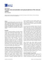
FIGURE 8.4 Change in the acoustic impedance recorded in both
ears simultaneously as a result of contraction of the stapedius
muscle elicited by tone bursts of different intensity. In the two left-
hand columns, one ear was stimulated. The solid lines are the
impedance change in the ipsilateral ear and the dashed lines are the
impedance change in the contralateral ear. The right-hand columns
show responses of both ears when both ears were stimulated simul-
taneously. The solid lines show contractions of the middle ear mus-
cles in the ipsilateral ear and the dashed lines are the responses in
the contralateral ear. The stimulus sound was 1.45 kHz pure tones
presented in bursts of 500 ms duration. The intensity of the sound is
given in dB SPL. The results were obtained in an individual with
normal hearing (reprinted from Møller, 1962, with permission from
the American Institute of Physics).
FIGURE 8.5 Typical stimulus response curves for the acoustic
middle ear reflex in an individual with normal hearing. Dashes show
the amplitude of the response to bilateral stimulation, solid lines
are the response to ipsilateral stimulation and the dots are the con-
tralateral response. Results from both ears are shown (right and left
graphs). The stimuli were 500 ms tone bursts. In these experiments the
stimulus intensity was first raised (in 2dB steps) from below threshold
to the maximal intensity used and then lowered again (in 2 dB steps)
to below threshold. The change in the ear’s impedance given is the
mean of two determinations, one when the stimulus was increased
from below threshold and the other when the stimulus intensity was
decreased from the maximal used intensity to the threshold. The
change in the ear’s acoustic impedance is given as a percentage of the
maximally obtained response at any stimulus frequency and situation
(usually bilateral stimulation) (reprinted from Møller, 1962, with
permission from the American Institute of Physics).
It is not known how the threshold of the acoustic
middle-ear reflex is set but it is interesting to note
that individuals whose auditory nerve is injured have
an elevated reflex threshold, and a poor growth of the
reflex response amplitude with increasing stimulus
intensity (see p. 291). Such injuries mainly affect the
synchronization of neural activity in the auditory
186 Section II The Auditory Nervous System
FIGURE 8.6 Stimulus responses curves similar to those in Fig. 8.5 showing the difference between
the response to tones of 500 ms duration (thin lines) and the responses to shorter tones (25 ms duration,
thick lines). Dots and dashes = bilateral stimulation; solid lines = ipsilateral stimulation; and dotted lines =
contralateral stimulation. The stimulus frequency was 0.525 kHz. Left-hand graph: stimulation of the left
ear; right-hand graph: stimulation of the right ear (reprinted from Møller, 1962, with permission from the
American Institute of Physics).
FIGURE 8.7 The sound level (in dB SPL) required to elicit an
impedance change of 10% of the maximal obtainable response
amplitude in the ear opposite to that which is stimulated is shown
as a function of the frequency of the tones used for stimulation. The
results were obtained in young individuals with normal hearing.
The thick line shows the sound levels (in dB SPL) that are 80 dB
above the threshold of hearing (80 dB HL) (reprinted from Møller,
1962, with permission from the Annals Publishing Company).
FIGURE 8.8 Similar graph as in Fig. 8.5 but showing the ampli-
tude of the response to each stimulus. The stimulus was increased
from below threshold to 115 dB SPL (in 2-dB steps and then reduced
in a 2 dB steps to below threshold) (reprinted from Møller, 1961).
nerve thus indicating that the function of the middle
ear reflex may depend on synchronization (temporal
coherence) of neural activity in many nerve fibers.
The latency of the earliest detectable response of
the acoustic middle ear reflex (recorded as a change in
the ear’s acoustic impedance) decreases with increas-
ing stimulus intensity. The shortest latency is approxi-
mately 25 ms and the longest is over 100 ms. The
individual variation is large. The latency of the
response to 1.5 kHz tones is shorter than the response
to 0.5 kHz tones [182]. The latency of the ipsilateral
and the contralateral responses are similar. The latency
of the change in the acoustic impedance is the sum
of the neural conduction time and the time it takes for
the stapedius muscle to develop sufficient tension to
cause a measurable change in the ear’s acoustic
impedance. Perlman and Case [229] recorded the EMG
response to “loud” tones and found a mean latency of
10.5 ms based on recordings from several patients.
This is a measure of the neural conduction time in
humans. The latency of the EMG response is shorter
than that of the change in the acoustic impedance,
which involves the time it takes to build up strength
of the contraction of the stapedius muscle.
The response of the acoustic middle-ear reflex is
affected by drugs such as alcohol (Fig. 8.10), and
sedative drugs such as barbiturates [16]. The threshold
of the reflex response increases as a function of the
concentration of alcohol in the blood. Blood alcohol
concentration of one tenth of one percent results in an
elevation of the reflex threshold of an average of 5 dB.
The individual variation is large.
4.2. Functional Importance of the
Acoustic Middle-ear Reflex
Many hypotheses about the functional importance
of the acoustic middle-ear reflex have been presented.
Perhaps the most plausible hypothesis is that it
keeps the input to the cochlea from steady sounds or
sounds with slowly varying intensity nearly constant
for sounds with intensities above the threshold of
the reflex, while allowing rapid changes in the sound
level to be preserved. The middle-ear reflex thus
acts as a relatively slow automatic volume control
that keeps the mean level of sound that reaches the
cochlea within narrow limits (amplitude compression)
[33, 194].
The functional importance of the acoustic middle-
ear reflex for speech discrimination has been studied
in individuals who have paresis of the stapedius
muscle in one ear (Bell’s Palsy [18]) and it was found
that discrimination of speech at high sound levels
is impaired when the acoustic middle-ear reflex is
not active (Fig. 8.11). These studies indicate that the
cochlea does not function properly at sound levels
above the normal threshold for the acoustic reflex.
Normally speech discrimination is nearly 100% in
the range of speech sound intensities from 60 dB to
120 dB SPL but when the stapedius muscle is para-
lyzed, speech discrimination deteriorates when the
sound intensity is above 90 dB SPL (Fig. 8.11).
Chapter 8 Acoustic Middle-ear Reflex 187
FIGURE 8.9 Illustration of the reproducibility of the responses
of the acoustic middle ear reflex. The changes in the ear’s impedance
expressed in percentage of the maximally obtainable response
amplitude are shown as a function in the stimulus intensity (dB SPL)
at two occasions, 2 months apart. The stimulus sounds were 0.5 kHz
tones applied to the contralateral ear (reprinted from Møller, 1961).
FIGURE 8.10 Mean value of the increase in stimulus intensity that
is necessary to obtain a reflex response that is 10% of the maximally
obtainable response as a function of blood alcohol concentration
for two different frequencies of the stimulus tones. Left hand
graph: stimulation with 0.5 kHz; right hand graph: stimulation with
1.45 kHz. Open circles are the ipsilateral response and closed circles
the contralateral response (reprinted from Borg and Møller, 1967,
with permission from Taylor & Francis).
188 Section II The Auditory Nervous System
Since the acoustic middle-ear reflex attenuates
the low frequency components of speech sounds
more than high frequency components it may reduce
masking from low frequency components of speech
sounds that may impair discrimination of speech of
high intensity. However, the high sound intensities
(above 90 dB SPL) where speech discrimination with-
out a functioning acoustic reflex becomes impaired do
not normally exist. The acoustic middle-ear reflex there-
fore seems to have little importance under normal
listening conditions.
When the acoustic middle-ear reflex is elicited by
complex sounds such as speech sounds the contraction
of the stapedius muscle will affect all low frequency
components of the sound, independent of whether or
not the spectral components contribute to activating
the reflex. Thus high frequency components of broad
band sounds will elicit contractions of the stapedius
muscle when the intensities of these components are
above the threshold of the reflex and that will cause
attenuation of low frequency components of sounds
even when these components are not sufficiently
intense to activate the reflex.
Contraction of the stapedius muscle that attenuates
low frequency sounds may help to separate specific
sounds from a noise background and may reduce
masking of high frequency components from strong
low frequency components, including one’s own
vocalizing and sounds from chewing. The ability of
the reflex to attenuate low frequency sounds of
high intensity has been referred to as the perceptual
theory of the action of the acoustic middle ear reflex
[15], and it relates to the proposal by Simmons [273].
These features may have exerted evolutionary pressure
to develop the acoustic middle-ear reflex.
Several studies have shown that the acoustic
middle-ear reflex gives some protection against noise
induced hearing loss. It is, however, questionable if
reduced noise induced hearing loss could have played
any role in the evolution of the acoustic middle-ear
reflex. The type of noise it would protect against, i.e.,
long duration, high intensity sounds, are not common
in nature.
The importance of being able to contract the middle-
ear muscles voluntarily is unknown. The acoustic
middle-ear reflex is well developed in mammals and
the threshold of the reflex is generally lower in animals
in which the acoustic reflex has been studied.
That the acoustic middle-ear reflex reduces the
input to the cochlea has been supported by a study
of the temporary threshold shift in response to expo-
sure to loud noise. It was shown that the resulting
FIGURE 8.11 Effect of speech discrimination from paralysis of the stapedius muscle. (A) Speech discrim-
ination’s dependence on the function of the stapedius muscle (the average of results obtained in 13 patients).
Speech discrimination scores (articulation scores in percentage) are shown as a function of the intensity for
monosyllables (maximal levels, in dB SPL), during paralysis of the stapedius muscle (from Bell’s Palsy) (thick
continuous line), and after recovery of the paralysis (thin line). The thick interrupted line shows the discrimi-
nation scores in the opposite (unaffected) ear during the paralysis. (B) Average difference in articulation
scores during and after paralysis of the stapedius muscle. The thick continuous line shows the difference
between the articulation scores when the sound was led to the unaffected ear and obtained when the sounds
were led to the affected ear at the time of paralysis. The thin interrupted line shows the difference between
the articulation scores in the affected ear at the time of paralysis and after recovery for 6 of the subjects who
participated in this study (reprinted from Borg and Zakrisson, 1973, with permission from the American
Institute of Physics).
Chapter 8 Acoustic Middle-ear Reflex 189
BOX 8.2
ACOUSTIC REFLEX AS A CONTROL SYSTEM
Contraction of the stapedius muscle reduces sound
transmission through the middle ear (Chapter 2). The
acoustic middle-ear reflex therefore functions as a control
system that makes the input to the cochlea vary less than
the sound that reaches the tympanic membrane, thus
amplitude compression. The compression of the input to
the cochlea is most effective for low frequency sounds
and it occurs with a latency that is equal to the time it
takes the stapedius muscle to contract after sound stimu-
lation. That means that the latency of the reduction in
sound transmission through the middle ear is at least 25
ms for sounds 20 dB or more above the threshold of the
reflex and it takes in the order of 100 ms for the stapedius
muscle to attain its full strength. The middle-ear reflex
therefore does not affect fast changes in sound intensity
and the amplitude compression is most effective for
steady-state sounds or sounds with slowly varying
amplitude.
The initial damped oscillation seen in the reflex
response to low frequency tone bursts (Fig. 8.12) is a sign
that the reflex regulates the input to the cochlea [194].
These oscillations occur because contractions of the
stapedius muscle reduce the input to the cochlea. The
attenuation caused by the stapedius muscle contraction
decreases the input to the cochlea and thereby decreases
the contraction of the stapedius muscle, and that in turn
causes the input to the cochlea to again increase, and that
increases the contraction of the stapedius muscle. This
sequence of events repeats but the amplitude of the oscil-
lations decay with time and the reflex response eventu-
ally becomes constant. The reflex response to tones above
approximately 0.8 kHz do not show such oscillations,
which is a sign that contraction of the stapedius muscle
does not affect the sound transmission through the
middle ear noticeably at that frequency, thus indicating
that the acoustic middle-ear reflex is a less efficient con-
trol system for sounds at 0.8 kHz and above.
Studies of individuals with Bell’s Palsy, in whom the
stapedius muscle was paralyzed on one side, also indi-
cated that low frequency sounds were more affected by
the reflex than high frequency sounds [17]. When the
reflex responses were elicited by stimulating the ear on the
paralyzed side with a low frequency tone, the impedance
change in the non-paralyzed side increased at a steeper
rate as a function of the stimulus intensity than it did
when the reflex was activated from the non-paralyzed
side (Fig. 8.13). No such difference in the slope of the
stimulus response curves was present when the reflex
was elicited by a tone of a higher frequency (1.45 kHz).
FIGURE 8.12 Recordings showing the change in the ear’s
acoustic impedance in response to stimulation of the ipsilateral
ear with tones of different frequencies. The duration or the
stimulus tones was 500 ms (reprinted from Møller, 1962, with
permission from the American Institute of Physics).
temporary threshold shift (TTS) was much greater in
an ear where the stapedius muscle is paralyzed than
it is in an ear with a normal functioning stapedius
muscle (Fig. 8.14) [327]. These studies were performed
in individuals with Bell’s Palsy, in whom the stapedius
muscle was paralyzed. The noise levels used caused
little TTS in the ear with the normally functioning
acoustic reflex. The TTS in the ear where the stapedius
muscle was paralyzed increased as a nearly linear
function of the level of the noise (Fig. 8.14). The indi-
vidual variations were considerable. The TTS after
exposure to noise centered at 2 kHz was not notice-
ably affected by the paralysis of the stapedius muscle
[327] in agreement with the findings of other studies
that have shown that the sound attenuation from
contraction of the stapedius muscle is small at frequen-
cies higher than 1 kHz.
Quantitative studies of the acoustic reflex as a
control system [17, 33] have shown that above its
threshold the reflex can keep the input to the cochlea
nearly constant for low frequency sounds with slowly
varying intensity despite the fact that the sound at
the tympanic membrane may vary.
4.3. Non-acoustic Ways to Elicit
Contraction of the Middle-ear Muscles
The tensor tympani muscle contracts normally
during swallowing. It can be brought to contract by
stimulating the skin around the eye, for instance
by air puffs [133]. The response was elicited by stimu-
lation of receptors in the skin that are innervated by
the trigeminal nerve. (These investigators believed
that it was the stapedius muscle that contracted while
it in fact most likely was the tensor tympani muscle.)
This response is similar to the blink reflex that is a nat-
ural protective reflex (see [187]), a test which is fre-
quently used in neurologic diagnosis.
4.4. Stapedius Muscle Contraction May Be
Elicited before Vocalization
Evidence that the stapedius muscle contracts a
brief period before vocalization has been presented in
studies in humans on the basis of EMG recording
from the stapedius muscle [19] and in the flying bat
where recordings of EMG potentials from the laryn-
geal muscles and the middle ear muscles have shown
that contractions of the middle ear muscles are
coordinated with the laryngeal muscles [90].
5. CLINICAL USE OF THE
ACOUSTIC MIDDLE-EAR
REFLEX
Recording of the acoustic middle-ear reflex response
can provide information about the function of the
190 Section II The Auditory Nervous System
BOX 8.2 (cont’d)
FIGURE 8.13 Stimulus response curves of the acoustic middle-ear reflex in an individual in whom the
stapedius muscle was paralyzed, elicited from the side of the paralysis (Bell’s Palsy) (reprinted from Borg,
1968, with permission from Taylor & Francis).
middle ear and it can help differentiate between hear-
ing loss caused by cochlear injury and that caused by
injury of the auditory nerve. The use of the acoustic
middle-ear reflex in diagnosis of middle-ear disorders
is based on the fact that contraction of the stapedius
muscle does not cause any noticeable change in
the ear’s impedance if the stapes is immobilized or if
the ossicular chain is interrupted (see Chapter 9). The
threshold of the acoustic middle-ear reflex is elevated
in patients with injuries to the auditory nerve but it
is nearly normal in patients with hearing loss of
cochlear origin (see Chapter 9). The acoustic middle-
ear reflex is therefore a valuable aid in diagnosis of
tumors of the auditory–vestibular nerve such as in
vestibular Schwannoma or other forms of injuries to
the auditory nerve (auditory nerve neuropathy) (see
Chapter 10). Testing the acoustic middle-ear reflex
may also help to identify malingering because it is
an objective test that does not require the patient’s
cooperation. The response of the acoustic middle-
ear reflex is now a routine test used in most clinics
involved in diagnosis of the auditory system.
Chapter 8 Acoustic Middle-ear Reflex 191
FIGURE 8.14 TTS in the affected ear during unilateral paralysis
of the stapedius muscle compared with the TTS in the other ear
(dashed line), as a result of exposure to band pass filtered noise
(centered at 0.5 kHz, 0.3 kHz wide), for 5 min. Mean values from
18 subjects and standard error of the mean are shown as a function
of the intensity of the noise. The TTS was measured 20 s after the
end of the exposure. In this study the noise exposure consisted of a
band of noise, centered at 0.5 kHz, and a width of 0.3 kHz. The
exposure time was 5 or 7 min. Hearing threshold was measured at
0.75 kHz before exposure and 20 s after the end of the exposure
using continuous pure tone (Békésy) audiometry (reprinted from
Zakrisson, 1975, with permission from Taylor & Francis).
192 Section II The Auditory Nervous System
BOX 8.3
EMG ACTIVITY IN THE STAPEOIUS MUSCLE
FOLLOWING VOCALIZATION
Recordings from the stapedius muscle in a patient in
whom the tympanic membrane had been deflected as a
part of a middle-ear operation have shown that EMG
potentials are present before the start of vocalization
(recorded by a microphone close to the patient’s mouth)
(Fig. 8.15). This means that the contractions of the
stapedius muscle are not a result of an acoustic reflex but
the muscle must have been brought to contract by activa-
tion of the facial motonucleus from the brain center that
is involved in controlling vocalization. Studies in humans
who have had laryngectomy do not show signs (change
in acoustic impedance) of contraction of middle ear mus-
cles during efforts to vocalize, thus contradicting the
hypothesis that middle-ear muscles are controlled by
CNS structures that are involved in generating com-
mands to vocalize [106].
FIGURE 8.15 Electrical activity (electromyographic [EMG]
potentials) recorded from the stapedius muscle during vocaliza-
tion (upper trace). The sound of the vocalization (lower trace)
was recorded near the patient’s mouth. The intensity of the
sound was 97 dB SPL. The timing impulses shown below have
intervals of 10 ms (reprinted from Borg and Zakrisson, 1975, with
permission from Taylor & Francis).
SECTION II REFERENCES
1. Achor L and Starr A. Auditory brain stem responses in the cat: I.
Intracranial and extracranial recordings. Electroenceph Clin
Neurophysiol 48: 154–173, 1980.
2. Agmon-Snir H, Carr CE, and Rinzel J. The role of dendrites in
auditory coincidence detection. Naure 393: 268–272, 1998.
3. Aitkin LM. The auditory midbrain, structure and function in the cen-
tral auditory pathway. Clifton, NJ: Humana Press, 1986.
4. Aitkin LM, Tran L, and Syka J. The responses of neurons in sub-
divisions of the inferior colliculus of cats to tonal, noise and
vocal stimuli. Exp Brain Res 98: 53–64, 1994.
5. Andersen P, Eccles JC, Schmidt RF, and Yokota T. Slow potential
wave produced by the cunate nucleus by cutaneous volleys and
by cortical stimulation. J Neurophys 27: 71–91, 1964.
6. Arthur RM, Pfeiffer RR, and Suga N. Properties of "two tone
inhibition" in primary auditory neurons. J Physiol (Lond) 212:
593–609, 1971.
7. Békésy von G. Experiments in hearing. New York: McGraw Hill,
1960.
8. Bieser A and Muller-Preuss P. Auditory responsive cortex in the
squirrel monkey: neural responses to amplitude–modulated
sounds. Exp Brain Res 108: 273–284, 1996.
9. Blauert J. Spatial hearing: The psychophysics of human sound local-
ization. Cambridge, MA: M.I.T, 1983.
10. Blauert J and Lindemann W. Auditory spaciousness: some fur-
ther psychoacoustic analyses. J Acoust Soc Am 80: 533–542, 1986.
11. Blum PS, Abraham LD, and Gilman S. Vestibular, auditory, and
somatic input to the posterior thalamus of the cat. Exp Brain Res
34: 1–9, 1979.
12. Boer de E. Correlation studies applied to the frequency resolution
of the cochlea. J Aud Res 7: 209–217, 1967.
13. Borg E. A quantitative study of the effect of the acoustic
stapedius reflex on sound transmission through the middle
ear in man. Acta Oto–laryng (Stockh) 66: 461–472, 1968.
14. Borg E. On the neuronal organization of the acoustic middle
ear reflex. A physiological and anatomical study. Brain Res 49:
101–123, 1973.
15. Borg E, Counter SA, and Roesler G. Theories of middle ear muscle
function. Orlando, FL: Academic Press, 1984.
16. Borg E and Møller AR. Effect of ethylalcohol and pentobarbital
sodium on the acoustic middle ear reflex in man. Acta
Otolaryngol (Stockh) 64: 415–426, 1967.
17. Borg E and Møller AR. The acoustic middle ear reflex in unanes-
thetized rabbit. Acta Otolaryngol (Stockh) 65: 575–585, 1968.
18. Borg E and Zakrisson JE. Stapedius reflex and speech features.
J Acoust Soc Am 54: 525–527, 1973.
19. Borg E and Zakrisson JE. The activity of the stapedius muscle in
man during vocalization. Acta Otolaryng (Stockh) 79: 325–333, 1975.
20. Boston JR and Ainslie PJ. Effects of analog and digital filtering
on brain stem auditory evoked potentials. Electroenceph Clin
Neurophysiol 48: 361–364, 1980.
21. Brodal P. The central nervous system. New York: Oxford Press,
1998.
22. Brownell WE. Observation on the motile response in isolated
hair cells. In: Mechanisms of hearing, edited by Webster WR and
Aitken LM. Melbourne: Monash University Press, 1983, p. 5–10.
23. Buchwald JS and Huang CM. Far field acoustic response:
Origins in the cat. Science 189: 382–384, 1975.
24. Casseday JH, Ehrlich D, and Covey E. Neural tuning for sound
duration: Role of inhibitory mechanisms in the inferior colliculus.
Science 264: 847–850, 1994.
25. Celesia GG and Puletti F. Auditory cortical areas of man. Neurology
19: 211–220, 1969.
26. Chiappa K. Evoked potentials in clinical medicine, 3rd ed.
Philadelphia: Lippincott-Raven, 1997.
27. Clarke SF, Ribaupierre de F, Bajo VM, Rouiller EM, and Kraftsik R.
The auditory pathway in cat corpus callosum. Exp Brain Res 104:
534–540, 1995.
28. Code RA and Winer JA. Commissural connections in layer III of
cat primary auditory cortex (AI): pyramidal and non-pyramidal
cell input. J Comp Neurol 242: 485–510, 1985.
29. Cody DT and Bickford RG. Averaged evoked myogenic
responses in normal man. Laryngoscope 79(3): 400–416, 1969.
30. Cooper NP, Robertson D, and Yates GK. Cochlear nerve fiber
responses to amplitude-modulated stimuli: variations with spon-
taneous rate and other response characteristics. J Neurophysiol
70: 370–386, 1993.
31. Covey E and Casseday JH. The monaural nuclei of the lateral
lemniscus in an echolocating bat: parallel pathways for analyzing
temporal features of sound. J Neurosci 11: 3456–3470, 1991.
32. Dallos P and Cheatham MA. Compound action potential tuning
curves. J Acoust Soc Am 59: 591–597, 1976.
33. Dallos PJ. Study of the acoustic reflex feedback loop. IEEE Trans
Bio-Med Eng 11: 1–7, 1964.
34. Davis H and Hirsh SK. A slow brain stem response for low-
frequency audiometry. Audiology: 441–465, 1979.
35. Diamond IT. The subdivisions of neocortex: a proposal to revise
the traditional view of sensory, motor, and association areas.
In: Progress in psychobiology and physiological psychology, edited by
Sprague JM and Epstein AN. New York: Academic Press, 1979,
p. 2–44.
36. Douek EE, Ashcroft PB, and Humphries KN. The clinical value
of the postauricular myogenic (crossed acoustic) response in
neuro-otology. In: Disorders of auditory function II, edited by
Stephens SDG. London: Academic Press 1976, p. 139–144.
37. Doyle DJ and Hyde ML. Bessel filtering of brain stem auditory
evoked potentials. Electroenceph Clin Neurophysiol 51: 446–448, 1981.
38. Druga R, Syka J, and Rajkowska G. Projections of auditory
cortex onto the inferior colliculus in the rat. Physiol Res 46:
215–222, 1997.
39. Dudley H. Remaking speech. J Acoust Soc Am 11: 169–177, 1939.
40. Eggermont JJ. Temporal modulation transfer functions for AM
and FM stimuli in cat auditory cortex. Effects of carrier type,
modulating waveform, and intensity. Hear Res 74: 51–66,
1994.
41. Eggermont JJ. Wiener and Volterra analyses applied to the
auditory system. Hear Res 66: 177–201, 1993.
42. Eggermont JJ, Johannesma PIM, and Aertsen AMH. Reverse-
correlation methods in auditory research. Quart Rev Biophys 16:
341–414, 1983.
43. Ehret G and Romand R. The central auditory pathway. New York:
Oxford University Press, 1997.
44. Engineer ND, Percaccio CR, Pandya PK, Moucha R, Rathbun
DL, and Kilgard MP. Environmental enrichment improves
response strength, threshold, selectivity, and latency of auditory
cortex neurons. J Neurophys 92: 73–82, 2004.
45. Erulkar SD, Nelson PG, and Bryan JS. Experimental and theoretical
approaches to neural processing in the central auditory pathway.
New York: Academic Press, 1968.
46. Evans EF. Frequency selectivity at high signal levels of single
units in cochlear nerve nucelus. In: Psycho physics and physiol-
ogy of heamy, edited by Evans EF and Wilson JP. New York:
Academic Press, 1977.
47. Evans EF. Normal and abnormal functioning of the cochlear
nerve. Symp Zool Soc Lond 37: 133–165, 1975.
48. Evans EF. The frequency response and other properties of single
fibers in the guinea pig cochlear nerve. J Physiol 226: 263–287, 1972.
Section II References 193
49. Evans EF. The sharpening of cochlear frequency selectivity
in the normal and abnormal cochlea. Audiology 14: 419–442,
1975.
50. Evans EF and Nelson PG. The responses of single neurons in the
cochlear nucleus of the cat as a function of their location and the
anaesthetic state. Exp Brain Res 17: 402–427, 1973.
51. Feddersen WE, Sandel TT, Teas DC, and Jeffress LA. Localization
of high frequency tones. J Acoust Soc Am 29: 988–991, 1957.
52. Ferber-Viart C, Soulier N, Dubreuil C, and Duclaux R.
Cochleovestibular afferent pathways of trapezius muscle
responses to clicks in humans. Acta Otolaryng (Stockh) 118: 6–10,
1998.
53. Fischer C, Bognar L, Turjman F, and Lapras C. Auditory
early- and middle-latency evoked potentials in patients with
quadrigeminal plate tumors. Neurosurgery 35: 45–51, 1994.
54. Fishman KE, Shannon RV, and Slattery WH. Speech recognition
as a function of the number of electrodes used in the SPEAK
cochlear implant speech processor. J Speech Lang Hear Res 40:
1201–1215, 1997.
55. Fitzpatrick DC, Kanwal JS, Butman JA, and Suga N.
Combination-sensitive neurons in the primary auditory cortex
of the mustached bat. J Neurosci 13: 931–940, 1993.
56. Frisina RD, Karcich KJ, Tracy TC, Sullivan DM, Walton JP, and
Colombo J. Preservation of amplitude modulation coding in
the presence of background noise by chinchilla auditory-nerve
fibers. J Acoust Soc Am 99: 475–490, 1996.
57. Frisina RD, Smith RL, and Chamberlain SC. Encoding of ampli-
tude modulation in the gerbil cochlear nucleus. I. A hierarchy of
enhancement. Hear Res 44: 99–122, 1990.
58. Frisina RD, Smith RL, and Chamberlain SC. Encoding of
amplitude modulation in the gerbil cochlear nucleus. II. Possible
neural mechanisms. Hear Res 44: 123–142, 1990.
59. Fullerton BC, Levine RA, Hosford Dunn HL, and Kiang NYS.
Comparison of cat and human brain stem auditory evoked
potentials. Hear Res 66: 547–570, 1987.
60. Galambos R and Hecox K. Clinical application of the brainstem
auditory evoked potentials. In: Auditory evoked potentials in man.
Psychopharmacology correlates of EPs prog clin neurophysiol Vol 2,
edited by Desmedt JE. Basel: Karger, 1977, p. 1–19.
61. Galambos R, Makeig S, and Talmachoff PJ. A 40 Hz auditory
potentials recorded from the humans scalp. Proc Natl Acad Sci
USA 78: 2643–2647, 1981.
62. Galambos R, Myers R, and Sheatz G. Extralemniscal activation
of auditory cortex in cats. Am J Physiol 200: 23–28, 1961.
63. Gardner MB. Historical background of the Haas/ or precedence
effect. J Acoust Soc Am 43: 1243–1248, 1968.
64. Garg BP, Markand ON, and Bustion PF. Brainstem auditory
evoked responses in hereditary motor sensory neuropathy : site
of origin of wave II. Neurology 32: 1017–1019, 1982.
65. Geisler CD, Frishkopf LS, and Rosenblith WA. Extracranial
responses to acoustic clicks in man. Science 128: 1210–1211, 1958.
66. Geisler CD, Rhode WS, and Kennedy DT. The responses to tonal
stimuli of single auditory nerve fibers and their relationship to
basilar membrane motion in the squirrel monkey. J Neurophysiol
37: 1156–1172, 1974.
67. Gelfan WR and Tarlov IM. Differential vulnerability of spinal
cord structures to anoxia. J Neurophysiol 18: 170–188, 1955.
68. Gerken GM, Moushegian G, Stillman RD, and Rupert AL.
Human frequency-following responses to monaural and
binaural stimuli. Electroencephalogr Clin Neurophysiol 38: 379–386,
1975.
69. Goldberg JM and Brown PB. Response of binaural neurons of
dog superior olivary complex to dichotic tonal stimuli: some
physiological mechanisms of sound localization. J Neurophysiol
32: 613–636, 1969.
70. Goldman-Rakic PS. Modular organization of prefrontal cortex.
Trends Neurosci 7: 419–429, 1984.
71. Graybiel AM. Some fiber pathways related to the posterior
thalamic region in the cat. Brain Behavior Evol 6: 363–393, 1972.
72. Guinan Jr. JJ, Warr WB, and Norris BE. Topographic organiza-
tion of the olivocochlear projections from the lateral and medial
zones of the superior olivary complex. J Comp Neurol 226: 21–27,
1984.
73. Guinan Jr. JJ, Norris BE, and Guinan SS. Single auditory units in
the superior olivary complex. II. Location of unit categories and
tonotopic organization. Int J Neurosci 4: 147–166, 1972.
74. Gummer M, Yates GK, and Johnstone BM. Modulation transfer
function of efferent neurons in the guinea pig cochlea. Hear Res
36: 41–52, 1988.
75. Hall JW. Handbook of auditory evoked responses. Boston, MA: Allyn
and Bacon, 1992.
76. Harrison JM and Howe ME. Anatomy of the descending
auditory system in auditory system. In: Handbook of sensory
physiology, edited by Keidel WD and Neff WD. Berlin: Springer-
Verlag, 1974, p. 363–388.
77. Harrison RV, Aran J-M, and Erre JP. AP tuning curves in normal
and pathological human and guinea pig cochlea. J Acoust Soc
Am 69: 1374–1385, 1981.
78. Hart HC, Palmer AR, and Hall DA. Different areas of human
non-primary auditory cortex are activated by sounds with spa-
tial and nonspatial properties. Hum Brain Mapp 21: 178–190,
2004.
79. Hashimoto I. Auditory evoked potentials from the humans mid-
brain: Slow brain stem responses. Electroenceph Clin Neurophysiol
53: 652–657, 1982.
80. Hashimoto I, Ishiyama Y, Yoshimoto T, and Nemoto S.
Brainstem auditory evoked potentials recorded directly from
human brain stem and thalamus. Brain 104: 841–859, 1981.
81. Haykin S and Chen Z. The cocktail party problem. Neural
Comput 17: 1875–1902, 2005.
82. Heil P and Irvine DR. Functional specialization in auditory
cortex: responses to frequency-modulated stimuli in the cat's
posterior auditory field. J Neurophysiol 79: 3041–3059, 1998.
83. Heil P, Rajan R, and Irvine DRF. Sensitivity of neurons in cat
primary auditory cortex to tones and frequency-modulated
stimuli: I. Organization of response properties along the "isofre-
quency" dimension. Hear Res 63: 135–156, 1992.
84. Hellige J. Hemispheric asymmetry: What's right and what's left.
Cambridge, MA: Harvard University Press, 1993.
85. Honrubia V and Ward PH. Longitudinal distribution of the
cochlear microphonics inside the cochlear duct (guinea pig).
J Acoust Soc Am 44: 951 958, 1968.
86. Irvine DRF. A comparison of two methods for measurement of
neural sensitivity to interaural intensity differences. Hear Res 30:
169–180, 1987.
87. Irvine DRF. Interaural intensity differences in the cat: Changes
in sound pressure level at the two ears associated with
azimuthal displacements in the frontal horizontal plane. Hear
Res 26: 267–286, 1987.
88. Jacobson JT. Principles and applications in auditory evoked potentials.
Boston: Allyn & Bacon, 1994.
89. Jeffress LA. A place theory of sound localization. J Comp Physiol
Psychol 41: 35–39, 1948.
90. Jen PH and Suga N. Coordinated activities of middle-ear
and laryngeal muscles in echolocating bats. Science 91: 950–952,
1976.
91. Jenkins WM, Merzenich MM, Ochs MT, Allard T, and Guic-
Robles E. Functional reorganization of primary somatosensory
cortex in adult owl monkeys after behaviorally controlled
tactile stimulation. J Neurophysiol 63: 82–104, 1990.
194 Section II References
92. Jepsen O. Studies of the acoustic stapedius refelx in man. Aarhus:
Universitetsforlaget 1955.
93. Jewett DL. The 3 channel Lissajous' trajectory of the auditory
brain stem response. IX. Theoretical aspects. Electroenceph Clin
Neurophysiol 68: 386–408, 1987.
94. Jewett DL, Deupree DL, and Bommannan D. Far field poten-
tials generated by action potentials of isolated frog sciatic
nerves in a spherical volume. Electroenceph Clin Neurophys 75:
105–117, 1990.
95. Jewett DL, Romano MN, and Williston JS. Human auditory
evoked potentials: possible brain stem components detected on
the scalp. Science 167: 1570–1571, 1970.
96. Jewett DL and Williston JS. Auditory evoked far fields
averaged from scalp of humans. Brain 94: 681–696, 1971.
97. Johnstone BM, Patuzzi R, and Yates GK. Basilar membrane
measurements and the traveling wave. Hear Res 22: 147–153,
1986.
98. Joris PX, Carney LH, Smith PH, and Yin TC. Enhancement
of neural synchronization in the anteroventral cochlear
nucleus. I. Responses to tones at the characteristic frequency.
J Neurophysiol 71: 1022–1036, 1994.
99. Joris PX, Schreiner CE, and Rees A. Neural processing of
amplitude-modulated sounds. Physiol Rev 84: 541–577, 2004.
100. Joris PX, Smith PH, and Yin TCT. Coincidence detection in the
auditory system: 50 years after Jeffress. Neuron 21: 1235–1238,
1998.
101. Joris PX and Yin TC. Envelope coding in the lateral superior
olive. III. Comparison with afferent pathways. J Neurophysiol
79: 253–269, 1998.
102. Joris PX and Yin TCT. Responses to amplitude modulated
tones in the auditory nerve of the cat. J Acoust Soc Am 91:
215–232, 1992.
103. Joseph MP, Guinan JJ, Fullerton BC, Norris BE, and Kiang NYS.
Number and distribution of stapedius motoneurons in cats.
J Comp Neurol 232: 43–54, 1985.
104. Kaas JH and Hackett TA. Subdivisions of auditory cortex
and processing streams in primates. Proc Nat Acad Sci USA 97:
11793–11799, 2000.
105. Katsuki Y, Sumi T, Uchiyama H, and Watanabe T. Electric
responses of auditory neurons in cat to sound stimulation.
J Neurophysiol 21: 569–588, 1958.
106. Kawase T, Ogura M, Kakehata S, and Takasaka T.
Measurement of stapedius contraction during vocalization
effort in patients after laryngectomy or tracheostomy. Hear Res
149: 248–252, 2000.
107. Kiang NY-S. The use of computers in studies of auditory
neurophysiology. Trans Am Acad Ophthal Otolaryngol 65:
735–747, 1961.
108. Kiang NY-S, Christ AH, French MA, and Edwards AG.
Postauricular electric response to acoustic stimuli in humans.
MIT Quart Prog Rep Research Laboratory of Electronics 68:
218–225, 1963.
109. Kiang NY-S and Peake WT. Cochlear Responses to
Condensation and Rarefaction Clicks. Biophys J 2: 23–34, 1962.
110. Kiang NYS, Watanabe T, Thomas EC, and Clark L. Discharge
patterns of single fibers in the cat's auditory nerve. Cambridge,
MA: MIT Press, 1965.
111. Kilgard MP and Merzenich MM. Cortical map reorganization
enabled by nucleus basalis activity. Science 279: 1714–1718, 1998.
112. Kilgard MP and Merzenich MM. Distributed representation of
spectral and temporal information in rat primary auditory
cortex. Hear Res 134: 16–28, 1999.
113. Klockhoff I and Anderson H. Recording of the stapedius reflex
elicited by cutaneous stimulation. Acta Otolaryngol (Stockh) 50:
451–454, 1959.
114. Knudsen EI, du Lac S, and Esterly SD. Computational maps in
the brain. Annu Rev Neurosci 10: 41–65, 1987.
115. Konishi M and Nielsen DW. The temporal relationship between
motion of the basilar membrane and initiation of nerve
impulses in auditory nerve fibers. J Acoust Soc Am 53: 325, 1973.
116. Kral A, Hartmann R, Tillrin J, Heid S, and Klinke R. Congenital
auditory deprivation reduces synaptic activity within the
auditory cortex in layer specific manner. Cerebral Cortex 10:
714–726, 2000.
117. Kraus N, Odzamar O, Hier D, and Stein L. Auditory middle
latency responses (MLRs) in patients with cortical lesions.
Electroencephalogr Clin Neurophys 54: 275–287, 1982.
118. Kreiman J and Van Lancker D. Hemispheric specialization for
voice recognition: evidence from dichotic listening. Brain &
Language 34: 246–252, 1988.
119. Kuroki A and Møller AR. Microsurgical anatomy around
the foramen of Luschka with reference to intraoperative
recording of auditory evoked potentials from the cochlear
nuclei. J Neurosurg: 933–939, 1995.
120. Kuwada S and Batra R. Coding of sound envelopes by inhibitory
rebound in neurons of the superior olivary complex in the
unanesthetized rabbit. J Neurosci 19: 2273–2287, 1999.
121. Kvasnak E, Popelar J, and Syka J. Discharge properties of
neurons in subdivisions of the medial geniculate body of
the guinea pig. Physiol Res 49: 369–378, 2000.
122. Kvasnak E, Suta D, Popelar J, and Syka J. Neuronal connec-
tions in the medial geniculate body of the guinea-pig. Exp Brain
Res 132: 87–102, 2000.
123. Lang J. Anatomy of the brainstem and the lower cranial nerves,
vessels, and surrounding structures. Am J Otol Suppl, Nov:
1–19, 1985.
124. Lang J. Clinical anatomy of the posterior cranial fossa and its foramina.
Stuttgart: Thieme Verlag, 1981.
125. Lang J. Facial and vestibulocochlear nerve, topographic
anatomy and variations. The cranial nerves, edited by
Samii M and Jannetta P. New York: Springer-Verlag, 1981,
p. 363–377.
126. Lang JJ, Ohmachi N, and Lang JS. Anatomical landmarks of the
rhomboid fossa (floor of the 4th ventricle), its length and its
width. Acta Neurochir (Wien) 113: 84–90, 1991.
127. Langner G and Schreiner CE. Periodicity coding in the inferior
colliculus of the cat. I. Neuronal mechanisms. J Neurophysiol 60:
1799–1822, 1988.
128. Langner GA. Review: periodicity coding in the auditory
system. Hear Res 60: 115–142, 1992.
129. Lazorthes G, Lacomme Y, Ganbert J, and Planel H. La constitu-
tion du nerf auditif. Presse Medicine 69: 1067–1068., 1961.
130. Le Prell CG, Dolan D, Schacht J, Miller JM, Lomax MI, and
Altschuler RA. Pathways for protection from noise induced
hearing loss. Noise Health 5: 1–17, 2003.
131. Leake PA, Snyder RL, Rebscher SJ, Moore CM, and Vollmer M.
Plasticity in central representation in the inferior colliculus
induced by chronic single- vs. two-channel electrical stimula-
tion by cochlear implant after neonatal deafness. Hear Res 147:
221–241, 2000.
132. LeDoux JE. Brain mechanisms of emotion and emotional
learning. Curr Opin Neurobiol 2: 191–197, 1992.
133. Lee YS, Lueders H, Dinner DS, Lesser RP, Hahn J, and Klem G.
Recording of auditory evoked potentials in man using chronic
subdural electrodes. Brain Res Brain Res Rev 107: 115–131, 1984.
134. Lepore F, Poirier P, Provencal C, Lassonde M, Miljours S, and
Guillemot JP. Cortical and callosal contribution to sound localization.
New York: Plenum, 1997.
135. Liberman MC. Auditory-nerve response from cats raised in
low-noise chamber. J Acoust Soc Am 63: 442–455, 1978.
Section II References 195
136. Liberman MC and Mulroy MJ. Acute and chronic effects of
acoustic traume: cochlear pathology and auditory nerve
pathology. In: New perspectives in noise-induced hearing loss,
edited by Hamernik RP, Henderson D and Salvi R. New York:
Raven Press, 1982.
137. Licklider JCR. Three auditory theories. New York: McGraw-Hill,
1959.
138. Liegeois-Chauvel C, Lorenzi C, Trebuchon A, Regis J, and
Chauvel P. Temporal envelope processing in the human left
and right auditory cortices. Cereb Cortex 14: 731–740, 2004.
139. Loizou PC. Introduction to cochlear implants. IEEE Signal
Processing Magazine September: 101–130, 1998.
140. Loizou PC. On the number of channels needed to understand
speech. J Acoust Soc Am 106: 2097–2103, 1999.
141. Loizou PC, Dorman M, and Fitzke J. The effect of reduced
dynamic range on speech understanding: implicatlons for
patients with cochlear implant. Ear Hear 21: 25–31, 2000.
142. Lorente de No R. Action potentials of the motoneurons of the
hypoglossus nucleus. J Cell Comp Physiol 29: 207–287, 1947.
143. Lorente de No R. Analysis of the distribution of action currents
of nerve in volume conductors. Studies of the Rockefeller Institute
for Medical Research 132: 384–482, 1947.
144. Lorente de No R. Anatomy of the eighth nerve III. General plan
of structure of the primary cochlear nuclei. Laryngoscope 43:
327–350, 1933.
145. Malhotra S, Hall, A.J., and Lomber, S.G. Cortical control of
sound localization in the cat: unilateral cooling deactivation of
nineteen cortical areas. J Neurophysiol 92: 1625–1643, 2004.
146. Markand ON, Farlow MR, Stevens JC, and Edwards MK. Brain
stem auditory evoked potential abnormalities with unilateral
brain stem lesions demonstrated by magnetic resonance
imaging. Arch Neurol 46: 295–299, 1989.
147. Martin WH, Pratt H, and Schwegler JW. The origin of the
human auditory brainstem response wave II. Electroenceph Clin
Neurophysiol 96: 357–370, 1995.
148. Mast TE. Binaural interaction and contralateral inhibition in
dorsal cochlear nucleus of chinchilla. J Neurophysiol 62: 61–70, 1973.
149. Mast TE. Short latency human evoked responses to clicks.
J Appl Physiol 20: 725–730, 1965.
150. McGee T, Kraus N, Littmanm T, and Nicol T. Contributions of
medial geniculate body subdivisions to the middle latency
response. Hear Res 61: 147–154, 1992.
151. Metz O. Studies of the contraction of the tympanic muscles
as indicated by changes in the impedance of the ear. Acta
Otolaryng (Stockh) 39: 397–405, 1951.
152. Metz O. The acoustic impedance measured on normal and
pathological ear. Acta Otolaryng (Stockh) 63, 1946.
153. Middlebrooks JC. Binaural mechanisms of spatial tuning in the
cat's superior colliculus distinguished using monaural occlusion.
J Neurophysiol 57: 688–701, 1987.
154. Mishkin M, Ungerleider LG, and Macko KA. Object vision
and spatial vision: two cortical pathways. Trends Neurosci
6: 415–417, 1983.
155. Mitani A and Shimokouchi M. Neural connections in the
primary auditory cortex: and electrophysiologic study in the
cat. J Comp Neurol 235: 417–429, 1985.
156. Moore BC. Psychoacoustics of normal and impaired hearing.
Br Med Bull 63: 121–134, 2002.
157. Moore JK. The human auditory brain stem as a generator for
auditory evoked potentials. Hear Res 29: 33–44, 1987.
158. Moore JK. The human auditory brain stem: a comparative
view. Hear Res 29: 1–32, 1987.
159. Morest DK. Structural organization of auditory pathways. In:
The nervous system, edited by Eagles EL. New York: Raven
Press, 1975, p. 19–29.
160. Morest DK. The neuronal architecture of the medial geniculate
body of the cat. J Anat (Lond) 98: 611–630, 1964.
161. Morest DK and Oliver DL. The neuronal architecture of the
inferior colliculus in the cat. J Comp Neurol 222: 209–236, 1984.
162. Mountain DC and Cody AR. Multiple modes of inner hair cell
stimulation. Hear Res 132: 1–14, 1999.
163. Mountain DC, Geisler C, D., and Hubbard AE. Stimulation of
efferents alters the cochlear microphonic and sound-induced
resistance changes measured in the scala media of the guinea
pig. Hear Res 3: 231–240, 1980.
164. Mountcastle VB. Modality and topographic properties of single
neurons of cat's somatic cortex. J Neurophysiol 20: 408–434, 1957.
165. Moushegian G, Rupert AL, and Stillman RD. Evaluation of
frequency following potentials in man: masking and clinical
studies. Electroenceph Clin Neurophysiol 45: 711–718, 1978.
166. Moushegian G, Rupert AL, and Stillman RD. Laboratory note.
Scalp-recorded early responses in man to frequencies in the
speech range. Electroenceph Clin Neurophysiol 35: 665–667, 1973.
167. Møller A. Unit responses in the cochlear nucleus of the rat to
sweep tones. Acta Physiol Scand 76: 503–512, 1969.
168. Møller AR. Auditory physiology. New York: Academic Press,
1983.
169. Møller AR. Bilateral contraction of the tympanic muscles in
man, examined by measuring acoustic impedance-change. Ann
Otol Rhinol Laryngol 70: 735–753, 1961.
170. Møller AR. Bilateral contraction of the tympanic muscles in man.
Royal Institute of Technology (KTH), Div of Telegraphy-Telephony,
Report No 18, Speech Transmission Laboratory 1–51, 1961.
171. Møller AR. Coding of amplitude and frequency modulated
sounds in the cochlear nucleus of the rat. Acta Physiol Scand 86:
223–238, 1972.
172. Møller AR. Coding of increments and decrements in stimuli
intensity in single units in the cochlear nucleus of the rat.
J Neurosci Res 4: 1–8, 1979.
173. Møller AR. Coding of sounds with rapidly varying spectrum in
the cochlear nucleus. J Acoust Soc Am 55: 631–640, 1974.
174. Møller AR. Dynamic properties of excitation and inhibition in
the cochlear nucleus. Acta Physiol Scand 93: 442–454, 1975.
175. Møller AR. Dynamic properties of primary auditory fibers
compared with cells in the cochlear nucleus. Acta Physiol Scand
98: 157–167, 1976.
176. Møller AR. Dynamic properties of the responses of single
neurons in the cochlear nucleus. J Physiol 259: 63–82, 1976.
177. Møller AR. Effect of tympanic muscle activity on movement of
the eardrum, acoustic impedance, and cochlear microphonics.
Acta Otolaryngol (Stockh) 58: 525–534, 1965.
178. Møller AR. Evoked potentials in intraoperative monitoring.
Baltimore, MD: Williams and Wilkins, 1988.
179. Møller AR. Frequency selectivity of phase–locking of complex
sounds in the auditory nerve of the rat. Hear Res 11: 267–284,
1983.
180. Møller AR. Frequency selectivity of single auditory nerve
fibers in response to broadband noise stimuli. J Acoust Soc Am
62: 135–142, 1977.
181. Møller AR. Frequency selectivity of the basilar membrane
revealed from discharges in auditory nerve fibers. In:
Psychophysics and physiology of hearing, edited by Evans EF and
Wilson JP. London: Academic Press, 1977, p. 197–205.
182. Møller AR. Intra-aural muscle contraction in man, examined
by measuring acoustic impedance of the ear. Laryngoscope
LXVIII: 48–62, 1958.
183. Møller AR. Intraoperative neurophysiologic monitoring. Luxem-
bourg: Harwood Academic Publishers, 1995.
184. Møller AR. Intraoperative neurophysiologic monitoring in
neurosurgery: Benefits, efficacy, and cost–effectiveness.
196 Section II References
In: Clinical neurosurgery, proceedings of the congress of neurological
surgeons' 1994 meeting, Chapter 12. Baltimore: Williams &
Wilkins, 1995, p. 171–179.
185. Møller AR. Intraoperative neurophysiologic monitoring, 2nd
edition: Humana Press Inc., 2006.
186. Møller AR. Latency of unit responses in the cochlear nucleus
determined in two different ways. J Neurophysiol 38: 812–821, 1975.
187. Møller AR. Neural plasticity and disorders of the nervous system.
Cambridge: Cambridge University Press (in press), 2006.
188. Møller AR. Origin of latency shift of cochlear nerve potentials
with sound intensity. Hear Res 17: 177–189, 1985.
189. Møller AR. Responses of units in the cochlear nucleus to sinu-
soidally amplitude modulated tones. Exp Neurol 45: 104–117, 1974.
190. Møller AR. Review of the roles of temporal and place coding of
frequency in speech discrimination. Acta Otolaryngol (Stockh)
119: 424–430, 1999.
191. Møller AR. Sensory systems: anatomy and physiology. Amsterdam:
Academic Press, 2003.
192. Møller AR. Similarities between severe tinnitus and chronic
pain. J Amer Acad Audiol 11: 115–124, 2000.
193. Møller AR. Statistical evaluation of the dynamic properties of
cochlear nucleus units using stimuli modulated with pseudo-
random noise. Brain Res 57: 443–456, 1973.
194. Møller AR. The acoustic reflex in man. J Acoust Soc Am 34:
1524–1534, 1962.
195. Møller AR. The sensitivity of contraction of the tympanic mus-
cles in man. Ann Otol Rhinol Laryngol 71: 86–95, 1962.
196. Møller AR. Unit responses in the cochlear nucleus of the rat to
pure tones. Acta Physiol Scand 75: 530–541, 1969.
197. Møller AR. Unit responses in the rat cochlear nucleus to repet-
itive transient sounds. Acta Physiol Scand 75: 542–551, 1969.
198. Møller AR. Unit responses in the rat cochlear nucleus to tones
of rapidly varying frequency and amplitude. Acta Physiol Scand
81: 540–556, 1971.
199. Møller AR. Use of stochastic signals in evaluation of the
dynamic properties of a neuronal system. Scand J Rehab Med 3:
37–44, 1974.
200. Møller AR and Burgess JE. Neural generators of the brain stem
auditory evoked potentials (BAEPs) in the rhesus monkey.
Electroenceph Clin Neurophysiol 65: 361–372, 1986.
201. Møller AR, Colletti V, and Fiorino F. Click evoked responses
from the exposed intracranial portion of the eighth nerve
during vestibular nerve section: bipolar and monopolar
recordings. Electroenceph Clin Neurophysiol 92: 17–29, 1994.
202. Møller AR, Colletti V, and Fiorino FG. Neural conduction
velocity of the human auditory nerve: Bipolar recordings from
the exposed intracranial portion of the eighth nerve during
vestibular nerve section. Electroenceph Clin Neurophysiol 92:
316–320, 1994.
203. Møller AR and Jannetta PJ. Auditory evoked potentials
recorded from the cochlear nucleus and its vicinity in man.
J Neurosurg 59: 1013–1018, 1983.
204. Møller AR and Jannetta PJ. Comparison between intracranially
recorded potentials from the human auditory nerve and scalp
recorded auditory brainstem responses (ABR). Scand Audiol
(Stockholm) 11: 33–40, 1982.
205. Møller AR and Jannetta PJ. Compound action potentials
recorded intracranially from the auditory nerve in man. Exp
Neurol 74: 862–874, 1981.
206. Møller AR and Jannetta PJ. Evoked potentials from the inferior
colliculus in man. Electroenceph Clin Neurophysiol 53: 612–620,
1982.
207. Møller AR and Jannetta PJ. Interpretation of brainstem audi-
tory evoked potentials: results from intracranial recordings in
humans. Scand Audiol (Stockh) 12: 125–133, 1983.
208. Møller AR and Jannetta PJ. Monitoring auditory functions
during cranial nerve microvascular decompression operations
by direct recording from the eighth nerve. J Neurosurg 59:
493–499, 1983.
209. Møller AR and Jannetta PJ. Neural generators of the brainstem
auditory evoked potentials. In: Evoked potentials II: the second
international evoked potentials symposium, edited by Nodar RH
and Barber C. Boston, MA: Butterworth Publishers, 1984,
p. 137–144.
210. Møller AR, Jannetta PJ, and Jho HD. Click–evoked responses
from the cochlear nucleus: a study in human. Electroenceph Clin
Neurophysiol 92: 215–224, 1994.
211. Møller AR, Jannetta PJ, and Sekhar LN. Contributions from the
auditory nerve to the brainstem auditory evoked potentials
(BAEPs): results of intracranial recording in man. Electroenceph
Clin Neurophysiol 71: 198–211, 1988.
212. Møller AR and Jho HD. Effect of high frequency hearing loss
on compound action potentials recorded from the intracranial
portion of the human eighth nerve. Hear Res 55: 9–23, 1991.
213. Møller AR and Jho HD. Late components in the compound
action potentials (CAP) recorded from the intracranial portion
of the human eighth nerve. Hear Res 45: 75–86, 1990.
214. Møller AR and Jho HD. Response from the exposed intracra-
nial human auditory nerve to low-frequency tones: Basic char-
acteristics. Hear Res 38: 163–175, 1989.
215. Møller AR and Jho HD. Responses from the exposed human
auditory nerve to pseudorandom noise. Hear Res 42: 237 252,
1989.
216. Møller AR, Jho HD, and Jannetta PJ. Preservation of hearing in
operations on acoustic tumors: an alternative to recording
BAEP. Neurosurgery 34: 688–693, 1994.
217. Møller AR, Jho HD, Yokota M, and Jannetta PJ. Contribution
from crossed and uncrossed brainstem structures to the
brainstem auditory evoked potentials (BAEP): A study in
human. Laryngoscope 105: 596–605, 1995.
218. Møller AR, Kern JK, and Grannemann B. Are the non-classical
auditory pathways involved in autism and PDD? Neurol Res
27: 625–629, 2005.
219. Møller AR, Møller MB, and Yokota M. Some forms of tinnitus
may involve the extralemniscal auditory pathway. Laryngoscope
102: 1165–1171, 1992.
220. Møller AR and Rollins P. The non-classical auditory system is
active in children but not in adults. Neurosci Lett 319: 41–44,
2002.
221. Müller M, Robertson D, and Yates GK. Rate-versus-level func-
tions of primary auditory nerve fibres: evidence of square law
behavior of all fibre categories in the guinea pig. Hear Res 55:
50–56, 1991.
222. Müsiek FE and Geurkink, N.A. Auditory brainstem response
and central auditory test findings for patients with brain stem
lesions: a preliminary report. Laryngoscope 92: 891–900, 1982.
223. Neff WD, Diamond JT, and Casseday JH. Behavioral studies
of auditory discrimination: central nervous system: central
nervous system. In: Handbook of sensory physiology, edited
by Keidel WD and Neff WD. Berlin: Springer-Verlag, 1975,
p. 307–400.
224. Oertel D and Young ED. What's a cerebellar circuit doing in the
auditory system? Trends Neurosci 27: 104–110, 2004.
225. Oliver DL and Morest DK. The central nucleus of the inferior
colliculus in the cat. J Comp Neurol 222: 237–264, 1984.
226. Ota CY and Kimura RS. Ultrastructural study of the human
spiral ganglion. Acta Otolaryngol (Stockh) 89: 53–62, 1980.
227. Palmer AR. Encoding of rapid amplitude fluctuations by
cochlear nerve fibers in the guinea pig. Arch Otorhinolaryngol
236: 197 202, 1982.
Section II References 197
228. Peake WT, Goldstein MH, and Kiang NYS. Responses of
the auditory nerve to repetitive stimuli. J Acoust Soc Am 34:
562–570, 1962.
229. Perlman HB and Case TJ. Latent period of the crossed stapedius
reflex in man. Ann Otol Rhin Laryngol 48: 663–675, 1939.
230. Pfeiffer RR. Classification of response patterns of spike dis-
charges for units in the cochlear nucleus: tone burst stimula-
tion. Exp Brain Res 1: 220–235, 1966.
231. Pickles JO. An introduction to the physiology of hearing, 2nd
edition. London: Academic Press, 1988.
232. Picton TW, Hillyard SA, Krausz HI, and Galambos R. Human
auditory evoked potentials. I. Evaluation of components.
Electroenceph Clin Neurophysiol 36: 176–190, 1974.
233. Portmann M, Cazals Y, Negrevergne M, and Aran JM.
Transtympanic and surface recordings in the diagnosis of
retrocochlear lesions. Acta Otolaryngol (Stockh) 89: 362–369, 1980.
234. Pratt H, Bleich N, and Martin WH. Three channel Lissajous'
trajectory of humans auditory brain stem evoked potentials.
I. Normative measures. Electroenceph Clin Neurophysiol 61:
530–538, 1985.
235. Pratt H, Martin WH, Schwegler JW, Rosenwasser RH, and
Rosenberg SJ. Temporal Correspondence of intracranial,
cochlear and scalp recorded humans auditory nerve action
potentials. Electroenceph Clin Neurophysiol 84: 447–455, 1992.
236. Rao KR and Ben-Arie J. Optimal head related transfer func-
tions for hearing and monaural localization in elevation: a
signal processing design perspective. IEEE Trans Biomed Eng
43: 1093–1105, 1996.
237. Rauschecker JP. Cortical processing of complex sounds. Curr
Opinion Neurobiol 8: 516–521, 1998.
238. Rauschecker JP and Harris LR. Auditory and visual neurons
in the cat's superior colliculus selective for the direction of
apparent motion stimuli. Brain Res 490: 56–63, 1989.
239. Rauschecker JP and Tian B. Mechanisms and streams for
processing of "what" and "where" in auditory cortex. Proc Nat
Acad Sci USA 97: 11800–11806, 2000.
240. Razak KA and Fuzessery ZM. Functional organization of the
pallid bat auditory cortex: emphasis on binaural organization.
J Neurophysiol 87: 72–86, 2002.
241. Razak KA, Fuzessery ZM, and Lohuis TD. Single cortical neu-
rons serve both echolocation and passive sound localization.
J Neurophysiol 81: 1438–1442, 1999.
242. Rees A and Møller AR. Responses of neurons in the inferior col-
liculus of the rat to AM and FM tones. Hear Res 10: 301–330, 1983.
243. Rees A and Møller AR. Stimulus properties influencing the
responses of inferior colliculus neurons to amplitude-modu-
lated sounds. Hear Res 27: 129–144, 1987.
244. Rhode WS and Greenberg S. Encoding of amplitude modulation
in the cochlear nucleus of the cat. J Neurophysiol 71: 1797–1825,
1994.
245. Rodrigues–Dagaeff C, Simm G, Ribaupierre de Y, Villa A,
Ribaupierre de F, and Rouiller EM. Functional organization of
the ventral division of the medial geniculate body of the cat:
evidence for a rostro–caudal gradient of response properties
and cortical projections. Hear Res 39: 103–126, 1989.
246. Roeder KD. Acoustic alerting mechanisms in insects. Ann New
York Acad Sci 188: 63–79, 1971.
247. Roeder KD and Treat AE. Detection and evasion of bats by
moths. American Scientist 49: 135–148, 1961.
248. Romanski LM, Tian B, Fritz J, Mishkin M, Goldman-Rakic PS,
and Rauschecker JP. Dual streams of auditory afferents target
multiple domains in the primate prefrontal cortex. Nature
Neurosci 2: 1131–1136, 1999.
249. Rose JE, Galambos R, and Hughes JR. Microelectrode studies
of the cochlear nuclei in the cat. Bull Johns Hopkins Hosp 104:
211–251, 1959.
250. Rose JE, Gross NB, Geisler CD, and Hind JE. Some neural
mechanisms in the inferior colliculus of the cat which may be
relevant to localization of a sound source. J Neurophysiol 29:
288–314, 1966.
251. Rose JE, Hind JE, Anderson DJ, and Brugge JF. Some effects of
stimulus intensity on response of auditory fibers in the squirrel
monkey. J Neurophysiol 34: 685–699, 1971.
252. Rouiller EM. Functional organization of the auditory system.
In: The central auditory system, edited by Ehret G and Romand R.
New York: Oxford University Press, 1997, p. 3–96.
253. Rouiller EM, Ribaupierre de Y, Toros-Morel A, and Ribaupierre
de F. Neural coding of repetitive clicks in the medial geniculate
body of cat. Hear Res 5: 81–100, 1981.
254. Rouiller EM, Rodrigues-Dagaeff C, Simm G, Ribaupierre de Y,
Villa A, and Ribaupierre de F. Functional organization of the
medial division of the medial geniculate body of the cat: tono-
topic organization, spatial distribution of response properties
and cortical connections. Hear Res 39: 127–142, 1989.
255. Ruben RJ and Walker AE. The VIIIth nerve action potential in
Ménière's disease. Laryngoscope 11: 1456–1464, 1963.
256. Ruggero MAand Rich NC. Peak splitting: intensity effects in cochlear
afferent responses to low frequency tones: New York: Plenum, 1989.
257. Ryugo DK. The auditory nerve: Peripheral innervation, cell
body morphology, and central projections. In: The mammalian
auditory pathway: neuroanatomy, edited by Webster DB, Popper
AN and Fay RR. New York: Springer-Verlag, 1992.
258. Sachs MB and Kiang NYS. Two tone inhibition in auditory
nerve fibers. J Acoust Soc Am 43: 1120–1128, 1968.
259. Sachs MB and Young ED. Encoding of steady-state vowels in
the auditory nerve: representation in terms of discharge rate.
J Acoust Soc Am 66: 470–479, 1979.
260. Sachs MB, Young ED, and Miller MI. Speech encoding in the
auditory nerve: implications for cochlear implants. Cochlear
Prostheses: An International Symposium 405: 94–113, 1983.
261. Scharf B, Magnan J, and Chays A. On the role of the olivo-
cochlear bundle in hearing: 16 case studies. Hear Res 103:
101–122, 1997.
262. Scherg M and von Cramon D. A new interpretation of the
generators of BAEP waves I V: results of a spatio temporal
dipole. Electroenceph Clin Neurophysiol 62: 290–299, 1985.
263. Schmidt RF and Thews G. Human physiology. Berlin: Springer-
Verlag, 1983.
264. Schreiner CE and Urbas JV. Representation of amplitude mod-
ulation in the auditory cortex of the cat. I. The anterior auditory
field (AAF). Hear Res 21: 227–242, 1986.
265. Schroeder M. Vocoders: Analysis and synthesis of speech. Proc
IEEE 54: 720–734, 1966.
266. Schucknecht HF. Pathology of the ear. Cambridge, MA: Harvard
University Press, 1974.
267. Sellick PM, Patuzzi R, and Johnstone BM. Measurement of
basilar membrane motion in the guinea pig using the
Mossbauer technique. J Acoust Soc Am 72: 131–141, 1982.
268. Sellick PM, Patuzzi R, and Johnstone BM. Modulation of
responses of spiral ganglion cells in the guinea pig cochlea to
low frequency sound. Hear Res 7: 199–221, 1982.
269. Shannon RV, Zeng F-G, Kamath V, Wygonski J, and Ekelid M.
Speech recognition with primarily temporal cues. Science 270:
303–304, 1995.
270. Shore SE, El Kashlan H, and Lu J. Effects of trigeminal ganglion
stimulation on unit activity of ventral cochlear nucleus neurons.
Neuroscience 119: 1085–1101, 2003.
271. Shore SE, Vass Z, Wys NL, and Altschuler RA. Trigeminal
ganglion innervates the auditory brainstem. J Comp Neurol 419:
271–285, 2000.
272. Silverstein H, Norrell H, Haberkamp T, and McDaniel AB. The
unrecognized rotation of the vestibular and cochlear nerves from
198 Section II References
the labyrinth to the brain stem: its implications to surgery of the
eighth cranial nerve. Otolaryngol Head Neck Surg 95: 543–549, 1986.
273. Simmons FB. Perceptual theories of middle ear muscle function.
Ann Otol Rhinol Laryngol 73: 724–739, 1964.
274. Sinex DG and Geisler CD. Auditory-nerve fiber responses to
frequency-modulated tones. Hear Res 4: 127–148, 1981.
275. Snyder RL, Rebscher SJ, Cao K, and Leake PA. Effects of
chronic intracochlear stimulation in the neonatally deafened
cat: I. Expansion of central spatial representation. Hear Res 50:
7–33, 1990.
276. Snyder RL and Schreiner CE. Forward masking of the auditory
nerve neurophonic (ANN) and the frequency following
response (FFR). Hear Res 20: 45–62, 1985.
277. Snyder RL and Schreiner CE. The auditory neurophonic: basic
properties. Hear Res 15: 261–280, 1984.
278. Sokolich WG, Hamernick RP, Zwislocki JJ, and Schmiedt RA.
Inferred response polarities of cochlear hair cells. J Acoust Soc
Am 59: 963–974, 1976.
279. Spangler KM, Cant NB, Henkel CK, Farley GR, and Warr WB.
Descending projections from the superior olivary complex to
the cochlear nucleus of the cat. J Comp Neurol 259: 452–465, 1987.
280. Spire JP, Dohrmann GJ, and Prieto PS. Correlation of brainstem
evoked response with direct acoustic nerve potential. New York:
Raven Press, 1982.
281. Spoendlin H and Schrott A. Analysis of the human auditory
nerve. Hear Res 43: 25–38, 1989.
282. Stainsby TH, Moore BC, and Glasberg BR. Auditory streaming
based on temporal structure in hearing-impaired listeners.
Hear Res 192: 119–130, 2004.
283. Stapells DR, Picton TW, and Smith AD. Normal hearing thresh-
olds for clicks. J Acoust Soc Am 72: 74–79, 1982.
284. Starr A and Zaaroor M. Eighth nerve contributions to cat audi-
tory brainstem responses (ABR). Hear Res 48: 151–160, 1990.
285. Stevens SS. The relation of pitch to intensity. J Acoust Soc Am 6:
150–154, 1935.
286. Suga N. Auditory neuroetology and speech processing: com-
plex-sound processing by combination-sensitive neurons. In:
Auditory function, edited by Edelman GM, Gall WE, and
Cowan WM. New York: John Wiley & Sons, 1988, p. 679–720.
287. Suga N. Multi-function theory for cortical processing of audi-
tory information: implications of single-unit and lesion data
for future research. J Comp Physiol 175: 135–144, 1994.
288. Suga N. Parallel-hierarchical processing of complex sounds for
specialized auditory function. In: Encyclopedia of acoustics,
edited by Crocker MJ. New York: John Wiley & Sons, 1997,
p. 1409–1418.
289. Suga N. Sharpening of frequency tuning by inhibition in the
central auditory system. Tribute to Yasuji Katsuki. Neurosci Res
21: 287–299, 1995.
290. Suga N, Yan J, and Zhang Y. Cortical maps for hearing and
egocentric selection for self-organization. Trends Cogn Sci 1:
13–19, 1997.
291. Suga N, Zhang Y, and Yan J. Sharpening of frequency tuning by
inhibition in the thalamic auditory nucleus of the mustached
bat. J Neurophysiol 77: 2098–2114, 1997.
292. Sullivan WE. Possible neural mechanisms of target distance
coding in auditory system of the echolocating bat Myotis
lucifugus. J Neurophysiol 48: 1033–1047, 1982.
293. Sunderland S. A classification of peripheral nerve injuries
producing loss of function. Brain 74: 491–516, 1951.
294. Syka J, Popelar J, and Kvasnak E. Response properties of
neurons in the central nucleus and external and dorsal cortices
of the inferior colliculus in guinea pig. Exp Brain Res 133:
254–266, 2000.
295. Szczepaniak WS and Møller AR. Interaction between auditory
and somatosensory systems: a study of evoked potentials in
the inferior colliculus. Electroencephologr Clin Neurophysiol 88:
508–515, 1993.
296. Terkildsen K. Acoustic reflexes of the human musculus tensor
tympani. Acta Otolaryng (Stockh) Suppl. 158, 1960.
297. Tian B and Rauschecker JP. Processing of frequency-modulated
sounds in the lateral auditory belt cortex of the rhesus monkey.
J Neurophysiol 95: 2993–3013, 2004.
298. Tian B, Reser D, Durham A, Kustov a, and Rauschecker JP.
Functional specialization in rhesus monkey auditory cortex.
Science 292: 290–293, 2001.
299. Tobias JV and Zerlin.S. Lateralization threshold as a function of
stimulus duration. J Acoust Soc Am 31: 1591–1594, 1959.
300. Tollin DJ. The lateral superior olive: a functional role in sound
source localization. Neuroscientist 9: 127–143, 2003.
301. Ungerleider LG and Mishkin M. Analysis of visual behavior.
In: Analysis of visual behavior, edited by Ingle DJ, Goodale MA,
and Mansfield RJW. Cambridge MA: MIT Press, 1982.
302. Wallach H, Newman EB, and Rosenzweig MR. The precedence
effect in sound localization. Am J Psychol 62: 315–336, 1949.
303. Warr WB. Organization of olivocochlear systems in mammals.
In: The mammalian auditory pathway: neuroanatomy, edited
by Webster DB, Popper AN, and Fay RR. New York: Springer-
Verlag, 1992.
304. Warren EH and Liberman MC. Effects of contralateral sound
on auditory-nerve responses. I. Contributions of cochlear
efferents. Hear Res 37: 89–104, 1989.
305. Webster DB. An overview of mammalian auditory pathways
with an emphasis on humans. In: The mammalian auditory
pathway: neuroanatomy, edited by Webster DB, Popper AN, and
Fay RR. New York: Springer-Verlag, 1992, p. 1–22.
306. Weinberg RJ and Rustioni A. A cuneocochlear pathway in the
rat. Neuroscience 20: 209–219, 1987.
307. Wever EG. Theory of hearing. New York: John Wiley & Sons, 1949.
308. Whitfield IC and Evans EF. Responses of auditory cortical
neurons to stimuli of changing frequency. J Neurophysiol 28:
655–672, 1965.
309. Williston JS, Jewett DL, and Martin WH. Planar curve analysis
of three–channel auditory brain stem response: a preliminary
report. Brain Res 223: 181–184, 1981.
310. Winer JA. The functional architecture of the medial geniculate
body and the primary auditory cortex. In: The mammalian
auditory pathway: neuroanatomy, edited by Webster DB,
Popper AN, and Fay RR. New York: Springer-Verlag, 1992,
p. 222–409.
311. Winer JA, Chernock ML, Larue DT, and Cheung SW.
Descending projections to the inferior colliculus from the
posterior thalamus and the auditory cortex in rat, cat, and
monkey. Hear Res 168: 181–195, 2002.
312. Winer JA, Diehl JJ, and Larue DT. Projections of auditory cortex
to the medial geniculate body of the cat. J Comp Neurol 430:
27–55, 2001.
313. Winer JA, Kelly JB, and Larue DT. Neural architecture of the rat
medial geniculate body. Hear Res 130: 19–41, 1999.
314. Winer JA, Larue DT, Diehl JJ, and Hefti BJ. Auditory cortical
projections to the cat inferior colliculus. J Comp Neurol 400:
147–174, 1998.
315. Winer JA and Prieto JJ. Layer V in cat primary auditory cortex
(AI): cellular architecture and identification of projection
neurons. J Comp Neurol 434: 379–412, 2001.
316. Winer JA, Sally SL, Larue DT, and Kelly JB. Origins of medial
geniculate body projections to physiologically defined zones of
rat primary auditory cortex. Hear Res 130: 42–61, 1999.
317. Winguth SD and Winer JA. Corticocortical connections of
cat primary auditory cortex (AI): laminar organization and
identification of supragranular neurons projecting to area AII.
J Comp Neurol 248: 36–56, 1986.
Section II References 199
318. Wise LZ and Irvine DRF. Topographic organization of interau-
ral intensity difference sensitivity in deep layers of cat superior
colliculus: implications for auditory spatial representation.
J Neurophysiol 54: 185–211, 1985.
319. Xiao Z and Suga N. Reorganization of the cochleotopic map in
the bat's auditory system by inhibition. Proc Natl Acad Sci 99:
15743–15748, 2002.
320. Yan J and Suga N. Corticofugal modulation of time-domain
processing of biosonar information in bats. Science 273:
1100–1103, 1996.
321. Yan J and Suga N. The midbrain creates and the thalamus
sharpens echo-delay tuning for cortical representation of
target-distance information in the mustached bat. Hear Res 93:
102–110, 1996.
322. Yin TCT. Neural mechansims of encoding binaural localization
cues in the auditory brainstem. In: Integrative functions in the
mammalian auditory pathway, edited by Oertel D, Fay RR, and
Popper AN. New York: Springer, 2002, p. 99–159.
323. Yin TCT and Chan JCK. Interaural time sensitivity in medial
superior olive of cat. J Neurophysiol 64: 465–488, 1990.
324. Yin TCT and Kuwada S. Neuronal mechanisms of binaural inter-
action. New York: Wiley & Sons, 1984.
325. York DH. Correlation between a unilateral midbrain–pontine
lesion and abnormalities of the brain–stem auditory evoked
potential. Electroenceph Clin Neurophysiol 65: 282–288, 1986.
326. Young ED and Sachs MB. Representation of steady-state
vowels in the temporal aspects of the discharge patterns of
populations of auditory nerve fibers. J Acoust Soc Am 66:
1381–1403, 1979.
327. Zakrisson JE. The role of the stapedius reflex in poststimula-
tory auditory fatigue. Acta Otolaryngol (Stockh) 79: 1–10, 1975.
328. Zanette G, Carteri A, and Cusumano S. Reappearance of
brainstem auditory evoked potentials after surgical treatment
of a brain-stem hemorrhage: contributions to the question of
wave generation. Electroencephalogr Clin Neurophys 77: 140–144,
1990.
329. Zappia M, Cheek JC, and Luders H. Brain-stem auditory
evoked potentials (BAEP) from basal surface of temporal lobe
recorded from chronic subdural electrodes. Electroencephalogr
Clin Neurophys 100: 141–151, 1996.
330. Zeki S. A vision of the brain. London: Blackwell Scientific
Publications, 1993.
331. Zhang M, Suga N, and Yan J. Corticofugal modulation of fre-
quency processing in bat auditory system. Nature 387: 900–903,
1997.
332. Zurek PM. The precedence effect. In: Directional hearing, edited
by Yost W and Gourevitch G. New York: Springer-Verlag, 1987.
333. Zwicker E. On a psychoacoustical equivalent of tuning curves.
In: Facts and models in hearing, edited by Terhardt E. Berlin:
Springer-Verlag, 1974, p. 132–141.
334. Zwislocki JJ. Are nonlinearities observed in firing rates of
auditory–nerve afferents reflections of a nonlinear coupling
between the tectorial membrane and the organ of Corti? Hear
Res 22: 217–222, 1986.
335. Zwislocki JJ. What is the cochlear place code for pitch? Acta
Otolaryngol (Stockh) 111: 256–262, 1991.
336. Zwislocki JJ and Sokolich WG. Velocity and displacement
responses in auditory nerve fibers. Science 182: 64–66, 1973.
200 Section II References
The signs and symptoms of disorders of the auditory system are decreased function
and abnormal function. Decreased function includes elevated threshold and decreased
speech discrimination, generally known as impairment of hearing. The most common
abnormal function is tinnitus, which is a sign of hyperactivity. Other examples of abnormal
function are recruitment of loudness, hyperacusis, distortion of sound, and phonophobia.
Such symptoms are mostly caused by changes in the function of the auditory nervous
system. Impairment of conduction of sound to the cochlea mainly causes elevation of
the hearing threshold with speech discrimination being unaffected provided that sound
is amplified to compensate for the threshold elevation. The symptoms and signs from
pathologies of the cochlea are similar but may in addition include symptoms such as
recruitment of loudness. While disorders of the ear normally are associated with mor-
phological abnormalities, such as loss of outer hair cells, disorders of the auditory the
nervous system often occur without any detectable morphological abnormalities. Injuries
to the auditory nerve and tumors, bleeding and ischemia are examples of morpholo-
gical changes in the auditory nervous system.
Speech discrimination can be predicted relatively well on the basis of the elevation
of the hearing threshold in disorders of the middle ear and the cochlea but the effect on
the function of the nervous system may affect speech discrimination. Impairment of
speech discrimination from injuries to the auditory nerve is less predicable from the
elevation in hearing threshold. Speech discrimination is typically more impaired than it
is in disorders of the ear with similar audiograms. Disorders of more central structures
of the auditory system are rare and give complex symptoms and signs.
Earlier, disorders of the auditory system have been divided into two broad groups:
conductive hearing loss and sensorineural hearing loss. Conductive hearing loss was
SECTION
III
DISORDERS OF THE AUDITORY
SYSTEM AND THEIR
PATHOPHYSIOLOGY
Chapter 9 Hearing Impairment
Chapter 10 Hyperactive Disorders of the Auditory System
Chapter 11 Cochlear and Brainstem Implants
defined as hearing loss caused by disorders of the apparatus that conduct sound to the
cochlea (or rather to the sensory cells), and sensorineural hearing loss was hearing loss
that was caused by pathologies of the cochlea (sensory cells) and the auditory nervous
system. While these broad divisions of causes of hearing impairment still serve as a
clinical useful division, it has become evident that the anatomical location of the phys-
iological abnormalities that cause hearing impairment is not localized only to the struc-
tures that have detectable morphological abnormalities. The old concept that hearing
loss that occurs in connection with impairment of sound conduction to the cochlea is
caused only by the effect of the morphological abnormalities in the conductive appara-
tus is no longer valid, and symptoms of such disorders cannot be completely described
by the pure tone audiogram. In a similar way, injuries to cochlear hair cells that impair
the function of the cochlea are often associated with abnormal function of the auditory
nervous system. Deficits in neural processing of sound may therefore affect individuals
with pathologies of the conductive apparatus and the cochlea. This means that the dis-
tinction between peripheral and central causes of symptoms is blurred and it is no
longer valid to divide disorders of the auditory system according to the anatomical
location of the detectable morphological abnormalities.
It is only relatively recently that it has become evident that the function of the audi-
tory nervous system can change without detectable changes in morphology. Such
changes occur as a result of expression of neural plasticity, which means that the func-
tion of specific parts of the nervous system changes more or less permanently as a result
of how it is activated.
The anatomical location of the physiological abnormalities that cause hyperactive
symptoms (tinnitus, hyperacusis and phonophobia) is the auditory nervous system.
Various forms of retraining can ameliorate symptoms of tinnitus and hyperacusis. Even
presbycusis may be affected by sound stimulation, and that opens a possibility for
reducing the risk of hearing loss with age.
Normal development of the auditory nervous system depends on appropriate stim-
ulation early in life. Deprivation from stimulation such as occurs from any form of hear-
ing deficit can severely affect the normal development of the auditory nervous system
during childhood and even change the function of the mature nervous system. Since
deficits from insufficient stimulation during childhood development are difficult to
reverse by sound exposure later in life, it is imperative that hearing of neonates is tested
and any hearing loss that is detected be compensated for in an early stage of life so that
appropriate stimulation of the auditory system is established.
Expression of neural plasticity plays a role in creating symptoms of disease. A typi-
cal example is some forms of tinnitus, but expression of neural plasticity may also be
involved in creating other abnormal functions such as hyperacusis, phonophobia and
perhaps even depression that occurs together with tinnitus. This has added importance
to understanding the pathophysiology of hearing disorders in general. Disorders
that are thought to have no biological basis are sometimes referred to as “functional
disorders”, meaning that the disorder is either psychological or psychiatric in nature.
We now believe that such disorders are also caused by biological changes, although
they may not have any detectable morphologic or physiological correlates.
While hearing loss that is caused by pathologies of the conductive apparatus
(ear canal and middle ear) can be successfully treated, little treatment is available for
treating hearing loss caused by pathologies of the cochlea and the nervous system, but
such patients can be helped by hearing aids and cochlear implants. Cochlear implants
now offer the possibility to restore some forms of hearing in people with profound
hearing loss due to injuries to cochlear hair cells, provided that the auditory nerve is
intact. More recently cochlear nucleus implants have been introduced to aid people in
whom the auditory nerve is severely injured or surgically removed such as is often the
case after operations for vestibular Schwannoma. Cochlear implants and auditory
brainstem implants are used for individuals with bilateral hearing deficits only.
Restoration of cochlear hair cells has not yet been done but extensive research efforts
are presently devoted to that task. Treatment of disorders that are caused by expression
of neural plasticity is in its infancy but the possibilities are there and future develop-
ment most likely will provide adequate treatment of such disorders as tinnitus and
hyperacusis.
This section will discuss the underlying pathologies of hearing impairment (Chapter 9)
and hyperactive hearing disorders (Chapter 10). The design and function of cochlear
and brainstem implants are discussed in Chapter 11.
This page intentionally left blank
205
HEARING: ANATOMY, PHYSIOLOGY, Copyright © 2006 by Academic Press, Inc.
AND DISORDERS OF THE AUDITORY SYSTEM Second Edition All rights of reproduction in any form reserved.
1. ABSTRACT
1. Disorders that affect conduction of sound to the
sensory cells in the cochlea cause elevation of the
hearing threshold and affect speech discrimination
in a similar way as reducing the intensity of the
sound that reaches the ear.
2. Common causes of conductive hearing loss are
obstruction of the ear canal by cerumen and
accumulation of fluid in the middle ear or the air
pressure in the middle-ear cavity being different
from the ambient pressure.
3. Various disorders and trauma can cause
interruption of the ossicular chain or perforation
of the tympanic membrane, resulting in
conductive hearing loss. Otosclerosis is
a disease that impairs sound conduction
through the middle ear by bone growth
around the stapes footplate, which ultimately
becomes immobilized.
4. Absence of the tympanic membrane and/or the
ossicles cause severe hearing loss (as much as
60 dB) because of loss of the transformer action
of the middle ear and because the sound reaches
both windows of the cochlea with nearly
the same intensity.
5. Diagnosis of conductive hearing loss is made from
pure tone audiometry, tympanometry and
recordings of the acoustic middle-ear reflex
response.
6. Some forms of conductive hearing loss reverse
without treatment. Other forms are treatable by
medicine or by surgery.
7. The most common cause of cochlear hearing loss
is injury to outer hair cells, which impairs the
cochlear amplifier. Injury to outer hair cells
causes elevation of the hearing threshold and it
is often accompanied by recruitment of loudness
and tinnitus. High frequencies are normally
affected more than low ones, and the hearing loss
rarely exceeds 50 dB.
8. Besides an elevation of the threshold of hearing,
injuries to outer hair cells causes the cochlear
filter to become broader and that may increase
masking and impair temporal coding of
broadband sounds such as vowels.
9. Amplitude compression is impaired from
injuries to outer hair cells causing recruitment
loudness.
10. Brainstem auditory evoked potentials (ABR) and
the acoustic middle-ear reflex are little affected by
injuries to outer hair cells.
11. Age-related changes are the most common cause
of cochlear hearing loss. Exposure to loud sounds
can cause injuries to cochlear hair cells, as can
drugs such as diuretics and aminoglycoside
antibiotics, trauma, and diseases. Some forms
of cochlear hearing loss are hereditary, and some
forms of hearing loss worsen in the first year
of life and may become severe.
12. Episodal cochlear hearing loss that is one of the
triad of symptoms that defines Ménière’s disease
is probably caused by an imbalance of pressure
(or rather volume) in the compartments of the
cochlea. In early stages of the disease, hearing
loss mostly affects low frequencies.
CHAPTER
9
Hearing Impairment
13. Impaired function of hair cells can reverse totally
or partially, such as occurs after noise exposure
(temporary threshold shift [TTS]) and after
administration of some ototoxic substances.
14. Permanent injury to hair cells cannot be restored
medically or surgically but can often be
compensated for by wearing a hearing aid.
15. Cochlear implants offer a possibility to restore
useable hearing in people with severe cochlear
damages as long as the auditory nerve is intact.
16. Disorders of the central auditory nervous system
are of two kinds, one that is associated with
detectable morphological changes and one that
is not associated with detectable morphologic
changes.
17. The most common disorder that affects the neural
conduction in the auditory nerve and which is
associated with detectable morphological changes
is vestibular Schwannoma. Injuries to the auditory
nerve may also be caused by surgically induced
injuries, by viral infections, and by vascular
compression.
18. Disorders of the auditory nerve cause hearing
loss with greater impairment of speech
discrimination than cochlear hearing loss of the
same magnitude, and the impairment cannot be
predicted from the threshold elevation for pure
tones. The audiogram often has irregular peaks
and dips, the ABR is abnormal and the threshold
of the acoustic middle ear reflex is elevated or
absent.
19. Patients with disorders of the auditory nerve
often have tinnitus (hearing meaningless sounds).
20. Disorders caused by lesions of brainstem
structures are rare and auditory signs are complex.
21. Lesions of the auditory cerebral cortex often
cause minimal threshold elevation and the speech
discrimination is often normal when tested using
standard audiological tests but such lesions can
be diagnosed by using low-redundancy speech
tests and by imaging techniques.
22. Disorders of sound conduction to the cochlea and
injuries to hair cells are often accompanied by
changes in the function of the auditory nervous
system that have no detectable morphological
correlates.
2. INTRODUCTION
Hearing impairment is a broad concept that com-
prises many disorders. It is mainly identified by pure
tone audiometry and speech discrimination tests but
there are other more specific tests in common use for
diagnosis of disorders that cause impaired hearing.
Hearing impairment had earlier been divided into two
large groups: conductive hearing loss and sensorineural
hearing loss. Conductive hearing loss was a group of
disorders with morphological changes in the middle
ear including obstruction of the ear canal. These disor-
ders have similar effects on hearing as reducing the
intensity of a sound (turning the volume of a loud-
speaker down). Many forms of disorders of the conduc-
tive apparatus will resolve on their own, as they often
do in the case of otitis media, or they can be success-
fully treated by surgery.
The definition of sensorineural hearing loss included
disorders where morphological changes could be shown
in the cochlea (mostly injuries to outer hair cells) and
many kinds of disorders where the morphological
abnormalities are located in the central auditory nerv-
ous system. This means that the old distinction between
causes of hearing impairment was based the anatomi-
cal location of detectable morphological abnormalities.
Diagnosis of disorders of the conductive apparatus
requires knowledge about the normal function of the
middle ear and what changes occur in function in
different kinds of pathologies. Recording otoacoustic
emission can assess the nature of injuries to outer hair
cells of the cochlea. Recent research has shown that
pathophysiology of hearing impairment is often far
more complex than detectable morphological changes
in the middle ear and the cochlea and functional and
even morphological changes in the central auditory
nervous system occur in such disorders. The changes
in function of the auditory nervous system that occur
concurrent with morphological changes in the middle
ear and the cochlea are more difficult to assess quantita-
tively and most of our knowledge about such changes
originates from studies in animals. Electrophysiological
tests such as auditory brainstem responses are valu-
able in assessing hearing loss caused by morphological
changes in the auditory nervous system such as that
caused by vestibular Schwannoma. This chapter will
discuss the pathophysiology of the middle ear, the
cochlea, and the auditory nervous system in disorders
that present with signs of impairment of hearing.
3. PATHOLOGIES OF THE
SOUND CONDUCTING
APPARATUS
The anatomical location of impairment of sound
transmission to the cochlea can be the ear canal, the
tympanic membrane, or the ossicular chain. Various
audiological tests can determine the anatomical loca-
tion of the pathology. Correct interpretation of such
206 Section III Disorders of the Auditory System and Their Pathophysiology
tests require knowledge and understanding about the
normal function of the sound conducting apparatus
(see Chapter 2) as well as knowledge about how vari-
ous disease processes and trauma can alter the func-
tion of the sound conducting apparatus.
3.1. Ear Canal
A build up of cerumen that blocks the ear canal
(impacted wax) causes the simplest and easiest treat-
able form of hearing loss. The obstruction of sound
conduction to the tympanic membrane caused by total
blockage of the ear canal results in a nearly flat hear-
ing loss that varies between 20 and 30 dB (Fig. 9.1)
Hearing is restored to normal by removal of the ceru-
men. The ear canal in frequent swimmers often nar-
rows because of formation of new bone (exostosis).
This makes it easier for the accumulation of cerumen
to obstruct the ear canal and it makes it more difficult
to clean the ear canal for cerumen. Cerumen may also
cover part of tympanic membrane.
With age, the outer (cartilaginous) portion of the ear
canal in many individuals changes from a nearly circu-
lar cross-section to an oval shape and consequently it
may become totally occluded. If the earphone used for
audiometry has a supra-aural cushion (AR/MX41) it
may cause a nearly collapsed ear canal to become totally
occluded due to assertion of pressure on the ear canal
by the earphone, thus providing erroneous results of
audiometry. The hearing loss is similar to that caused
by impacted cerumen (approximately 25 dB). Placing
a short plastic tube in the ear canal during hearing tests
can solve the problem. These problems do not exist
when insert earphones are used for audiometry.
Ear-canal atresia is a condition where one or both
ear canals have not opened during prenatal life. The
mild form of this congenital malformation is charac-
terized by a small ear canal and a nearly normal
middle ear. In a more severe form the ear canal is
totally occluded (or actually missing) and the ossicular
chain is malformed. In the most severe form the middle
ear space is small or absent in addition to the ear canal
being occluded. Ear-canal atresia impairs transmission
of airborne sound to the tympanic membrane and the
function of the middle ear may be impaired. Hearing
loss of 55–70 dB (Fig. 9.2) occurs in ear canal atresia.
If the atresia is bilateral such hearing loss implies a
listening distance of less than 10 cm (4 inches) (hearing
loss of 60 dB in the speech frequency range results in
a listening distance of 10 cm from the ear in order to
obtain speech communication.) A person with such
a condition will require a hearing aid. The bone con-
duction is little affected (Fig. 9.2) and therefore bone
conduction hearing aids have been used to help such
patients as an alternative to surgical intervention.
Ear-canal atresia on one side will allow a person to
hear with one ear, but such a person will have difficul-
ties in determining the direction of a sound source and
have difficulties in the discrimination of speech in noisy
environments and where more than one speaker is
present.
3.2. Middle Ear
The middle ear is the site of most disorders that
affect sound transmission to the cochlea. The air pres-
sure in the middle ear cavity being different from the
ambient pressure is probably the most common cause
of impairment of sound transmission to the cochlea.
Chapter 9 Hearing Impairment 207
FIGURE 9.1 Effect of blocking the ear canal from impacted ceru-
men (data from Sataloff and Sataloff, 1993).
FIGURE 9.2 Hearing loss from congenital ear canal atresia (data
from Lidén, 1985).
Accumulation of fluid in the middle-ear cavity and
when the pressure in the middle-ear cavity is different
from the ambient pressure are the causes of some of
the most common disorders that can impair sound
transmission to the cochlea. More serious pathologies
of the middle ear include perforation of the tympanic
membrane and interruption or fixation of the ossicular
chain. Each one of these conditions affects sound
transmission through the middle ear in specific ways.
Sound transmission to the cochlea is impaired when
the air pressure in the middle-ear cavity is different
from that in the ear canal (the ambient pressure) as
was discussed in Chapter 2. The effect is a decrease
in transmission that is greatest for low frequencies.
A negative pressure in the middle-ear cavity causes
larger hearing loss than the same value of positive
pressure. Negative pressure in the middle-ear cavity
can be caused by malfunction of the Eustachian tube
and often occurs in connection with middle-ear infec-
tions. If the Eustachian tube does not open normally,
oxygen absorption by the mucosa in the middle ear
will cause the pressure to decrease. Positive pressure in
the middle-ear cavity may occur in the ascending phase
of flying because of a decrease in the ambient pressure
but it usually equalizes spontaneously, even with a
partly functioning Eustachian tube. Negative pressure
that occurs during descent is more difficult to equalize
because the higher ambient pressure exerts a closing
pressure on the opening of the Eustachian tube in the
pharynx. Therefore, a person is more likely to have
problems equalizing pressure in the middle ear on
landing than after take-off.
The mucosa of the middle-ear cavity has attracted
attention because it is involved in a common disorder
known as otitis media with effusion (OME), which is
an inflammation of the lining of middle-ear cavity, the
mastoid cell system and the Eustachian tube. It has
been estimated that approximately 90% of children
within the first 3 years of life acquire OME [324, 325]
see also Bernstein [22]. OME at an early age may also
disturb the pneumatization of mastoid air cells [324,
325]. Small mastoid cell systems promote middle-ear
infections later in life. The incidence of middle-ear
infections decreases rapidly with age and it is usually
over around the age of 7 years. OME occurs rarely in
adults.
The inflammation of the middle-ear mucosa prevents
the Eustachian tube from opening normally, which
creates a negative air pressure in the middle-ear cavity
because of absorption of oxygen by the mucosa. Clear
fluid may effuse from the mucosa of the middle ear
and this fluid accumulates in the middle-ear cavity.
Viscous fluid may accumulate as a result of inflamma-
tory processes. The major reasons that OME is more
frequent in children than adults are that the Eustachian
tube is shorter in children up to age 5–6 years than in
adults and that the direction of the Eustachian tube is
nearly horizontal rather than pointing 45 degrees
downwards as it does in adults (see Chapter 1, and
Fig. 1.6B).
The hearing loss from fluid in the middle ear depends
on how much air remains in the middle ear and the
location of the air [265]. The presence of clear fluid in
the middle-ear cavity affects sound conduction only
when it covers the tympanic membrane. Fluid that
covers the tympanic membrane in the middle-ear
cavity impairs its movement. When the entire tympanic
membrane is covered with fluid it resembles a situa-
tion where sound is transferred to a fluid, and is thus
very inefficient, as was discussed in Chapter 2. Hearing,
however, is likely to be essentially unaffected by fluid
that fills the middle-ear cavity incompletely as long as
there is air behind the tympanic membrane. Clear (low
viscosity) fluid that covers the ossicles has minimal
effect on their movement and fluid covering the round
window of the cochlea will not affect the motion of the
cochlear fluid noticeably. Since hearing loss depends
on how large a portion of the tympanic membrane is
covered with fluid, the resulting hearing loss will
depend on the head position, provided that the fluid
has a low viscosity so that it can move freely in the
middle-ear cavity. Hearing loss may only be evident
when a patient is lying down with the head turned to
the side of the fluid in the ear. In that body position
hearing loss may become evident even when the
amount of fluid in the middle-ear cavity is small. The
air behind the tympanic membrane acts as a cushion
that adds stiffness to the middle ear and impedes the
motion of the tympanic membrane for low frequencies.
The stiffness of that air cushion increases when the
volume decreases but it causes only slight hearing
loss at low frequencies and it therefore often escapes
detection in audiometric testing. The tympanogram
will have a small peak in an ear where fluid does
not completely cover the backside of the tympanic
membrane and it is unlikely that a response of the
acoustic middle-ear reflex can be recorded in such
an ear.
Hearing loss is independent of the head position in
patients whose middle ear is totally fluid filled or in
patients with highly viscous fluid. Fluid with the con-
sistency of a gel, as often is present in the middle ear
in patients with middle-ear infections, may impair
hearing noticeably even without covering the tym-
panic membrane because it impedes the motion of the
middle-ear ossicles. Such “glue ears” thus typically
present with hearing loss that is independent of the
position of the head.
208 Section III Disorders of the Auditory System and Their Pathophysiology