Báo cáo y học: " Wound trauma mediated inflammatory signaling attenuates a tissue regenerative response in MRL/MpJ mice" ppsx
Bạn đang xem bản rút gọn của tài liệu. Xem và tải ngay bản đầy đủ của tài liệu tại đây (669.92 KB, 9 trang )
Zins et al. Journal of Inflammation 2010, 7:25
/>Open Access
RESEARCH
© 2010 Zins et al; licensee BioMed Central Ltd. This is an Open Access article distributed under the terms of the Creative Commons At-
tribution License ( which permits unrestricted use, distribution, and reproduction in any
medium, provided the original work is properly cited.
Research
Wound trauma mediated inflammatory signaling
attenuates a tissue regenerative response in
MRL/MpJ mice
Stephen R Zins
1
, Mihret F Amare
1
, Khairul Anam
1
, Eric A Elster
1,2
and Thomas A Davis*
1
Abstract
Background: Severe trauma can induce pathophysiological responses that have marked inflammatory components.
The development of systemic inflammation following severe thermal injury has been implicated in immune
dysfunction, delayed wound healing, multi-system organ failure and increased mortality.
Methods: In this study, we examined the impact of thermal injury-induced systemic inflammation on the healing
response of a secondary wound in the MRL/MpJ mouse model, which was anatomically remote from the primary site
of trauma, a wound that typically undergoes scarless healing in this specific strain. Ear-hole wounds in MRL/MpJ mice
have previously displayed accelerated healing and tissue regeneration in the absence of a secondary insult.
Results: Severe thermal injury in addition to distal ear-hole wounds induced marked local and systemic inflammatory
responses in the lungs and significantly augmented the expression of inflammatory mediators in the ear tissue. By day
14, 61% of the ear-hole wounds from thermally injured mice demonstrated extensive inflammation with marked
inflammatory cell infiltration, extensive ulceration, and various level of necrosis to the point where a large percentage
(38%) had to be euthanized early during the study due to extensive necrosis, inflammation and ear deformation. By day
35, ear-hole wounds in mice not subjected to thermal injury were completely closed, while the ear-hole wounds in
thermally injured mice exhibited less inflammation and necrosis and only closed partially (62%). Thermal injury resulted
in marked increases in serum levels of IL-6, TNFα, KC (CXCL1), and MIP-2α (CXCL2). Interestingly, attenuated early ear
wound healing in the thermally injured mouse resulted in incomplete tissue regeneration in addition to a marked
inflammatory response, as evidenced by the histological appearance of the wound and increased transcription of
potent inflammatory mediators.
Conclusion: These findings suggest that the observed systemic inflammatory response of a severe thermal injury
undoubtedly has an adverse effect on wound healing and tissue regeneration.
Background
Wound healing is a complex process involving many cell
types and mediators that regulate tissue repair. Successful
wound healing and tissue regeneration depends on tightly
regulated hemostasis, inflammation, matrix synthesis,
proliferation, wound contraction and tissue remodeling
to restore tissue function and integrity [1,2]. A thermal
injury is among the most severe forms of trauma with
effects both locally and systemically [3]. Patients with
large burn injuries have a multitude of immunological
alterations and impaired functions of multiple effector
cells of innate immunity and acquired immunity (includ-
ing macrophages, dendritic cells (DC), natural killer (NK)
cells, and T cells) at the wound site or a systemic change
in circulating inflammatory mediators [3-8].
Systemic inflammation can lead to profound suppres-
sion of the innate and adaptive immune system [4-8]
resulting in increased sepsis, wound healing complica-
tions, multi-system organ failure, and remote organ
injury at sites such as the lung, liver, small intestines, and
brain, representing major causes of morbidity and mor-
tality in burn trauma patients [3,9]. These thermally
* Correspondence:
1
Regenerative Medicine Department, Operational and Undersea Medicine
Directorate at the Naval Medical Research Center Silver Spring, MD 20910-
7500, USA
Full list of author information is available at the end of the article
Zins et al. Journal of Inflammation 2010, 7:25
/>Page 2 of 9
induced organ injuries appear to be caused by toxic
inflammatory mediators produced by infiltrating acti-
vated neutrophils early after thermal injury that are asso-
ciated with increased chemokine levels [10-13].
The complex balance between innate and adaptive
immune cell function after a severe injury is vital in
determining wound healing outcome [4]. Innate immune
cells show a progressive increase in the production of
pro-inflammatory immune regulatory molecules (IL-1β,
IL-6, TNFα and PGE
2
), while cells of the adaptive
immune system display counter-inflammatory responses
such as IL-10 and TGFβ [13-15]. The interplay between
pro- and anti-inflammatory mechanisms is key for avoid-
ing further tissue damage beyond that of the primary
insult and a systemic inflammatory response [4,6-
8,13,16].
Mice of the MRL/MpJ strain have been reported to
have a unique capacity for limited regenerative wound
healing, as shown by the complete closure of 2-mm ear-
hole wounds [17-19]. Excised tissue is quickly replaced
with normal tissue architecture that retains its full func-
tionality. In contrast, others have shown that small, open,
excisional cutaneous wounds in MRL/MpJ mice heal with
marked scarring and no evidence of tissue regeneration
[17,20-22]. Recently, our laboratory reported that a
severe thermal wound on the dorsum of MRL/MpJ mice
heal with scar formation and a delay in two critical
wound healing events: wound closure and myofibroblast
development [22]. The mechanism(s) involved are
unclear, but it appears that the anatomical site of the
injury, the severity of the injury, and the milieu of pro- or
anti-inflammatory cytokines are all critical factors in
determining whether a wound heals with or without a
scar [20,21,23-25]. Moreover, in the MRL/MpJ mouse
model we have demonstrated that the systemic response
to a severe thermal injury can trigger a lethal autoim-
mune response within weeks-to-months following severe
traumatic injury [26]. Understanding the dichotomous
role of innate immune responses and inflammation on
tissue regeneration versus delayed healing and scar for-
mation may ultimately lead to innovative approaches for
treatment of severe wounds to promote accelerated and
scarless healing as well as tissue regeneration.
In this study we show in wild-type MRL/MpJ mice that
scarless ear-hole healing does not occur following a
severe thermal injury at an anatomically remote site. Dur-
ing the early inflammatory phases of healing, we
observed marked pathological cutaneous skin lesions on
the ear pinnae, including hyperkeratosis, acanthosis,
mononuclear cell infiltration and necrosis in close prox-
imity to ear-hole wound margins. In addition, we
observed a significantly augmented inflammatory
response in the serum, lung, ear wound, and burn wound
margin tissue by analyzing various chemokine/cytokine
expression levels. These findings underscore the pro-
found importance of the systemic inflammatory response
following peripheral tissue injury which can modulate
other cellular events critical in wound healing, as evi-
denced by the impediment of an otherwise normal and
complete wound healing-tissue regenerative response in
MRL/MpJ mice.
Methods
Animals
Age matched (8-12 weeks) female MRL/MpJ mice were
purchased from the Jackson Laboratory (Bar Harbor, ME)
and housed at the Walter Reed Army Institute of
Research/Naval Medical Research Center (WRAIR/
NMRC) animal facility, which is certified by the Associa-
tion for the Assessment and Accreditation of Laboratory
Animal Care International. All procedures were con-
ducted using facilities and protocols approved by the Ani-
mal Care and Use Committee of WRAIR (protocols
#K06-05 and K01-08). The experiments reported herein
were conducted in compliance with the Animal welfare
Act and in accordance with the principles set forth in the
current edition of the Guide for Care and Use of Labora-
tory Animals, Institute for Laboratory Animal Resources,
National Research Council, National Academy Press,
1996. Animals were housed 5 to a cage until study initia-
tion, and individually housed thereafter using standard
micro-isolator polycarbonate caging. Animal rooms were
kept at 21 ± 2°C with 50 ± 10% humidity on a 12-hr light/
dark cycle. Commercial rodent ration (Harlan Teklad
Rodent Diet 8604) was available freely, as was acidified
(pH = 2.5) water to control opportunistic infections.
Burn wound model
Mice were anesthetized with an intraperitoneal injection
of ketamine (75 mg/kg), xylazine (15 mg/kg), and acepro-
mazine (2.5 mg/kg). After shaving the dorsum, the
exposed skin was washed gently with room temperature
sterile water and prepped with Betadine (a 10% povi-
done-iodine solution for skin disinfection). The Betadine
solution from the prep area was wiped off using 3 series
of sponge gauzes containing 70% isopropyl alcohol. A sin-
gle full thickness circular burn (15 mm diameter; ~15%
total body) was introduced with electrocautery Bovie
(370-400°C for 1.5 sec; Bovie Aaron Medical, St. Peters-
burg, FL) on the mid-dorsum of each mouse. This proto-
col produces a histologically proven well demarcated, full
thickness, anesthetic injury that is non-lethal (< 0.5%
mortality). Wounds were treated with bacitracin (Phar-
maderm, Melville, NY) (applied topically) immediately
after wounding, left uncovered, and allowed to desiccate.
Once mice recovered from anesthesia, they were placed
alone in separate cages and maintained under standard
conditions in the animal facility (as described above).
Zins et al. Journal of Inflammation 2010, 7:25
/>Page 3 of 9
Buprenorphine (Reckitt Benckiser Pharmaceuticals,
Richmond, VA) 0.1 mg/kg SC BID was given on post-
operative days 1 through 3 for pain management. No
additional therapeutic intervention was provided, as
administration of anti-inflammatory or analgesic drugs
may introduce complications into the assessment of
inflammatory responses. Wounds became covered with
inflammatory eschar, and no infection was evident mac-
roscopically. At various time points post injury, cohorts
of mice were killed by carbon dioxide inhalation and sub-
sequent cervical dislocation. At the time of killing, car-
diac blood was collected and serum was obtained and
aliquoted and stored at -80°C until use. Lungs and wound
edge/margin tissue from the ear and skin were collected
and stored in RNALater (Ambion, Austin, TX). All sam-
ples were kept at 4°C until use.
Ear wound and ear-hole-closure measurements
A 2-mm through-and-through circular hole was made in
the center of the cartilaginous part of each ear using a
standard surgical biopsy punch (Acuderm, Inc, Ft. Lau-
derdale, FL) immediately following thermal injury. Ear-
hole diameter size was determined using a 7X Bausch &
Lomb ocular magnifier (Fisher Scientific) immediately
following the wounding procedure (day 0) and at frequent
intervals over a 31-day evaluation period. Mice with ear-
holes that were not cleanly cut or altered in configuration
by the mice through scratching within a few days of injury
were excluded from the study. Typically, in non-injured
mice, the circular wounds healed in a circular fashion
whereby the mean hole diameter was calculated by taking
the average of the longest and the corresponding perpen-
dicular measurement. Wound size was calculated based
on the formula A = (d/2)
2
(π), where A is the area of the
wound in square millimeters and d is the mean ear-hole
diameter.
Cytokine and chemokine measurements
Quantification of murine IL-1α, IL-1β, IL-2, IL-3, IL-4,
IL-5, IL-6, IL-7, IL-8, IL-10, IL-12(p40), IL-12(p70), IL-
13, IL-15, IP-10, Eotaxin, IFN-γ, GM-CSF, MCP-1, MIP-
1α, RANTES, TNFα and MIP-2α in mouse serum was
determined in duplicative measurements using Luminex-
100 technology (Austin, TX) with Fluorokine MAP Mul-
tiplex Mouse Cytokine Panels (R&D Systems, Minneapo-
lis, MN) and Mouse Proinflammatory (7-plex) Arrays
(Mesoscale Discovery, Gaithersburg, MD) in accordance
to manufacturer's instructions. Sample concentrations
were interpolated from standard curves, and results were
expressed in pg/ml.
RNA extraction
Total RNA was extracted from skin, lung and ear tissue
and stored in RNALater (Ambion, Austin, TX). Briefly,
tissue was homogenized using Trizol reagent (Invitrogen,
Carlsbad, CA) and total RNA was isolated using Qiagen
RNeasy Lipid Tissue Mini Kit (QIAGEN Inc. Valencia,
CA) according to manufacturer's instructions. Sample
quantity, and quality was assessed by determining the
A
260/280,
A
260/230
ratio on a Nanodrop 2000 Spectropho-
tometer (NanoDrop Technologies Inc. Wilmington, DE)
and by measuring 28S/18S ribosomal RNA ratios and
RNA Integrity Number (RIN) using an Agilent 2100 Bio-
Analyzer (Agilent Technologies Inc., Santa Clara, CA).
Agilent RNA integrity values for all sampled wound spec-
imens in this study were ≥ 8.5. Reverse transcription was
performed with Roche 1
st
Strand Synthesis kit (Roche
Diagnostics Corporation, Indianapolis, IN). Briefly, 1.0 μg
sample RNA was added to a master mix containing 1x
reaction buffer, 5 mM MgCl
2
, 1 mM deoxynucleotide
mix, 6.4 μg random primers, 100 units RNase inhibitor,
and 40 units AMV reverse transcriptase. 10 mM Tris buf-
fer, pH 7.5 was used to reach 40 μl final reaction volume.
Then, final reaction mixture was subjected to a single
reverse-transcription cycle of 25°C for 10 min, 42°C for
60 min, 99°C for 5 min, and 4°C for at least 10 min.
Real-Time quantitative PCR (RT-PCR) gene profiling for pro-
inflammatory transcripts
Quantitative real-time polymerase chain reaction (RT-
PCR) was performed using the ABI Prism 7900 HT
Sequence Detection System (Applied Biosystems, Foster
City, CA). Custom designed 'Wound Repair' TaqMan
®
Low Density Array (TLDA) cards (Applied Biosystems,
Foster City, CA) and catalogued "Primer Assays" (SABio-
sciences, Rockville, MD) were used to assess gene expres-
sion of a small cohort of pro-inflammatory and/or anti-
inflammatory cytokines: (IL-1α, IL-1β, IL-6, TNFα,
MCP-1, MIP-2α, IL-10, PGE-2, MIP-1α, MIP-1β, PF-4,
ENA-78, IP-10, I-TAC, and iNOS). Amplification param-
eters were as follows: one cycle of 50°C for 2 min and
95°C for 10 min followed by 40 cycles of 95°C for 30 sec
and 60°C for 1 min.
RT-PCR data analysis
RT-PCR data were analyzed using the Sequence Detec-
tion System version 2.3 included with the ABI Prism 7900
HT RT-PCR system or using Microsoft Excel. The thresh-
old cycle (C
t
) for each sample was manually set to 0.2 and
the baseline was set between 3 and 15 cycles. 18 S ribo-
somal RNA was used as an endogenous housekeeping
control for normalization and the comparative C
t
method
was used to calculate the relative fold expression by
. Assays with C
t
values greater than 35 cycles were
excluded from analysis [27,28].
Statistics
Statistical analysis of variance was used to analyze the
data and a nonparametric Mann-Whitney U test was
2
−ΔΔC
T
Zins et al. Journal of Inflammation 2010, 7:25
/>Page 4 of 9
used to determine the level of significance of differences
in sample means (GraphPad PRISM 4.0). A p value < 0.05
was considered significant.
Results
Impaired ear-hole wound closure in the MRL/MpJ mouse
following a severe thermal injury at a remote injury site
Full-thickness ear-hole biopsies were made in the pinnae
of both ears within 1 hr after burn wounds (~177 mm
2
)
were created on the mid-dorsum of each mouse. As
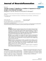
shown in Figure 1A, ear-hole closure was significantly
impaired in thermally injured mice. The average wound
closure response was delayed and reduced to 65% of that
of sham-treated mice. By 1-4 days post wounding, the
wound edge and adjacent tissue surrounding the ear-
holes became transiently inflamed (observed macro-
scopic reddening) in the majority of the mice (Figure 1B).
Moreover, in 50-60% of the thermally injured mice the
inflammatory response gradually became more progres-
sive resulting in an augmented inflammatory cell infiltra-
tion (data not shown), which lead to extensive necrosis
and fibrosis development (Figure 1B-C). Within 10-21
days wounding a large percentage (38%) had to be eutha-
nized early during the study due to extensive necrosis,
inflammation and ear deformation. The wound closure
measurements for these mice were not included in the
results depicted in Figure 1A. In contrast, no such ear
lesions were observed in mice that were sham-injured or
burned only (no ear wounds).
Cytokine and chemokine mRNA expression in wound
margin tissue from full-thickness burns
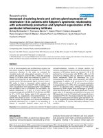
mRNA transcript expression levels were determined by
RT-PCR and normalized to gene transcript levels in nor-
mal uninjured naïve skin (Figure 2). The expression of
mRNA transcripts for IL-1α, IL-1β, TNFα, PGE
2
, MCP-1
Figure 1 Severe thermal injury at a remote distal site attenuates ear-hole wound closure and tissue regeneration in MRL/MpJ mice. (A) Time
course of ear-hole closure in thermally injured and solely ear-hole wounded MRL/MpJ mice. Data represent mean percent ear-hole closure ± 1 SD of
ten animals in each group. (B) Macroscopic changes in ear-hole healing control and thermally injured mice. Representative photographs of the ear-
hole wound sites at the indicated time post injury. The arrow points to a ear-hole wound that is 85% closed. (C) Representative histological sections
of ear wound margin tissue 9 and 21 days after full-thickness third degree thermal injury (original magnification, × 40)
0
10
20
30
40
50
60
70
80
90
100
0 4 8 121620242832
Days post burn injury
Percent ear-hole closure
D-4
D-9
D-21
D-9 D-21
C
A
B
No thermal
injury
Thermal
injury
Zins et al. Journal of Inflammation 2010, 7:25
/>Page 5 of 9
(CCL2), MIP-1α (CCL3), MIP-1β (CCL4), MIP-2α
(CXCL2), PF4 (CXCL4), and ENA-78 (CXCL5) was sig-
nificantly increased at days 1 and 3 post thermal injury.
By contrast, IL-6, IP-10 (CXCL10) and I-TAC (CXCL11)
expression peaked at day 1 post injury and then gradually
declined over the subsequent 6 days. Compared with
burn wounds on Balb/c mice (data not shown), MRL/MpJ
burn wounds had increased and prolonged transcription
of IL-6, IL-1β and decreased transcription of IL-10 and
chemokines MIP-2α, ENA-78, IP-10 and I-TAC in burn
wound margin tissue [26].
Severe thermal injury induces a systemic inflammatory
response which profoundly augments the local
inflammatory wound healing response as evidenced by
increased soluble mediator production in the serum
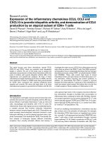
At 2, 6 and 24 hours post burn injury, the levels of various
inflammatory mediators were quantified in the serum.
Four cytokines/chemokines (IL-6, TNFα, KC (CXCL1),
and MIP-2α (CXCL2)) were significantly elevated within
the first 24 hr post wounding compared with unburned
(sham-treated) mice (Figure 3). A similar pattern was
observed in dually injured (ear punch and burn) mice. IL-
1β, and IL-10 were not detectable in thermally injured
mice at any of the studied time points, (data not shown)
and measurement of MIP-2α levels was not performed in
the dually-injured group.
Severe cutaneous thermal injury induces systemic pro-
inflammatory/anti-inflammatory mediator expression in
tissues remote from the wound site
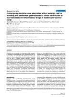
Significantly upregulated expression of mRNA tran-
scripts for proinflammatory cytokine/chemokines IL-6,
IL-1β, TNFα, MIF-1α, MIP-2α and MCP-1 as well as the
anti-inflammatory cytokine IL-10 and inducible nitric
oxide synthase (iNOS) were measured in the wounded
lung and ear at 6 and 24 hrs post thermal injury (Figures 4
and 5). Marked inflammation, as evidenced by the gross
and histological appearance of the wound (Figures 1B-C)
and increased global expression of potent inflammatory
mediators (Figure 5), was seen in the early ear wound
healing in the thermally injured mouse.
Discussion
Numerous studies have shown that severe traumatic
injury can lead to systemic pro-inflammatory responses
and cellular immune dysfunction [4-8,12,13,16]. In this
study, we demonstrate that systemic wound trauma
inflammatory signaling, mediated by acute thermal
injury, attenuates normal ear-hole closure and scarless
Figure 2 Cutaneous dermal burn injury induces a significant local inflammatory response as evidenced by soluble mediator production.
Chemokine, proinflammatory cytokine and PGE
2
synthase gene transcript levels at the wound edge/margin were assessed using quantitative RT-PCR
as described in the Material and Methods. Representative results from two independents experiments are shown. The expression of transcripts nor-
malized to 18 s at the wound margin at days 1, 3 and 7 post thermal injury were determined. Each value represents the mean ± SEM fold increase in
gene expression versus naïve uninjured skin from control MRL/MpJ mice. (n = 10 mice/time point, all data points have P values < 0.05 as compared
with naïve skin).
1
10
100
1000
10000
d-1
d-3
d-7
IL-1D IL-1E IL-6 TNFD IL-10 PGE2 MCP-1 MIP-1D MIP-1E MIP-2D PF4 ENA-78 IP-10 I-TAC
Gene
Gene expression (fold increase)
Zins et al. Journal of Inflammation 2010, 7:25
/>Page 6 of 9
healing in MRL/MpJ mice. Based on our previous find-
ings that MRL/MpJ mice exhibit a delay in wound closure
and myofibroblast development following sever thermal
trauma, and that these mice show a propensity to develop
the autoimmune state lupus following significant tissue
injury [22,26], we hypothesized that the activation of spe-
cific cell types and the production of cytokines and other
wound healing reparative mediators may be detrimental
to a remote ear-hole tissue regenerative response in
MRL/MpJ mice following traumatic tissue injury. Several
other groups have examined the link between local injury
and a systemic response. Similar to our findings Schwa-
cha et al [29], reported a significant inflammatory
response after burn wounds in small animals and delayed
wound healing distant to the site of injury.
In the present study, we show attenuated ear-hole clo-
sure and tissue regeneration in a large percentage of
MRL/MpJ mice following a remote, severe, full-thickness,
cutaneous thermal injury. Pathological examinations of
ear-hole wounds demonstrated excessive sequestration
and infiltration of macrophage and neutrophils. The
majority of the MRL/MpJ ear wounds healed with histo-
logical evidence of fibrosis and scar formation or became
chronically inflamed and necrotic to the point where
some of the animals had to be euthanized. The precise
reasons for aberrant ear-hole healing (wound closure) in
these mice following thermal injury are unclear, but may
be related to heightened systemic inflammation, higher
fibrotic cytokine signaling and the overproduction of
danger signals (presence of pathogens, pathogen-derived
molecules, or even self-derived molecular danger signals,
which arise from tissue damage) [30] which lead to
uncharacteristic healing and scar formation.
An essential feature of scarless healing/tissue regenera-
tion in adults and in fetal tissues appears to be a dimin-
ished cytokine response to injury [1,31,32]. On the other
hand, cytokines introduced into the fetal environment
evoke heightened inflammatory responses and tissue
fibrosis [33,34]. Similarly, PGE
2
stimulates leukocyte
accumulation, fibroblast proliferation, and collagen depo-
sition resulting in delayed wound healing and scar forma-
tion when introduced into early fetal wounds [35].
Elevated levels of IL1-β, IL-6, TNFα, PGE
2
, iNOS and
various chemokines are associated with areas of local as
well as systemic inflammation [13]. We observed a
heightened inflammatory response at the site of a remote
secondary injury following severe burn trauma; a patho-
logical picture suggestive of a global systemic immune
response. In contrast, blunted expression and production
of IL-1β, IL-6, TNFα, iNOS correlated with complete ear-
hole closure and scarless tissue healing. Pathological
Figure 3 Serum IL-6, TNFα, KC, and MIP-2α levels at 0, 2, 6 and 24 hr post thermal injury and post thermal injury plus ear punch. Thermal
injury induced a significant systemic inflammatory response, as did the dual-injury model. Serum levels were determined as described in the Material
and Methods section. MIP-2α measurement not performed for thermal injury plus ear punch group. Data are mean ± SEM (n = 5-7 mice/time point, *
P < 0.05 as compared with baseline time 0-hr levels)
0
10
20
30
40
50
60
70
80
90
100
0-hr
2-hr
6-hr
24-hr
IL-6 TNF-D KC MIP-2D IL-6 TNF-D KC
picograms/ml
*
*
*
*
*
*
*
*
*
*
*
*
*
*
*
*
*
*
Thermal injury only
Thermal + ear-hole injury
Zins et al. Journal of Inflammation 2010, 7:25
/>Page 7 of 9
examinations of ear-hole wounds in thermally injured
mice demonstrated excessive sequestration and infiltra-
tion of macrophage and neutrophils. These observations
lead us to speculate that macrophage hyperactivity after
thermal injury may play a critical role in altered ear-hole
healing response, as primed hyperactive macrophages
might contribute to the increased recruitment and
sequestration of leukocytes, tissue inflammation, and
damage-tissue necrosis precipitated through excessive
cytokine and chemokine production [36].
Conclusion
Collectively, the findings from this study have important
implications in defining how immune responses induced
following a severe traumatic injury regulate other cellular
events critical in distal wound healing. Although the
mechanism(s) mediating scarless healing are not fully
understood, an absence of local as well as systemic
inflammation seems to be particularly important for scar-
less healing in the MRL/MpJ mouse [21,23-25]. We spec-
ulate that optimal ear-hole healing requires activation of
inhibitory signals that suppress chemokine and cytokine
synthesis, resulting in resolution of the inflammatory
infiltrate. The effects of trauma on healing and tissue
regeneration responses may depend upon the stimuli for
its induction, its anatomical site, the degree of immune
cell activation and the time post burn at which the injury
is induced. Our findings are not altogether surprising;
extensive tissue damage can trigger potent cellular activa-
tion signals that prime innate immune system to sense
"danger" [30,37] and that tissue regeneration and scarring
are tightly regulated by inflammation. Additional studies
are warranted and we believe that this is an appropriate
model to investigate the pathomechanisms of normal and
aberrant wound healing.
Figure 4 Systemic inflammation in the lung following a local full-thickness cutaneous thermal injury. Lungs from MRL/MpJ mice with ear-hole
wounds ± thermal injury were evaluated for inflammatory gene expression. Severe thermal trauma induced significant elevation of lung inflammatory
chemokine, and iNOS transcript levels at 6 and 24 hr post injury in the lung. Gene levels were assessed using quantitative RT-PCR as described in the
Material and Methods. The expression of transcripts normalized to 18 s in the lung at 6 and 24 hrs post thermal injury were determined. Each value
represents the mean ± SEM fold increase in gene expression versus lung tissue from uninjured control MRL/MpJ mice (n = 5-7 mice/time point, * P <
0.05 as compared with baseline time 0-hr levels)
Time (hr) and Gene
0
5
10
15
20
25
624624624624624624624624
*
*
*
*
*
*
*
*
*
Gene expression (fold increase)
IL-6 IL-1E TNF-D MIF-1 MIP-2D iNOS MCP-1 IL-10
No thermal injury
Thermal injury
Zins et al. Journal of Inflammation 2010, 7:25
/>Page 8 of 9
List of abbreviations used
CCL-: chemokine ligand with cysteine-cysteine motif;
CXC-: chemokine with cysteine-amino acid-cysteine
motif; ENA-78: epithelial cell-derived neutrophil-activat-
ing peptide-78; GM-CSF: granulocyte/macrophage col-
ony stimulating factor; IFN-γ: interferon gamma; IL-
interleukin-; iNOS: inducible nitric oxide synthase; IP-10:
interferon inducible protein 10; I-TAC: interferon induc-
ible t cell alpha; KC: cytokine induced neutrophil
chemoattractant; MCP-1: monocyte chemoattractive
protein-1; MIP-: macrophage inflammatory protein-;
NMRC: Naval Medical Research Center; PF-4: platelet
factor 4; PGE
2
: prostaglandin E2; RANTES: regulated
upon activation, normal T cell expressed and secreted;
RT-PCR: real-time polymerase chain reaction; TGF-:
transforming growth factor-; TNF-: tumor Necrosis Fac-
tor-; USUHS: Uniformed Services University of the
Health Sciences; WRAIR: Walter Reed Army Institute of
Research.
About the Authors
The authors are employees of the U.S. Government. This
work was prepared as part of their official duties. Title 17
U.S.C. §105 provides that 'Copyright protection under
this title is not available for any work of the United States
Government.' Title 17 U.S.C §101 defines a U.S. Govern-
ment work as a work prepared by a military service mem-
ber or employees of the U.S. Government as part of that
person's official duties. The opinions or assertions con-
tained in this paper are the private views of the authors
and are not to be construed as reflecting the views, policy
or positions of the Department of the Navy, Department
of Defense nor the U.S. Government.
Competing interests
The authors declare that they have no competing interests.
Authors' contributions
TAD conceived and designed the research. TAD, MFA, KA and SRZ carried out all
the experimental work and data collection. TAD, MFA, KA, EAE and SRZ con-
ducted the data analysis and interpretation. TAD, EAE, and SRZ wrote the man-
uscript and/or made critical revisions. All authors read and approved the final
version of the manuscript.
Acknowledgements
This work was supported by BUMED work units 601153N.04508.5180.A0801,
601153N.04508.519.A0508, and ONR work unit 602236N.42237.W160.A0806
Author Details
1
Regenerative Medicine Department, Operational and Undersea Medicine
Directorate at the Naval Medical Research Center Silver Spring, MD 20910-
7500, USA and
2
Department of Surgery, Uniformed Services University of the
Health Sciences, Bethesda, MD 20814, USA
Received: 12 March 2010 Accepted: 25 May 2010
Published: 25 May 2010
This article is available from: 2010 Zins et al; licensee BioMed Central Ltd. This is an Open Access article distributed under the terms of the Creative Commons Attribution License ( .0), which permits unrestricted use, distribution, and reproduction in any medium, provided the original work is properly cited.Journal of Inflammation 2010, 7:25
Figure 5 Severe local thermal injury markedly heightens the remote ear-hole inflammatory processes resulting in attenuated tissue heal-
ing. MRL/MpJ mice with ear-hole wounds ± thermal injury were evaluated. Gene transcript levels obtained from tissue collected at the edge/margin
of the ear-hole wounds were assessed using quantitative RT-PCR as described in the Material and Methods. The expression of transcripts normalized
to 18 s in ear tissue at 6 and 24 hrs post thermal injury were determined. Each value represents the mean ± SEM versus ear tissue from uninjured con-
trol MRL/MpJ mice (n = 5-7 mice/time point, * P < 0.05 as compared with baseline time 0-hr levels).
0.1
1
10
100
1000
624624624624624624624624
*
Time (hr) and Gene
*
*
*
Gene expression (fold increase)
IL-6 IL-1E TNF-D MIF-1 MIP-2D iNOS MCP-1 IL-10
No thermal injury
Thermal injury
Zins et al. Journal of Inflammation 2010, 7:25
/>Page 9 of 9
References
1. Singer AJ, Clark RA: Cutaneous wound healing. N Engl J Med 1999,
341:738-746.
2. Menke NB, Ward KR, Witten TM, Bonchev DG, Diegelmann RF: Impaired
wound healing. Clinics in Dermatology 2007, 25:19-25.
3. Gibran NS, Heimbach DM: Current status of burn wound
pathophysiology. Clin Plast Surg 2000, 27:11-22.
4. Mannick JA, Rodrick ML, Lederer JA: The immunologic response to
injury. J Am Coll Surg 2001, 193:237-244.
5. Schwacha MG: Macrophages and post-burn immune dysfunction.
Burns 2003, 29:1-14.
6. Lederer JA, Rodrick ML, Mannick JA: The effects of injury on the adaptive
immune response. Shock 1999, 11:153-159.
7. Kell MR, Kavanaugh EG, Goebel A, Soberg CC, Lederer JA: Injury primes
the immune system for an enhanced and lethal T-cell response against
bacterial superantigen. Shock 1999, 12:139-144.
8. Kelly JL, O'Suilleabhain CB, Soberg CC, Mannick JA, Lederer JA: Severe
injury triggers antigen-specific T-helper cell dysfunction. Shock 1999,
12:39-45.
9. Moore FA, Moore EE: Evolving concepts in the pathogenesis of
postinjury multiple organ failure. Surg Clin North Am 1995, 75:257-277.
10. Piccolo MT, Wang Y, Verbrugge S, Warner RL, Sannomiya P, Piccolo NS,
Piccolo MS, Hugli TE, Ward PA, Till GO: Role of chemotactic factors in
neutrophil activation after thermal injury in rats. Inflammation 1999,
23:371-385.
11. Botha AJ, Moore FA, Moore EE, Kim FJ, Banerjee A, Peterson VM: Postinjury
neutrophil priming and activation: an early vulnerable window.
Surgery 1995, 118:358-364. discussion 364-355
12. Dovi JV, Szpaderska AM, DiPietro LA: Neutrophil function in the healing
wound: adding insult to injury? Thromb Haemost 2004, 92:275-280.
13. Ipaktchi K, Mattar A, Niederbichler AD, Hoesel LM, Vollmannshauser S,
Hemmila MR, Su GL, Remick DG, Wang SC, Arbabi S: Attenuating burn
wound inflammatory signaling reduces systemic inflammation and
acute lung injury. J Immunol 2006, 177:8065-8071.
14. Lyons A, Goebel A, Mannick JA, Lederer JA: Protective effects of early
interleukin 10 antagonism on injury-induced immune dysfunction.
Arch Surg 1999, 134:1317-1323. discussion 1324
15. Ogle CK, Guo X, Szczur K, Hartmann S, Ogle JD: Production of tumor
necrosis factor, interleukin-6 and prostaglandin E2 by LPS-stimulated
rat bone marrow macrophages after thermal injury: effect of
indomethacin. Inflammation 1994, 18:175-185.
16. Paterson HM, Murphy TJ, Purcell EJ, Shelley O, Kriynovich SJ, Lien E,
Mannick JA, Lederer JA: Injury primes the innate immune system for
enhanced Toll-like receptor reactivity. J Immunol 2003, 171:1473-1483.
17. Clark LD, Clark RK, Heber-Katz E: A new murine model for mammalian
wound repair and regeneration. Clin Immunol Immunopathol 1998,
88:35-45.
18. Davis TA, Longcor JD, Hicok KC, Lennon GG: Prior injury accelerates
subsequent wound closure in a mouse model of regeneration. Cell
Tissue Res 2005, 320:417-426.
19. Metcalfe AD, Willis H, Beare A, Ferguson MW: Characterizing
regeneration in the vertebrate ear. J Anat 2006, 209:439-446.
20. Colwell AS, Krummel TM, Kong W, Longaker MT, Lorenz HP: Skin wounds
in the MRL/MPJ mouse heal with scar. Wound Repair Regen 2006,
14:81-90.
21. Beare AH, Metcalfe AD, Ferguson MW: Location of injury influences the
mechanisms of both regeneration and repair within the MRL/MpJ
mouse. J Anat 2006, 209:547-559.
22. Davis TA, Amare M, Naik S, Kovalchuk AL, Tadaki D: Differential cutaneous
wound healing in thermally injured MRL/MPJ mice. Wound Repair
Regen 2007, 15:577-588.
23. Heber-Katz E, Gourevitch D: The relationship between inflammation
and regeneration in the MRL mouse: potential relevance for putative
human regenerative(scarless wound healing) capacities? Ann N Y Acad
Sci 2009, 1172:110-114.
24. Ueno M, Lyons BL, Burzenski LM, Gott B, Shaffer DJ, Roopenian DC, Shultz
LD: Accelerated wound healing of alkali-burned corneas in MRL mice is
associated with a reduced inflammatory signature. Invest Ophthalmol
Vis Sci 2005, 46:4097-4106.
25. Li X, Mohan S, Gu W, Baylink DJ: Analysis of gene expression in the
wound repair/regeneration process. Mamm Genome 2001, 12:52-59.
26. Anam K, Amare M, Naik S, Szabo KA, Davis TA: Severe tissue trauma
triggers the autoimmune state systemic lupus erythematosus in the
MRL/++ lupus-prone mouse. Lupus 2009, 18:318-331.
27. Hoffmann SC, Pearl JP, Blair PJ, Kirk AD: Immune profiling: molecular
monitoring in renal transplantation. Front Biosci 2003, 8:e444-462.
28. Livak KJ, Schmittgen TD: Analysis of relative gene expression data using
real-time quantitative PCR and the 2(-Delta Delta C(T)) Method.
Methods 2001, 25:402-408.
29. Schwacha MG, Nickel E, Daniel T: Burn injury-induced alterations in
wound inflammation and healing are associated with suppressed
hypoxia inducible factor-1alpha expression. Mol Med 2008, 14:628-633.
30. Akira S, Uematsu S, Takeuchi O: Pathogen recognition and innate
immunity. Cell 2006, 124:783-801.
31. Buchanan EP, Longaker MT, Lorenz HP: Fetal skin wound healing. Adv
Clin Chem 2009, 48:137-161.
32. Linares HA: From wound to scar. Burns 1996, 22:339-352.
33. Krummel TM, Michna BA, Thomas BL, Sporn MB, Nelson JM, Salzberg AM,
Cohen IK, Diegelmann RF: Transforming growth factor beta (TGF-beta)
induces fibrosis in a fetal wound model. J Pediatr Surg 1988, 23:647-652.
34. Haynes JH, Johnson DE, Mast BA, Diegelmann RF, Salzberg DA, Cohen IK,
Krummel TM: Platelet-derived growth factor induces fetal wound
fibrosis. J Pediatr Surg 1994, 29:1405-1408.
35. Wilgus TA, Bergdall VK, Tober KL, Hill KJ, Mitra S, Flavahan NA, Oberyszyn
TM: The impact of cyclooxygenase-2 mediated inflammation on
scarless fetal wound healing. Am J Pathol 2004, 165:753-761.
36. Alexander M, Chaudry IH, Schwacha MG: Relationships between burn
size, immunosuppression, and macrophage hyperactivity in a murine
model of thermal injury. Cell Immunol 2002, 220:63-69.
37. Matzinger P: The danger model: a renewed sense of self. Science 2002,
296:301-305.
doi: 10.1186/1476-9255-7-25
Cite this article as: Zins et al., Wound trauma mediated inflammatory signal-
ing attenuates a tissue regenerative response in MRL/MpJ mice Journal of
Inflammation 2010, 7:25