Báo cáo y học: "Differential induction of inflammatory cytokines by dendritic cells treated with novel TLR-agonist and cytokine based cocktails: targeting dendritic cells in autoimmunity" potx
Bạn đang xem bản rút gọn của tài liệu. Xem và tải ngay bản đầy đủ của tài liệu tại đây (1.77 MB, 12 trang )
RESEARC H Open Access
Differential induction of inflammatory cytokines
by dendritic cells treated with novel TLR-agonist
and cytokine based cocktails: targeting dendritic
cells in autoimmunity
Simon S Jensen
1*
, Monika Gad
2
Abstract
Background: Dendritic cells (DC) are main gate-keepers of the immune system, bridging the innate and
adaptive immune system. DCs are able to mature into inflammatory DCs at sites of inflammation in both
autoimmune and allergic disease, thereby sustaining a continuous activation of the adaptive immune
system at sites of inflammation. This function of DCs makes them attractive target cells for therapeutic
intervention in inflammatory diseases. We have designed a DC-based screening model by which drug
candidates can b e evaluated for their ability to suppress DC maturation into an inflammatory and disease
promoting phenotype.
Methods: Human monocyte derived DCs were differentiated using IL-4 and GM-CSF to immature DCs (imDCs).
The imDCs were treated with various combinations of TLR-agonists and pro-inflammatory cytokines to identify
cocktails with ability to mature imDCs into inflammatory DCs. The effect of the cocktails on DC maturation was
evaluated using ELISA and cytokine arrays to measure secreted cytokines and chemokines. FACS analysis was used
to assess expression of maturation markers, and functional studies were carried out using naïve allogeneic T-cells
to assay for a Th1-promoting DC phe notype.
Results: Nine cocktails were designed with potent ability to induce secretion of the Th1-promoting cytokines
IL-12p70 and TNFa from imDCs, and three were able to induce the Th17-promoting cytokine IL-23. The cocktails
were further characterized using cytokine arrays, showing induction of inflammation related cytokines and
chemokines like CXCL10, CCL2, CCL4, CCL8, CCL15, CCL20 and IL-8, of which some are present in a range of
autoimmune pathologies. Prostaglandin E2 secretion was identified from DCs treated with TLR agonists poly I:C
and peptidoglycan, but not LPS. The cocktails were able to induce DC maturation markers like HLA-DR, CD40,
CD80, CD83 and CD86, except the TLR7/8 agonist R848. Functional end-points made by co-culture of allogeneic
CD4
+
T cells with the cocktail treated DCs, showed that five cocktails in particular could induce a classical
Th1-phenotype with ability to secrete high amounts of the hall-mark cytokine IFNg. The model was validated using
dexamethasone and two COX-inhibitors, which were able to suppress the cocktail driven pro-inflammatory DC
maturation.
Conclusions: The identification of novel Th1-promoting cocktails allows screening of anti-inflammatory
drug candidates by assessing the ability to suppress the activation and differentiation of imDCs into
inflammatory DCs with a specific Th1-promoting phenotype. The model thus provides a screening tool,
which can identify potential anti-inflammatory effects on the natural regulator of th e immune response,
the dendritic cell.
* Correspondence:
1
Department of immune targeting, Bioneer A/S, Kogle Allé 2, Hørsholm, DK-
2970, Denmark
Jensen and Gad Journal of Inflammation 2010, 7:37
/>© 2010 Jensen and Gad; licensee BioMed Central Ltd. This is an Open Access article distributed under the terms of the Creative
Commons Attribution License ( which permits unrestricted use, distribution, and
reproduction in any medium, provided the original work is properly c ited.
Background
Dendritic cells (DCs) are central in the pathogenesis of
immune disorders, where they respond towards envir-
onmental factors by regulating the adaptive immune
system through activation and expansion of T cells. In
autoimmunity the immature DCs develop into inflam-
matory DCs, which present self-antigens to T cells,
which are activated towards the self-antigen, causing
an autoimmune response. The inflammatory DCs are
responsible for secretion of cytokines with a pro-
inflammatory function like TNFa, IL-12p70, IL-23, and
several other inflammatory mediators like nitric oxide,
prostaglandins and chemokines [1,2]. During inflam-
mation, immature DCs are attracted to the site of
inflammation by chemokines in the microenvironment,
and a large number of DCs are often present at sites
of inflammation. Aft er antigen uptake at the inflamma-
tory site and maturation by inflammatory cytokines
and chemokines, the DCs differentiate into inflamma-
tory DCs which migrate to the lymph nodes and sti-
mulate T cell function and proliferation. Since DCs
have a short half-life under inflammatory conditions
and are upstream in the inflammatory process, they
are attractive target cells for therapeutic intervention
of inflam matory disease s [1]. D Cs express a unique
repertoire of receptors essential for the innate immune
response, termed pattern recognition receptors (PRRs),
including the Toll Like Receptors (TLRs), nucleotide
binding and oligomerization domain-like receptors
(NLRs) and C-type lectin-like receptors (CLRs) [3].
These receptors and their corresponding signalling
pathways are involved in the pathology of autoimmune
diseases like e.g. psoriasis and rheumatoid arthritis
(RA), and in particular inflammatory bowel disease
(IBD) defining Crohns disease and ulcerative colitis [4].
DCs also express receptors involved in cytokine
responsesaswellaschemokinereceptorsinvolvedin
cell migration, and are thus responsive to various types
of environmental factors [5]. Pro-inflammatory activa-
tion of DCs can be naturally counterbalanced by inhi-
bitory molecules believed to regulate and fine-tune
T-cell responses, and particular the B7-H1 and H4
receptors provide negative signals that suppress effec-
tor T-cell responses [6]. Finally, DCs are the source of
secretion of very potent pro-inflammatory cytokines
and chemokines. The hall-mark cytokines involved in
initiation of the adaptive Th1 immune responses are
IL-12p70 whereas IL-6, TGFb and IL-23 are involved
in initiation and sustainment of Th17 differentiation.
Both types of immune responses are known to be
involved in chronic autoimmune disorders like
e.g. Crohns disease [7,8]. Essential chemokines secreted
from DCs are IL-8, CCL17, CCL18, CCL22 and APRIL,
involved in both Th1 and Th2 type responses [8,9].
Theuniqueandverycomplexsignallingnetworkin
DCs involving PRRs and secretion of early triggers of
inflammation, which are mainly associated with or
restricted to DC function, opens a window of opportu-
nities for DC-specific therapeutics in treatment of
inflammatory disease [1]. The aim of this work was to
mimic the development in vitro,ofimmatureDCsinto
inflammatory DCs as seen in autoimmune condi tions
like e.g. Crohns disease, arthritis and psoriasis. We
used human monocyte derived imDCs for evaluation
of various combinations of TLR agonists and pro-
inflammatory cytokines, in the attempt to design cock-
tails able to stimulate an inflammatory DC phenotype,
mimicking the situation in vivo, where immature DCs
migrate to sites of inflammation, after subsequent
exposure to TLR agonists and inflammatory cytokines
as seen in the inflammatory tissue. To mimic the
potential therapeutic situation in vivo,wheremono-
cytes and imDCs are exposed to a drug prior to migra-
tion into the t issue and towards the inflammatory site,
imDCs are in our model exposed to the drug candidate
prior to exposure to the cocktail which mimics the
microenvironment present at the inflammatory site.
The ability o f the drug and cocktail treated DCs to sti-
mulate an immune response are then determined after
18-24 h, at a time point where inflammatory DCs in
vivo are in the process of migration to and activation
of the adaptive immune response in the lymph nodes.
We suggest that DC-based in v it ro models of inflam-
matory conditions as described here are suitable models
for screening of compounds or i mmune modulating
agents like microorganisms specifically targeting
immune disorders.
Methods
DC development from monocytes
PBMC were purified from buffy coats from healthy
donors above the age of 18, which did not suffer from
immune disorders or had been on recent medication.
PBMCs were purified by centrifugation over a Ficoll-
pague (GE Healthcare limited, Buckinghamshire, UK)
gradient, and monocytes were isolated from PBMCs by
positive selection of CD14
+
cells (specific for monocytes)
by magnetic beads (Dynal, Invitrogen, Carlsbad, CA,
USA) according to the manufacturer’s instructions. The
CD14
+
monocytes were cultured in 6-well plates in a
conc. at 2 × 10
6
cells/ml (3 mL/well) in RPMI/5% FCS
supplemented with recombinant human GM-CSF (20
ng/mL) and IL-4 (20 ng/mL), (PeproTech, London, UK).
The medium was changed after 2 and 3 days. After 6
days of culture the immature DCs were re-cultured into
96-well plates at 10
6
cells/well.
Jensen and Gad Journal of Inflammation 2010, 7:37
/>Page 2 of 12
ELISA and PGE2 measurements
Human ELISA kits were used from the following manu-
facturers: IL-12p70, TNFa, (R&D Systems, Minneapolis,
MN, USA), IFNg, IL-23 (eBioscience, San Diego, CA,
USA). Prostaglan din E2 was measured using the Prosta-
glandin E2 EIA Kit - Monoclonal (Cayman Chemical,
Ann Arbor. MI, US A). DCs were setup in 96 well plates
with 100.000 cells/well and cocktails tested in triplicates.
After 24 h incubation with cocktails, the conditioned
media was removed from the wells and stored at -80°C
until analyses. The media was diluted to reach the linear
range for each ELISA assay, and the amount of cytokine
for each sample was determined in duplicate. Using a
standard curve provided in the kit, the concentration of
cytokines was determined for each sample.
Cytokine array
Human inflammation antibody based cytokine arrays
were used from RayBiotech, (Norcross GA, USA)
according to manufacturers instructions, using condi-
tioned media from DCs from four different donors trea-
ted with the individual cocktails and mixed in equal
amounts. The membranes were developed using west
pico luminescence reagent from Pierce ( Rockford, IL,
USA) and exposed on Amersham hyperfilm ECL, (GE
Healthcare limited, Buckinghamshire, UK). Exposures
which allowed identification of most abundantly
secreted cytok ines and chemokines were scanned and
quantitated using the ImageJ-software from NIH, and
induced cytokines and chemokines for each cocktail
compare d to the level seen for non-treated DCs. Induc-
tion up to 10 fold of the level seen for non-treated DCs
was termed weakly induced, between 10-100 fold was
termed modest induction, and above 100 fold induction
termed strong induction. The induced cytokines and
chemokines are shown in table 1. Constitutively secreted
proteins as seen for non-treated DCs included IL-8,
CCl2, CCL5, CCL13, TGFb, TIMP1 and 2.
DC cocktail screening and validation
Potent IL-12p70 and TNFa stimulating cocktails were
designe d by series of combinations of pro-infl ammatory
cytokines and TLR agonists. The cytokines TNFa, IFNg,
IFNa IL-6 and IL-1b were from Peprotech (London,
UK) and diluted in cell media to reach the indicated
concentrations in table 1. TLR agonists were of TLR
grade and from the following suppliers: lipopolysacchar-
ide (LPS), polyinosinic polycytidylic acid (poly I:C),
(Sigma, St. Louis, MI, USA). Peptidoglycan and R848
(Resiquimod) were from Alexis biochemicals (Axxora,
San Dieg o, CA, USA). Cocktails wer e com bined in a 5×
working stock and added to immature DCs 30-60 min
after counting and plating in 96 well plates. Cell density
were 100.000 cells/96 well. For inhibition experiments,
dexamethasone was added (0.01-0.1-1 uM) either 6 or
24 h prior to addition of cocktail to allow equilibrium
and inhibition of inflammatory targets within the cells
prior to addition of cocktails. The unspecific Cox inhibi-
tor indomethacin (Sig ma) was solubilized in ETOH, the
specific Cox2 inhibitor NS398 (Cayman Chemical) was
solubilizedinDMSOandbothaddedtwohpriorto
cocktails. The DCs were routinely tested for cell viabi-
lity, and no significant differences were seen for dexa-
methasone and/or cocktail treated cells (data not
shown).
Flow cytometric analysis
Harvested DCs were washed twice with PBS supplemen-
ted with 1% FBS. Fc receptors were blocked with excess
human IgG (Sigma) on ice for 10 min. Immunofluores-
cence staining was performed by incubation of DCs for
30 min at 4°C with each mAb diluted to the optimal
concentration according to the manufacturer’sinstruc-
tions. The follo wing mAbs were used: anti-CD1a-APC,
anti-CD14-Pe, anti-HLA-DR-Pe, anti- CD80-Pe, anti-
CD83-Pe, anti-CD86-Pe, anti-CD40-Pe (all antibodies
were from Becton Dickinson Pharmingen, NJ, USA).
Relevant isotype controls were always used. Samples
were acquired on a FACSArray (Becton Dickinson, NJ).
At least 5000 mononuclear cells were gated using a
combination of forward-angle and side scatter to
exclude dead cells and debris. Data were analysed with
FACSDiva software.
Western blot
Cox2, and actin expression was analysed by Western
blot as described by the supplier (Invitrogen). Briefly,
DCs were seeded in 6 well plates at a density of 3-4 ×
10
6
cells/well. 24 h after addition of cocktail, adherent
and non-adherent cells were washed in ice cold PBS.
Adhe rent cells were scraped off using a cell scraper and
spun down. Loading buffer was added to the cell pellet
and lysates processed as described by the manuf acturer
using Tris-Glycine based SDS-PAGE gels ( Invitrogen).
Exposure was made on ECL fil ms as for cytokine arrays.
Antibodies against Cox2 and actin were from S anta
Cruz (Santa Cruz, CA, USA).
MLR
In a MLR, DCs and peripheral blood lymphocytes from
two different (allogeneic) individuals are mixed together
in tissue culture round bottom wells for several days.
DCs will stimulate lymphocy tes from an i ncompatible
individual to proliferate significantly whereas those from
compatible individuals will not. CD4
+
T cells were iso-
lated directly from PBMCs with anti-CD4 Dynabeads
and Detach-aBead (Dynal, Invitrogen,) according to the
manufacturer’s instructions. Mixed lymphocyte cultures
Jensen and Gad Journal of Inflammation 2010, 7:37
/>Page 3 of 12
were performed in quadruplicates in 96-well round-
bottom microtiter plates. A fixed number of 10
5
respond-
ing CD4
+
T cells were added to a 2 fold titrated number
of allogeneic mitomycin-C treated (25 μg/ml for 30 min
by 37°C) DCs starting from 10
4
(DC:T cell ratio:
1:10-1:640). The cells were cultured for 5 days and prolif-
eration was measured after addition of 0.5 μCi/well of
[
3
H]thymidine (Amersham, Little Chalfont, UK) for the
last 18-24 h. The cells were harvested on a Filtermate
196 (Packard instruments, CT, USA) and incorporation
was determined by liquid scintillation counting (Top-
count, Packard Instruments, CT, USA).
Analyses of cytokines secreted from T-cells in MLR
Supernatants from the mixed lymphocyte cultures w ere
selected on day 5 and measured for levels o f TNFa,IL-
13 (R&D Systems) and IFNg by ELISA (e-Bioscience).
To further boost the secretion of cytokines, primed CD4
+
Tcellsfroma7dayMLRculturewerecollectedand
washed. For detection of cytokine production in the cul-
ture supernatants, the T cells were restimulated with
plate-bound anti -CD3 (OKT3, 5 ug/mL) and soluble
anti-CD28 (1 ug/mL) (both from BD Biosciences, NJ) at
a concentration of 10
6
cells/mL for 24 h. The ELISA
was performed in triplicates.
Statistical analyses
Data were analyzed using unpaired, two sided t-test,
(***P < 0.005, **P < 0.01, *P < 0.05).
Results
Identification of cocktails with the capability to induce
secretion of the Th-1 promoting cytokines IL-12p70 and
TNFa from human dendritic cells
The two pro-inflammatory cytokines IL-12p70 and
TNFa were chosen as end-points for our present DC
based screening model. After several rounds of optimi-
zation in order to select for potent combinations of
TLR-agonists and cytokines, a range of cocktails were
designed, which were able to induce IL-12p70 and
TNFa secretion when added to immature DCs. The 9
most potent cocktails are seen in table 1, listed 1-9. In
order to determine the variation in donor r esponse for
each cocktail, a series of 15 different donor-derived
batches of DCs were tested for their individual response
to the range of optimized cocktails from table 1. The
Table 1 Cocktail compositions and the cocktail stimulated DC expression pattern for selected cytokines and
chemokines on cytokine arrays
Cocktail Composition Secreted cytokines and chemokines
Weak
induction
(1-10 fold)
Modest
induction
(10-100 fold)
Strong
induction
(> 100 fold)
LPS LPS (0,1ug/mL) IL-8, CCL2, CCL4, CXCL5,
CXCL10, GM-CSF
IL-10, CCL20, CXCL1-3 IL-6, IL-12, TNFa, VEGF, CCL8,
CCL15, CXCL1
Cocktail 1 IFNg (20ng/mL)+ TNFa (50ng/mL)+
Poly I:C 12,5 ug/mL + IL-1b (10ng/
mL) + IFNa (6ng/mL)
IL-8, CCL2, CCL4, CCL5, CXCL5,
CXCL10
IL-10, CCL20, CXCL1-3, GM-CSF IL-6, IL-12, TNFa, VEGF, CCL8,
CCL15, CXCL1
Cocktail 2 IFNg (20ng/mL)+ Poly I:C 12,5 ug/mL
+ IL-1b (10 ng/mL) + IFNa (6ng/mL)
IL-8, CCL2,
CCL4, CCL5, CXCL5, CXCL10
IL-10, CCL20, CXCL1-3, GM-CSF IL-6, IL-12, TNFa, VEGF, CCL8,
CCL15, CXCL1
Cocktail 3 IFNg (20ng/mL)+TNFa (50ng/mL)+
peptidoglycan (10ug/mL)
IL-8, CCL2, CCL4, CCL5, CXCL5 IL-10, CCL20, CXCL1-3, CXCL10,
GM-CSF
IL-6, IL-12, TNFa, VEGF, CCL8,
CCL15, CXCL1
Cocktail 4 LPS (0,1ug/mL)+ IFNg (20ng/mL) IL-8, CCL2, CCL4, CCL5, CXCL5,
GM-CSF
IL-10, CCL20, CXCL1-3, CXCL10 IL-6, IL-12, TNFa, VEGF, CCL8,
CCL15, CXCL1
Cocktail 5 LPS (0,1ug/mL)+ IFNg (20ng/mL)+
TNFa (50ng/mL)
IL-8, CCL2, CCL4, CCL5, CXCL5,
GM-CSF
IL-10, CCL20, CXCL1-3, CXCL10 IL-6, IL-12, TNFa, VEGF, CCL8,
CCL15, CXCL1
Cocktail 6 R848 (1,0ug/mL) IL-8, CCL2, CCL4, CCL5, CXCL10 IL-10, CCL15, CCL20, CXCL1-3,
CXCL5, GM-CSF
IL-6, IL-12, TNFa, VEGF, CCL8,
CXCL1
Cocktail 7 R848 (1,0ug/mL) + IFNg (20ng/mL) IL-8, CCL2, CCL4, CCL5, CCL20,
CXCL5, GM-CSF
IL-10, CCL15, CCL20, CXCL1-3,
CXCL10
IL-6, IL-12, TNFa, VEGF, CCL8,
CXCL1
Cocktail 8 R848 (1,0ug/mL) + poly I:C
(10ug/mL)
IL-8, CCL2, CCL4, CCL5, CXCL5,
CXCL10
IL-10, CCL20, CXCL1-3, GM-CSF IL-6, IL-12, TNFa, VEGF, CCL8,
CCL15, CXCL1
Cocktail 9 IFN
g (20ng/mL) + IL-1b (10ng/mL) IL-8, CCL2, CCL4, CCL5, CXCL5,
GM-CSF
IL-10, CCL15, CCL20, CXCL1-3,
CXCL10
IL-6, IL-12, TNFa, VEGF, CCL8,
CXCL1
Table 1 shows the composition of cocktails combined by TLR-agonists and cytokines. TLR agonists were peptidoglycan (TLR2), Poly I:C (TLR3), LPS (TLR4/CD14/
MD2), R848 (TLR7/8) and cytokines IFNg,TNFa,IL-1b, IFNa. Secreted cytokines and chemokines were determined by combination of conditioned media for
treatment of immature DCs from four different donors in order to obtain information for a general response and reduce single donor-variations. Signal intensities
were semi-quantitatively calculated using the ImageJ software, and the fold of induction compared to untreated cells. Induced cytokines and chemokines were
classified into weakly induced proteins (1-10 fold induced vs untreated), modestly induced proteins (10-100 fold induced vs untreated controls), and strongly
induced proteins (more than 100 fold vs untreated controls).
Jensen and Gad Journal of Inflammation 2010, 7:37
/>Page 4 of 12
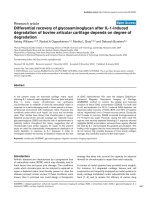
secretion of IL-12p70 was determined for all DC-batches
and the exact p rotein levels shown for each cocktail in
figure 1A. LPS was included as a control in these experi-
ments, but induced only slightly IL-12p70 compared to
the optimized cocktails. The addition of peptidoglycan
and poly I:C alone showed similar effects, with very
weak induction of IL12-p70 (data not shown). The aver-
age IL-12p70 secretion ranged from approximately 5 ng/
mL for cocktail 6 (R848), to above 20 ng/mL for cocktail
2(IFNg,PolyI:C,IL-1b,IFNa). The most potent IL-
12p70 inducing cocktails are cocktail 1, 2, 4, 5 and 8.
A similar analysis was made for secretion of TNFa,
where the total amount of TNFa secreted for each cock-
tail treated DC batch is shown in figure 1B. The variation
in total amounts secreted from each DC-batch was lower
than for IL-12p70 secretion, indicating that the donor
derived DCs are more responsive for induction of TNFa
secretion than for IL-12p70 using these cocktails. LPS
was able to induce TNFa secretion to significant levels in
all DC -ba tches compared to untreated cells, in contrast
to LPS induced IL-12p70 secretion. Cocktail 1 to 5 and 8
stimulated TNFa secretion significantly better than LPS,
where cocktail 6, 7 and 9 did not.
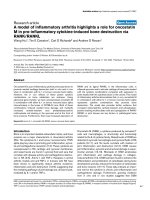
Cytokine array
In order to determine if the cocktails induce a cytokine
and chemokine profile which corresponds to the pattern
seen in tissue from patients suffering from Th1-directed
immune disorders we performedacytokinearrayon
conditioned media from cocktail treated DCs mixed
from four different donors (figure 2). LPS induced a
range of pro-inflammato ry proteins like IL-6, IL-8,
TNFa, CCL2, 5, 8, 15, 20, CXCL1-3 and 10 (figure 2
and table 1). The cocktails all induced inflammation
associated cytokines like IL-6 (> 100 fold), IL-12p70 (>
100 fold) and TNFa (> 100 fold). The cocktails also sti-
mulated secretion of chemokines like IL-8, CXCL1-3, 5
and 10, CCL2, 4, 5, 8, 15, 20, which mainly are involved
in recruitment of leukocytes like neutrophils, basophils,
monocytes, DCs, Th1 and NK cells to sites of inflamma-
tion[10],aswellastheangiogenicstimulatorVEGF.
Also the tolerance inducing cytokine IL-10 was induced
by the cocktails, although not as strongly as IL-6, IL-
12p70 and TNFa.
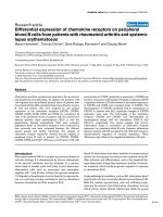
Model validation using the anti-inflammatory drug
dexamethasone
The screening model was validated using dexametha-
sone (dex) to suppre ss the maturation of imDCs into
inflammatory DCs. Dexamethasone was added to the
imDCs for 6 or 24 h prior to addition of selected cock-
tails for another 24 h, with subsequent measurements
of IL-12p70 and TNFa in the conditioned media (fi g-
ure 3). Dexamethasone was able to prevent IL-12p70
secretion stimulated by cocktails 3, 4, 6, 7 and 8 in a
dose-dependent manner. The suppressive function was
seen after both 6 and 24 h pre-incubation, but stron-
gest after 24 h pre-incubation (figure 3A and 3B). In
thesameexperiment,TNFa expression was also sup-
pressed in a dose dependent manner by dexametha-
sone, (figure 3C and 3D). After 6 h pre-incubation a
weak suppression of TNFa secretion was seen for
cocktail 4, 6, 7 and 8, but after 24 h pre-incubation
the suppression was stronger. The suppressive effect of
dexamethasone on cocktail 3 induced TNFa secret ion
was less prominent due to the addition of TNFa as a
component of cocktail 3. How ever, cocktail 3 is useful
for screening using other end-points like IL-12p70 a nd
other cytokines, chemokines, maturation markers or
ability to induce a MLR.
Figure 1 Cocktail screening and donor variation for IL-12p70
and TNFa secretion. A total of 15 different donor-derived imDCs
were treated with LPS (0.1 μg/mL) or the 9 cocktails as indicated in
table 1. After 24 h of incubation, IL-12p70 and TNFa levels in the
conditioned media was determined by ELISA. A) The amount of IL-
12p70 protein in each donor is indicated by a dot, and the average
of all 15 donors indicated by a horizontal bar. B) Amount of TNFa
protein was determined similarly in the 15 donors. Data were
analyzed using unpaired, two sided t-test, (***P < 0.005, **P < 0.01,
*P < 0.05).
Jensen and Gad Journal of Inflammation 2010, 7:37
/>Page 5 of 12
Cocktail stimulated Prostaglandin E2 secretion and its
suppression using unspecific and specific COX inhibitors
Prostaglandin E2 (PGE2) is a well known mediator of
inflammation, and its secretion from dendritic cells trea-
ted with cocktails could serve as a rel evan t end-point in
screening of anti-inflammatory compounds specifically
targeting DCs. We determined the cocktail induced
PGE2 secretion into the conditioned media from two dif-
ferent imDC batches. PGE2 secretion w as low or weakly
inducedbyLPSandcocktail4,5,6,7and9,whereas
DCs treated with cocktail 1, 2, 3 and 8 showed higher
PGE2 secretion (fi gure 4A). The higher levels of PGE2
stimulated by cocktail 1, 2, 3 and 8 was reflected in
increased expression of COX2 compared to untreated
cells, however, cocktails which did not stimulate PGE2
secretion to levels above untreated cells like LPS and
cocktail 6, 7 and 9 also caused COX2 induction, shown
by western blot of total lysates from DCs treated with the
respective cocktails (figure 4B). The unspecific COX-1
and 2-inhibitor indom ethazin, and the speci fic COX-2
inhibitor NS398 were both able to inhibit the secretion of
PGE2 into the c onditioned media, when added to the
imDCs 2 h prior to addition of the most potent PGE2
Neg ctrl
Cocktail 1
Cocktail 2
Cocktail 3
Cocktail 4
Cocktail 5
Cocktail 6
Cocktail 7
Cocktail 8
Cocktail 9
Pos ctrl
IL-8
TGF 2
Pos ctrl
CCL13
CCL2
CXCL1-3
IL-6
CXCL1
IL-10
CCL8
TNF
CCL5
Oncostatin M
CXCL10
CCL20
IL-12p40/70
IL-1
IFN
TNF
VEGF
Leptin
IL-1
IFN
TNF
IL-1
IFN
IFN
TNF
IFN
IFN
IFN
CCL4
GM-CSF
CCL15
CXCL5
TIMP1 TIMP2
LPS
Cytokine or chemokine
induced by the cocktail
Cytokine added as part
of the cocktail
Figure 2 Cytokine array using conditioned media from cocktail
treated DCs. Cytokine array membranes were incubated with
conditioned media from DCs treated with cocktails for 24 h. To
compensate for donor variations the conditioned media from four
donors was mixed. Upper left four spots and lower right two spots
serves as positive controls on each membrane. On the figure
constitutively secreted cytokines can be seen on the picture
indicated Neg. ctrl, which were untreated cells. Each induced
protein is marked only once, squares mark DC-produced cytokines
and chemokines, and a circle marks a cytokine which is added as
part of the cocktail (TNFa, IL-1b and IFNg).
Figure 3 Dexamethasone prevents cocktail induction of IL-
12p70 and TNFa. Immature DCs from a single donor shows a
dexamethasone-mediated dose dependent suppression of IL-12p70
and TNFa secretion. Dexamethasone was pre-incubated with imDCs
for 6 hours (A and C) or 24 hours (B and D) with increasing
concentration of dexamethasone at 0-0.01-0.1 and 1.0 μM.
Dexamethasone treatment without cocktail did not induce IL-12p70
or TNFa (first 4 bars). The cocktails used are indicated below each
set of data, and their exact composition is seen in table 1. This
shows one representative example out of three. Cell viability was
not significantly affected by treatment with cocktail and/or
dexamethasone (not shown).
Jensen and Gad Journal of Inflammation 2010, 7:37
/>Page 6 of 12
stimulating cocktail 8 (figure 4C). Indomethazin and
NS398 were not able to influence the DC mediated secre-
tion of IL-6, TNFa or IL-12p70 (data not shown) into the
conditioned media, indicating that these three cytokines
are not induced in a PGE2 autocrine fashion, and that
thedrugsdidnotinfluencecellsurvivalleadingto
decreased PGE2 production. Hence, the screeni ng model
is able to identify immuno modulating compounds which
can influence COX-activation and PGE2 generation.
Cocktail induced IL-23 secretion
IL-23 is a cytokine known to be involved in sustainment
of Th17-typer responses implicated in chronic autoim-
munity, and in particular IBD [7,8] and psoriasis
[11-13]. We analysed the ability of the nine cocktails to
induce secretion of IL-23 into the conditioned media,
and found that the TLR7/8 agonist R848 was able to
induce the IL-23 heterodimer (figure 4D). R848 induced
IL-23 more than 4 fold above levels for untreated cells
(figure 4D, cocktail 6), and when R848 was combined
with IFNg (cocktail 7) or poly I:C (cocktail 8) the IL-23
secretion was reduced. The remaining cocktails except
cocktail 9, induced IL-23 moderately above the level
seen for untreated cells.
Maturation of cocktail treated DCs
A phenotypic analysis was performed by flow cytometry
in order to investigate the expression of relevant
maturation markers on the DCs after LPS and cocktail
stimulation. LPS and all cocktails except cocktail 6
induce a high expression of the activation markers
CD40, CD80, CD83, CD86 and HLA-DR (figure 5A).
Selected cocktails with ability to stimulate these markers
potently were selected for validation using dexametha-
sone pre-treatment for 6 h prior to addition of cocktail.
Dexamethasone was able to lower the expression of a
majority of the activation markers HLA-DR, CD40,
CD80 and CD86 induced by the cocktails (figure 5B).
The strongest effe ct of dexamethas one was found for
coc ktail 8 as the expression of activation markers CD80
and CD86 was found to be below the immature state.
Allogeneic T-cell proliferation induced by cocktail treated
DCs
The mixed lymphocyte reaction (MLR) was used as a
functional endpoint to assess the in vitro T lymphocyte
proliferation in response to DCs treated with the 9
cocktails. Cocktail treated DCs and CD4
+
T cells from
allogeneic individuals were mixed together in a one-way
primary MLR and T cell proliferation was measured by
incorporation of
3
H-thymidine (figure 6A). DCs were
treated with cocktails alone or pre-treated with dexa-
methasone 6 h prior to addition of the cocktail. As seen
in figure 6A, LPS and all cocktails stimulated T-cell pro-
liferation, although cocktail 6 and 7 only stimulated
approximately 2 fold higher proliferation than untreated
cells. Pre-treatment of DCs with dexamethasone was
able to suppress proliferation for all cocktails and LPS.
Cocktail treated DC with ability to induce IFNg producing
T-cells
The ability of cocktail treated DCs to induce T-cell pro-
liferation shows that these DCs have become immuno-
genic (figure 6A). However, increased proliferation does
Figure 4 Cocktail induced stimulation of inflammatory markers.
Prostaglandin E2 was determined in conditioned media from two
representative donor derived DC batches as described in M&M (A).
PGE2 secretion was highest when imDCs were treated with cocktail
1, 2, 3 and 8 for 24 hours. B) COX2 protein levels were analysed by
western blot of lysates from DCs treated with cocktails for 24 h.
Actin was used as loading control, and results shown are
representative blot from two donors. C) Inhibition of PGE2 secretion
induced by cocktail 8 was shown by pre-incubation of imDCs from
two different donors for 2 h with the unspecific COX inhibitor
indomethazine (indo) at 10 μM or the specific COX-2 inhibitor
NS398 at 10 μM. D) The ability of cocktails to induce secretion of IL-
23, shown as an average of measurements on 3 different donors.
Bars show standard deviation between the three donors.
Jensen and Gad Journal of Inflammation 2010, 7:37
/>Page 7 of 12
not indicate if the T-cells has differentiated into Th1,
Th2 or Th17 T-cells when the DCs are cocultured with
CD4
+
T cells. In order to evaluate whether the cockta ils
were truly Th1-inducing, we measured IFNg in the con-
ditioned media from T-cells restimulated with anti-CD3
and anti CD28. Cocktails 1-5 all induced high levels of
IFNg secretion, whereas LPS and cocktail 9 did not
induce IFNg above the level seen for untreated cells.
Cocktail 6-8 induced IFNg slightly above the control
level (figure 6B). All cocktails showed reduced IFNg
secretion when the DCs were pretreated with dexa-
methasone as expected from the proliferation data.
Cocktail 3, 4 and 5 were potent inducers of T-cell
secreted TNFa, whereas the other cocktails were less
potent in T -cell stimulated TNFa (figure 6C). Finally, in
Figure 5 FACS profile on cocktail stimulated DCs.Surface
staining by flow cytometric analysis of immature (untreated), LPS
and cocktail stimulated DCs. The surface expression of relevant
activation markers was analyzed on day 7. A) A total of 5000 events
were collected by gating hDCs defined by forward (FSC) and side-
scatter (SSC) characteristics. All histograms were gated for CD1a cells
(70-95%). All our cells were CD14 negative (data not shown). Flow
cytomeric analysis of maturation markes were done for DCs from
three donors stimulated with LPS or cocktails, and results
normalized to the untreated DCs (average value of Mean
florescence intensity for untreated DCs from the three donors was
set to 100%). The vertical bars indicate standard deviation (SD)
values. B) Phenotypic surface analysis of the suppressive effect of
dex on cocktail treated human DCs from two donors. Pre-treatment
of immature DCs with dex for 6 h before addition of selected
cocktails reduced the expression of activation markers. Results for
the different treatments have been normalized in proportion to the
untreated DC. The vertical bars indicate (SD) values.
Figure 6 T-cell proliferation and secreted cytokines stimulated
by cocktail treated DCs. MLR performed on CD4
+
T cells and
allogeneic DCs. Mature cocktail stimulated DCs were more potent
inducers of T cell proliferation in the MLR than immature DCs. A
pretreatment of DCs with dexamethasone for 6 h before addition of
cocktails, significantly prevent CD4
+
T cell proliferation to a level
similar to immature DCs. The results are representative of three
donors. A) CD4
+
T cells were cultured with allogeneic DCs for 5
days, and mitomycin-C treated in order to inhibit their proliferation.
Proliferation of CD4
+
T cells was determined in the last 18-24 h of
culture. Each column represents the mean cpm of four replicates.
Vertical bars represent the SD. B) The amount of IFNg production by
T cells was measured in the supernatants after a restimulation with
platebound anti-CD3 and soluble anti-CD28 mAbs for 24 h. In the
conditioned media was also measured C) TNFa, and D) IL-13 by
ELISA. Each column represents the mean of triplicate wells. Vertical
bars represent the SD values.
Jensen and Gad Journal of Inflammation 2010, 7:37
/>Page 8 of 12
order to exclude t hat the cocktails induced a Th2
response, IL-4, IL-5 and IL-13 were measur ed in the T-
cell conditioned media. IL-13 was highest in the media
from untreated control cells, showing that in a MLR
reaction by itself, a certain pool of the T-cells develop
into Th2-cells. However, none of the cocktails induced
IL-4, IL-5 or IL-13 above the level seen for untreated
cells, showing that none of the cocktails induce a Th2-
response (figure 6D, showing IL-13 as representative,
IL-4 and IL-5 data not shown).
Discussion
During recent years the role of DCs in immune disor-
ders has been substantiated and the potential targeting
of DCs for treatment of autoimmune and allergic dis-
eases been suggested [14]. One of the interesting advan-
tages of targeting DCs is their role as key initiators of
adaptive immunity, t hereby positioned upstream of the
effector cells in e.g. autoimmunity [1,8]. Furthermore
DCs have a relatively short life span compared to other
primary cell types. At steady state conditions, immature
DCs are quiescent until approached by a pathogen
invading the tissue, or until tissue factors like cytokines
or chemokines stimulate DC activation [2]. At s ites of
inflammation chemoattractants are produced by
immune and epithelial cells, and will promote immature
DC migration to the site of inflammation. After expo-
sure to maturation stimuli at the inflammator y site,
likely through stimulation with inflammatory chemo-
kines and cytokines and/or combined with TLR agonists
present at the site, DCs migrate to the lymph nodes and
activate the adaptive immune response, and subse-
quently undergo apoptosis once they have activated a
number of T-cells [15,16]. Thus, DCs at inflammatory
sites have a higher flux through the inflammatory tissue.
We have approached the use of dendritic cells in tar-
geting of immune disorders by establishment of a DC
based in vitro screening model by mimicking this
in vivo function of DCs. Immature DCs are developed
from monocytes using conventional methods. The treat-
ment with a drug candidate in our setting correlates to
the potential treatment at the monocyte or steady state
immature DCs level in the periphery of the patient,
prior to chemoattractio n of these cells from the circula-
tion into the site of inflammation. The treatment in our
in vitro model with cocktails containing Th1 inducing
or inflammatory cytokines and TLR agonists mimics the
in vivo situation where immature DCs are matured by
factors at the site of inflammation. The cocktail matures
the DCs towards a Th1-inducing phenotype, which
mimics the maturation seen in many autoimmune con-
ditions. By measuring drug induced changes in expres-
sion of maturation markers and secreted cytokines and
chemokines , inflammatory lipids, intracellular signalling
molecules and ability to induce T-cell responses, the
effect on drug treated DCs in vivo can be predicted.
An interesting f eature of DCs is their relatively com-
plex expression of pattern re cognition receptors and
their corresponding signalling pathways. Some of the
PRRs like e.g. TLR4 are expressed on antigen present-
ing cells in general, including monocytes, macrophages
and B-cells, however, other TLRs are mainly or exclu-
sively expressed on myeloid or plasmacytoid DCs.
Monocyte derived myeloid DCs are known to express
TRL1-11, except TLR9 which is expressed in plasmacy-
toid DCs [17,18]. TLR ligation has shown to be
involved in several autoimmune diseases, where
increased TLR ligands are present at the diseased site
as well as in patient serum. One example is seen in
patients suffering from RA, where TLR3, 4 and 7/8
expression is increased in the synovium, and where
TLR ligation further encreases expression of inflamma-
tory mediators in DCs from the RA patients compared
to DCs from healthy controls [19]. W e have in our
present Th1-inducing cocktails utilized TLR ligation
with ligands towards these 4 TLRs, by combining poly
I:C (TLR3), LPS (TLR4) and R848 (TLR7/8) with
proinflammatory cytokines in order to mimic the sti-
muli from autoimmune conditions.
TheuniqueexpressionpatternofTLRsonantigen
presenting cells and in particular on DCs, supports the
idea that DCs are promising therapeutic target cells for
treatment of inflammation and autoimmune disorders,
since TLRs and their corresponding signalling pathways
can be explored for more diverse target molecules, and
in some cases targets that are exclusively expressed on
DCs [10,20].
The cocktail treated DCs express intracellular inflam-
matory proteins like COX2, and membrane associated
markers involved in regulation of adaptive immunity
like HLA-DR, CD40, CD80, CD83 and CD86. Five of
the defined cocktails potently stimulated development
of Th1-cells, shown by their secretion of IFNg in an
allogeneic MLR.
Cocktail 1 was designed as in a previous reported
cocktail [21], where the combination of poly I:C, IFNa,
IFNg,TNFg and IL-1b showed very potent IL-12p70 sti-
mulating properties compared to the standard DC cock-
tail used for DC based cancer vaccines, containing IL-6,
TNFa,IL-1b and PGE2. The latter cocktail is slightly
more potent in inducing DC migration, which is impor-
tant for the DCs to reach local lymph nodes. In con-
trast, cocktail 1 was superio r in inducing CTL-mediate d
cancer cell lysis and in vivo tumor antigen responses
[21]. Our data show that cocktail 1 indeed induces
PGE2 in itself, which could account for the migratory
capabilities of cocktail 1 treat ed DCs as shown by Milli-
ard and colleagues [21].
Jensen and Gad Journal of Inflammation 2010, 7:37
/>Page 9 of 12
Ouranalysesofthe9differentcocktailsin15donors
showed that cocktail 3, 6 and 9 showed the greatest
donor variation in IL-12p70 secretion. Although all
cocktails induced significantly higher IL-12p70 secretion
than LPS, the level of significance was lower for cocktail
6 and 9. The variation on TNFa secretion was slightly
lower, where all cocktails and LPS significantly induced
TNFa secretion compared to control treated cells, and
with cocktail 1, 2, 3, 4, 5 and 8 significantly higher than
LPS.
In most donors cocktail 6, which contains R848, was
partly impaired in stimulation of IL-12p70, with an aver-
age level at 5 ng/mL. Taken in consideration that R848
only slightly stimulate IFNg in the MLR assay, and is
the most potent stimulator of IL-23, R848 seems to be a
good candidate for induction of differentiation to a DC
phenotype with ability to induce naïve CD4
+
T cells
towards the Th17 lineage [22]. The secretion of IL-23
by R848 was reduced when the Th1-promoti ng cytokine
IFNg and the TLR3 agonist poly I:C were added
together with R848. In particu lar the IL-23 suppressing
ability of IFNg supports the finding, where hall-mark
Th1-promoting cytokines have the ability to suppress
the Th17 promoting phenotype, a po tential cross-talk
dis cussed extensively for inflammatory diseases particu-
lar for intestinal inflammation [6]. Our data show that
LPS alone was able t o stimulate secretion of only minor
amounts of both IL-12p70, IL-23 and IFNg compared to
cocktail treated DCs, and in particular in comparison to
the two LPS containing cocktails 4 and 5, which con-
tains IFNg and IFNg and TNFa respectively. This find-
ing clearly shows that the identified cocktails are
superior to the use of LPS alone as an immunogenic sti-
muli, and that our present model is superior as a DC
based screening model for screening og drug candidates
with potential Th1-suppressive function.
Combining LPS with IFNg in our model (cocktail 4)
preferentially increased IL-12p70 and not IL-23, which
is in contrast to the findings by Roses et al. [22]. This
difference can be explained by the fact that our imDCs
were matured using IL-4 and GM-CSF for the whole
culture period, where presence of IL-4 for the last 24 h
of incubation reduces IL-12p70 and IL-23 production
[22]. The finding by Roses et al., strongly indicates that
in vitro culture conditions for both DCs and T-cells is
influencing the ability of the model system to induce IL-
23 and IL-17. Surp risingly, R848 was the only TLR ago-
nist/cocktail that did not stimulate maturation marker
expression HLA-DR and CD40, 80, 83 and 86.
The lower capacity of cocktail 9 to stimulate IL-12p70
could be explained by the lack of a TLR agonist in this
cocktail, and indicates that the presence of IFNg is not
enough to drive potent in duction of IL-12p70. However,
in presence of a TLR agonist like LPS, IFNg was able to
potently polarize the DCs towards IL-12p70 induction
(LPS alone vs cocktail 4), which is in line with other
observations [21,22]. LPS and all cocktails more readily
induced TNFa secretion from nearly all donor derived
DCs, although the variation s were also high for some
cocktails, in particular for cocktail 6, 8 and 9. The most
suitable cocktails for a DC based screening model
should preferably induce the chosen end-point in as
many donors as possible. However, for the ability of a
compound to suppress cytokine secretion, the observed
donor variation is not critical unless a certain amount of
donors do not respond at all, as seen for e .g. LPS
induced IL-12p70. In this regard, LPS is not a suitable
maturation factor if one wishes to identify IL-12p70 or
IL-23 suppressing compounds.
The DC responses of the cocktails were correlated to
the pattern of chemokines and cytokines seen in auto-
immune diseases. Using cytokine antibody arrays and
ELISA we showed secretion of hall mark cytokines and
chemokines like TNFa, IL-6, IL-10, IL-12p70, IL-23, IL-
8, CCL2, -4, -5, -8, -15, -20, and CXCL1, 5 and 10.
Interestingly, the cocktail treated DCs also induced
secretion of particular VEGF and for some cocktails also
GM-CSF.
Themimicryofthesecytokineandchemokinepat-
terns with specific disease pathologies is complex, in
particular since t issue samples from patients suffering
from these conditions contains facto rs secreted by a
broad repertoire of leucocytes, fibroblasts and epithelial
cells, and not DCs alone. However, certain comparisons
are striking, where coc ktail induced factors are similar
to the ones identified in the pathological conditions.
Biopsies from Crohn disease patients show increased
expression of cyto kines like T NFa,IL-1b IL-6, IL-
12p70, IL-23 [7,8] and in particular the IL-12/IL-23 sub-
unit p40 seems to be important for disease development
since monoclonal antibodies towards this subunit is able
to reduce symptoms and expression of both IL-12p70
and IL-23, but also IL-6 and IL-17 [23]. Chemokines
implicated in Crohns disease are numerous, some
includesCCL2,4,5,8and20,andCXCL1,2,3,8,10,
which are expressed by several of our cocktail treated
DCs [24]. To this end, the cocktails mimicking the early
phases of Crohns disease are cocktails 1, 2, 4 and 5,
since they induce key factors like TNFa, IL-6, IL-12p70,
CCL2, 4, 8, CXCL1-3 and upon T-cell co-culture also
IFNg. However, the later and chronic phase of Crohns
disease involves activation of the IL-23/IL-17 axis, which
might be better represented using R848 in the cocktails,
as seen for cocktail 6, 7 and 8 [7,8,25]. In psoriatic
lesions, cytokines l ike IL-6, IL-12p70, IFNg and TNFa
are identified, together with chemokines like CCL2-5
and CXCL1 [12,13], which shows that several of the
cocktails stimulate cytokines and chemokines that to a
Jensen and Gad Journal of Inflammation 2010, 7:37
/>Page 10 of 12
large extent overlap with the ones expressed in different
autoimmune conditions.
The tolerance inducing cytokine IL-10 was modestly
induced by all cocktails, as well as LPS alone. This is a
paradox compared to the ability of the cocktails to
induce high IFNg secretion, in particular for cocktail 1-
5. However, the fact that both pro-inflammatory and
anti-inflammatory cytokines are produced in the same
pool of T-cells, is most likely due to the fact that the
pool of T-cells to some extent are heterogeneous, and
cocktail 1-5 most potently skew the T-cel ls towards the
Th1 response, whereas other cocktails stimulate a more
heterogeneous response [22].
Similarly, the pathogenesis of psoriasis involves key
Th1 and Th17 inducing cytokines like TNFa,IL-1b,IL-
2, IL-6, IL-10, IL-12p70, IL-23, CCL2, 3, 4, 5, 20,
CXCL1, 8, 9 and 10, many of which are induced by
cocktail treated DCs. The question whether TLR ago-
nists are invo lved in the pathoge nesis of psoriasis is
unclear, but the finding that a TLR7 agonist, imiquimod,
induces psoriasis like plaques in mice indicates that
TLR7 induction could play a role [26]. In this respect,
R848 or alternative cocktails using imiquimod could be
the basis for design of cocktails with an optimal mimicry
of the immune pathology seen in psoriasis. In addition,
plasmacytoid DCs (pDC) also seems to play an impor-
tant role in psoriasis, pointing to a po tential pDC-based
screening platform which could be generated using both
TLR7 agonists and inflammatory cytokines. Other auto-
immune diseases like RA and multiple sclerosis similarly
involves combinations of Th1 and Th17 responses, sug-
gesting that our present cocktails induce DC-phenotypes
mimicking responses of DCs seen in these diseases
[27-29].
Conclusion
Nine cocktails were identified with potent ability to sti-
mulate DC development into Th1-promoting inflamma-
tory DCs with slightly different profiles mimicking DCs
in autoimmune conditions as seen in Crohns disease,
psoriasis, multiple sclerosis and RA. In particular 5
cocktails were able to mature DCs into potent Th1-
inducing DC phenotypes, which showed high secretion
of IL-6, TNFa and IL-12p70. When cocultured with
CD4
+
T-cells, the cocktail martured DCs were able to
stimulate development of Th1-cells which possessed a
high proliferative capacity, and induced high levels of
the hall-mark Th1 cytokine IFNg.
The present scre ening model provides a novel precli-
nical screening tool, where Th1-suppressing compounds
can be examined at several stages of immune activation;
1) at the innate receptor level on DCs, 2) at the innate
TLR mediated signalling response in DCs, 3) at the level
of DC-T cell interaction at receptor level or secretion of
inflammatory mediators, 4) at the T-cell level targeting
intracellular signalling and secretion of inflammatory
mediators.
Acknowledgements
This work was supported by the Danish ministry for science, technology and
innovation. We wish to thank Trine Møller and Bente M. Tolstrup for
valuable technical assistance.
Author details
1
Department of immune targeting, Bioneer A/S, Kogle Allé 2, Hørsholm, DK-
2970, Denmark.
2
Bioneer A/S, Kogle Allé 2, Hørsholm, DK-2970, Denmark.
Authors’ contributions
SJ drafted the manuscript, designed and evaluated cocktails and made data
analysis to figures 1, 2, 3, 4. MG prepared and analysed data for figures 5
and 6. All experiments were carried out with technical help from Trine
Møller and Bente M. Tolstrup. SJ and MG both read, commented and
approved the final manuscript.
Competing interests
The authors discloses that part of the work described in the present
manuscript has been filed for a patent application. Furthermore, Bioneer A/S
provides DC based screening services for customers.
Received: 8 June 2009 Accepted: 27 July 2010 Published: 27 July 2010
References
1. Steinman RM, Banchereau J: Taking dendritic cells into medicine. Nature
2007, 449(27):419-426.
2. Shortman K, Shalin HN: Steady-state and inflammatory dendritic-cell
development. Net Rev Immunol 2007, 7:19-30.
3. Roy CR, Mocarski ES: Pathogen subversion of cell-intrinsic innate
immunity. Nat Immunol 2007, 8(11):1179-1187.
4. Bouma G, Strober W: The immunological and genetic basis of
inflammatory bowel disease. Nat Rev Immunol 2003, 3:521-533.
5. Bachmann MF, Kopf M, Marsland BJ: Chemokines: more than just road
signs. Nat Rev Immunol 2006, 6:159-164.
6. Zou W, Chen L: Inhibitory B7-family molecules in the tumor
microenvironment. Nat Rev Immunol 2008, 8:467-477.
7. Neurath MF: IL-23: a master regulator in Crohns disease. Nat Med 2007,
13(1):26-28.
8. McGovern D, Powrie F: The IL-23 axis plays a key role in the
pathogenesis of IBD. Gut 2007, 56:1333-1336.
9. Hammad H, Smits HH, Ratajczak C, Nithiananthan A, Wierenga EA,
Stewart GA, Jacquet A, Tonnel AB, Pestel J: Monocyte-derived dendritic
cells exposed to Der p 1 allergen enhance the recruitment of Th2 cells:
major involvement of the chemokines TARC/CCL17 and MDC/CCL22. Eur
Cytokine Netw 2003, 14(4):219-28.
10. Mantovani A, Bonecchi R, Locati M: Tuning inflammation and immunity
by chemokine sequestration: decoys and more. Nat Rev Immunol 2006,
6:907-918.
11. Lee E, Trepicchio WL, Oestreicher JL, Pittman D, Wang F, Chamian F,
Dhodapkar M, Krueger JG: Increased expression of interleukin 23 p19 and
p40 in lesional skin of patients with psoriasis vulgaris. J Exp Med 2004,
199(1):125-30.
12. Nickoloff BJ, Xin H, Nestle FO, Qin JZ: The cytokine and chemokine
network in psoriasis. Clin Dermatol 2007, 25(6):568-73.
13. Torti DC, Feldman SR: Interleukin-12, interleukin-23, and psoriasis: current
prospects. J Am Acad Dermatol 2007, 57(6):1059-68.
14. Hackstein H, Thomson AW: Dendritic cells: emerging pharmacological
targets of immunosuppressive drugs. Nat Rev Immunol 2004, 4(1):24-34.
15. Langenkamp A, Messi M, Lanzavecchia A, Sallusto F:
Kinetics of dendritic
cell activation: impact on priming of TH1, TH2 and nonpolarized T cells.
Nature Immunology 2000, 1:311-316.
16. Pradhan S, Genebriera J, Denning WL, Felix K, Elmets CA, Timares L: CD4 T
cell-induced, bid-dependent apoptosis of cutaneous dendritic cells
regulates T cell expansion and immune responses. J Immunol 2006,
177:5956-67.
Jensen and Gad Journal of Inflammation 2010, 7:37
/>Page 11 of 12
17. Akira S, Takeda K: Toll-like receptor signalling. Nat Rev Immunol 2004,
4:499-511.
18. Hopkins PA, Sriskandan S: Mammalian Toll-like receptors:to immunity and
beyond. Clin Exp Immmunol 2005, 140:395-407.
19. Roelofs MF, Joosten AB, Abdollahi-Roodsaz S, van Lieshout AWT, Sprong T,
van den Hoogen FH, van den Berg WB, Radstake TRDJ: The expression of
toll-like Receptors 3 and 7 in Rheumatoid Arthritis Synovium Is
Increased and Costimulation of Toll-like Receptors 3, 4 and 7/8 Results
in Synergistic Cytokine Production by Dendritic Cells. Arhtitis &
Rheumatism 2005, 52(8):2313-2322.
20. Coombes JL, Powrie F: Dendritic cells in intestinal immune regulation.
Nature Immunology 2008, 8:435-446.
21. Mailliard RB, Wankowicz-Kalinska A, Cai Q, Wesa A, Hilkens CM,
Kapsenberg ML, Kirkwood JM, Storkus WJ, Kalinski P: Alpha-type-1
polarized dendritic cells: a novel immunization tool with optimized CTL-
inducing activity. Cancer Res 2004, 64(17):5934-7.
22. Roses RE, Xu S, Xu M, Koldovsky U, Koski G, Czerniecki BJ: Differential
production of IL-23 and IL-12 by myeloid-derived dendritic cells in
response to TLR agonists. J Immunol 2008, 181(7):5120-7.
23. Fuss IJ, Becker C, Yang Z, Groden C, Hornung RL, Heller F, Neurath MF,
Strober W, Mannon PJ: Both IL-12p70 and IL-23 are synthesized during
active Crohns disease and are down-regulated by treatment with anti-
IL-12p40 monoclonal antibody. Inflamm Bowel Dis 2006, 12(1):9-15.
24. Zhong W, Kolls JK, Chen H, McAllister F, Oliver PD, Zhang Z: Chemokines
orchestrate leukocyte trafficking in inflammatory bowel disease. Front
Biosci 2008, 13:1654-64.
25. Hanauer SB: Inflammatory Bowel Disease: Epidemiology, pathogenesis
and therapeutic opportunities. Inflamm Bowel Dis 2006, 12(1):S3-S9.
26. Gilliet M, Conrad C, Geiges M, Cozzio A, Thürlimann W, Burg G, Nestle FO,
Dummer R: Psoriasis triggered by toll-like receptor 7 agonist imiquimod
in the presence of dermal plasmacytoid dendritic cell precursors. Arch
Dermatol 2004, 140(12):1490-5.
27. Iwamoto G, Okamoto H, Toyama Y, Momohara S: Molecular aspects of
rheumatoid arthritis:chemokines in joints of patients. FEBS Journal 2008,
275(18):4448-55.
28. McFarland HF, Martin R: Multiple sclerosis: a complicated picture of
autoimmunity. Nat Immunol 2007, 8(9):913-9.
29. Annunziato F, Cosmi L, Liotta F, Romagnani S: Typer 17 T helper cells-
origins, features and possible roles in rheumatic disease. Nat Rev
Rheumatol 2009, 5(6):325-31.
doi:10.1186/1476-9255-7-37
Cite this article as: Jensen and Gad: Differential induction of
inflammatory cytokines by dendritic cells treated with novel TLR-
agonist and cytokine based cocktails: targeting dendritic cells in
autoimmunity. Journal of Inflammation 2010 7:37.
Submit your next manuscript to BioMed Central
and take full advantage of:
• Convenient online submission
• Thorough peer review
• No space constraints or color figure charges
• Immediate publication on acceptance
• Inclusion in PubMed, CAS, Scopus and Google Scholar
• Research which is freely available for redistribution
Submit your manuscript at
www.biomedcentral.com/submit
Jensen and Gad Journal of Inflammation 2010, 7:37
/>Page 12 of 12