Báo cáo y học: "Insulin alleviates degradation of skeletal muscle protein by inhibiting the ubiquitin-proteasome system in septic rats" pps
Bạn đang xem bản rút gọn của tài liệu. Xem và tải ngay bản đầy đủ của tài liệu tại đây (463.36 KB, 8 trang )
RESEARC H Open Access
Insulin alleviates degradation of skeletal muscle
protein by inhibiting the ubiquitin-proteasome
system in septic rats
Qiyi Chen, Ning Li, Weiming Zhu, Weiqin Li, Shaoqiu Tang, Wenkui Yu
*
, Tao Gao, Juanjuan Zhang and Jieshou Li
Abstract
Hypercatabolism is common under septic conditions. Skeletal muscle is the main target organ for hypercatabolism,
and this phenomenon is a vital factor in the deterioration of recovery in septic patients. In skeletal muscle, activation
of the ubiquitin-proteasome system plays an important role in hypercatabolism under septic status. Insulin is a vital
anticatabolic hormone and previous evidence suggests that insulin administration inhibits various steps in the
ubiquitin-proteasome system. However, whether insulin can alleviate the degradation of skeletal muscle protein by
inhibiting the ubiquitin-proteasome system under septic condition is unclear. This paper confirmed that mRNA and
protein levels of the ubiquitin-proteasome system were upregulated and molecular markers of skeletal muscle
proteolysis (tyrosine and 3-methylhistidine) simultaneously increased in the skeletal muscle of septic rats. Septic rats
were infused with insulin at a constant rate of 2.4 mU.kg
-1
.min
-1
for 8 hours. Concentrations of mRNA and proteins of
the ubiquitin-proteasome system and molecular markers of skeletal muscle proteolysis were mildly affected. When
the insulin infusion dose increased to 4.8 mU.kg
-1
.min
-1
, mRNA for ubiquitin, E2-14 KDa, and the C2 subunit were all
sharply downregulated. At the same time, the levels of ubiquitinated proteins, E2-14KDa, and the C2 subunit protein
were significantly reduced. Tyrosine and 3-methylhistidine decreased significantly. We concluded that the ubiquitin-
proteasome system is important skeletal muscle hypercatabolism in septic rats. Infusion of insulin can reverse the
detrimental metabolism of skeletal muscle by inhibiting the ubiquitin-proteasome system, and the effect is
proportional to the insulin infusion dose.
Introduction
Muscle catabolism, resulting in muscle wasting and fati-
gue, is a characteristic metabolic response to sepsis [1-3].
Sepsis-induced muscle catabolism is mainly c aused by
increased protein breakdown, in particular myofibrillar
protein breakdown, although reduced protein synthesis
and inhibited amino acid tra nsport contribute to the
metabolic response. Muscle breakdown may impair the
recovery in septic patients and increase the risk for pul-
monary and thrombo-embolic complications when
respiratory muscles and ambulation are affected [3-6]
Previous studies provided evidence that sepsis-induced
muscle proteolysis is caused by increased protein break-
down, through the ubiquitin (Ub)-proteasome pathway
[4-6]. In this pathway, Ub, which contains 76 amino
acids, is conjugated to proteins destine d for degradation
by Ub-activating enzyme (E1), Ub-conjugating enzyme
(E2), and Ub-ligase (E3) [1,7]. The 14-kDa ubiquitin-
conjugating enzyme E2 has been proposed to be a regu-
lation site for the Ub-proteasome proteolytic pathway in
skeletal muscle [8]. This process is repeated as multiple
Ub molecules are added to form a Ub chain. Ub-protein
conjugates are recognized by a 26S proteasome complex,
composed of two subproteasome complexes, a 19S regu-
latory particle, and a 20S catalytic particle. Ubiquitinated
proteins are rapidly degraded by the proteasome in an
ATP-dependent manner [2,9].
Insulin is an anabolic and anticatabolic hormone.
When administered to healthy volunteers, it stimulates
muscle protein synthesis and inhibits protein metabo-
lism at the whole body level [10]. In addition, several
studies have repeatedly demonstrated the beneficial
effects of insulin treatment on protein wasting caused
by different pathological conditions. For example, in
* Correspondence:
Department of General Surgery, Jinling Hospital, Medical College of Nanjing
University, Nanjing 210002, Jiangsu Province, China
Chen et al. Journal of Inflammation 2011, 8:13
/>© 2011 Chen et al; licensee BioMed Central Ltd. This is an Open Access article distributed under the terms of the Creative Comm ons
Attribution License ( censes/by/2.0), which permits unrestricted use, distribu tion, and reproduction in
any medium, provided the original work is properly cited.
burn-injured patients and in animal experiments, nitro-
gen balance is partially restored after continuous infu-
sion of insulin [11]. In diabetic patients and diabetic
rats, administration of insulin decreases or completely
prevents the release of urinary 3-methylhistidine (3MH)
[10]. In patients in intensive care unit, insulin adminis-
tration reduces morbidity by preventing organ failure, as
evidenced by a reduction in duration of mechanical ven-
tilation [12]. However, the molecular mechanism by
which insulin suppresses protein degradation remains
poorly understood.
Previous evidence suggests that insulin deficiency results
in activity of the Ub-proteasome system [13-15]. Insulin
resistance causes muscle wasting by mechanisms that
involve activation of the Ub-proteasome proteolytic path-
way, cau sing muscle pro tein degradation [15,16]. Ins ulin
administration can inhibit various steps of the Ub-protea-
some system; for example, a hyperinsulinaemic euglycae-
mic clamp significantly reduced mRNA expression for
theubiquitin system in rat skeletal muscle [17]. Fouzia
Sadiqetal.showedthat,inC2C12myotubes,insulin
administration was associated with downregulated expres-
sion of the Ub-proteasome pathway [18]. However, no
reports on the influence of insulin on theUb-proteasome
system under sepsis currently exist. In this study,
we hypothesize that infusion of insulin would a lleviate
degradation of skeletal muscle protein by inhibiting the
Ub-proteasome system in septic rats.
Materials and methods
Animals
This study used 44 adult male Sprague-Dawley rats,
weighing 200 ± 20 g, from the animal center of Jinling
Hospital. The Institutio nal Animal Care Committee
approved the study protocol. T he Association a ccredits
the animal care facility for Assessment and Accredita-
tion of Laboratory Animal Care. Rats were housed in
mesh cages in a 25°C room, illuminated in 12:12-h
light-dark cycles and acclimatized to their environment
for 7 d before the study. They were provided with stan-
dard rodent chow and water ad libitum.
Animal preparation
Rats were anesthetized intraperitoneally with phenobar-
bital sodium (60 mg/kg), and catheters (PE-50, PE-10;
Becton-Dickinson, Sparks, MD) were implanted into the
right jugular vein and the left carotid artery, as
described previously [19]. The right jugular vein was
used to infuse insulin and dextrose solution by micro-
pump (proved by the Research Center for Analytica l
Instrument, Zhejiang University) and the left carotid
artery was used to monitor blood glucose with an Elite
glucometer (Bayer, Elkha rt, IN). The catheters were
filled with saline containing heparin sodium.
Group distribution and insulin infusion strategy
After 5-6 days recovery, rats were fasted for 12 h and
divided randomly into fo ur groups as follows: control
group (n = 12) LPS group (n = 12), low-dose insulin group
(n = 12) and high-dose insulin group (n = 12). The sepsis
model was mimicked by intraperitoneal injection with
10 mg/kg LPS (Escherichia coli serotype 055:B5, Sigma, St.
Louis, MO). The low- or high-dose insulin group rats
received a continuous infusion of insulin (Humulin R, Eli
Lilly & Co., Indianapolis, IN) at a constant rate of 2.4
(low) or 4.8 (high) mU
-1
min
-1
kg
-1
for 8 hours after LPS sti-
mulation. Blood glucose was maintained betwe en 4.4-6.1
mmol/L by varying the infusion rate of a 50% dextrose
solution. The LPS group was injected intraperitoneally
with 10 mg/kg LPS only. The control group received an
intraperitoneal injection with an equal volume of sterile
saline only. Experiment were performed while the rats
were awake and unrestrained.
At the end of the infusion, rats were killed with pheno-
barbital sodium. The extensor digitorum longus (EDL)
was immediately excised to measure the proteolytic rate,
and the gastrocnemius muscle was harvested and frozen
in -80°C.
Rate of protein turnover
To measure protein breakdown rates, freshly EDL muscle
was fixed via the tendons to aluminum wire supports at
resting length, and preincubated in oxygenated medium
(95% O
2
-5% CO
2
): Krebs-Henseleit bicarbonate buffer (pH
7.4) containing 5 Mm glucose, 0.1 U/ml insulin, 0.17 mM
leucine, 0.1 mM isoleucine, and 0.20 mM valine. After a
1 h preincubation, muscles were transferred to fresh med-
ium of identical composition and incubated for a further
2 h with 0.5 mM cycloheximide. The degradation rates of
total and myofibrillar proteins were determined by release
in the medium of free tyrosine and 3-MH, respectively,
and expressed as nanomoles of tyrosine/methylhistidine in
medium per 2·h
-1
g·muscle
-1
. Muscle was also homoge-
nized in 0.4 mM perchlori c acid to determine tissue free
tyrosine and 3-MH. The net production of free tyrosine
was calculated as the amount of tyrosine released into the
mediumplustheincreaseintissuefreetyrosineduring
incubation. Net 3-MH production was cal culated as the
amount of 3-MH in the medium minus the decrease in
tissue free 3-MH before and after incubation. Levels of
both tyrosine and 3-MH in medium or tissue samples
were determined by high-performance liquid chromato-
graphy (HPLC).
RT-PCR RNA preparation and analysis of expression of
ubiquitin-proteasome system genes
Total mRNA was extracted from gastrocnemius with
TRIzol reagents (Life Technologies), and the mRNA
concentration determined by ultraviolet light absorbency
Chen et al. Journal of Inflammation 2011, 8:13
/>Page 2 of 8
at 260 nm. Measurement of mRNA of the Ub-protea-
some system components ubiquitin, 14-kDa Ub-conju-
gating enzyme (14-kDa E2), and proteasome subunit C2
was performed by semi-quantitative reverse transcrip-
tase-polymerase chain reaction (RT-PCR). Before reverse
transcription, 1-5 μg of total RNA was reverse tran-
scribed at 42°C for 1 h using standard reagents of 50 μl
of 1×RT, reverse transcriptase (RTase), and poly(dT)12-
18 primer. RT mixtures were heated at 100°C for
10 min to inactivate RTase. PCR reactions were 50 μlof
1×PCR buffer with 5 μlofRTtemplate,200nMeach
sense and antisense primers, 1 unit of Taq polymerase,
and 200 μM each dNTPs. The reaction was covered
with 30 μl of mineral oil, and PCR was performed in a
DNA Thermal Cycler 480 (Perkin-Elmer, Norw alk, CT).
After 94°Cfor 5 min, cycles were 94°C for 30 s, anneal-
ing at a product-specific temperature for 1 min, and
72°C for 1 mi n. The last cycle was foll owed by 5 min at
72°C. The number of amplification cycles was optimized
for primer pairs to produce a densitometric result that
correlated closely with the template. To determine the
relative quantities of mRNA, 10 μl each of Ub, 14-kDa,
proteasome subunit C2, and b-actin PCR products
amplified from the same RT template were combined
and electrophoresed on 2% agarose gel in Tris-acetate-
EDTA buffer for 30 min. After ethidium bromide stain-
ing for 15 min, bands were measured for densitometry
using Quantity One Analysis software. Relative Ub
mRNA levels in the original RNA extracts fromthe ske-
letal muscle preparations were obtained by normalizaion
to b-actin expression. Primer sequences were: Ubiquitin
(200 bp): forward, 5’-TCTTCGTGAAGACCCTGACC-
3’ ; reverse,5’-CAGGTGCAGGGTTGACTCTT-3’;E2-
14KDa(221): forward, 5’ -GTGCACCATCTGAAAA-
CAA-3’;reverse, 5’-ATCGGTTCTGCAGGAT GTCT-3’;
C2(256):forward, 5’-GGCTGCTCATTGCTGGTTAG-3’;
reverse,5’-CCAACAATCCCAATGGAAAC-3’ ; b-actin
(180): forward, 5’-TCCTGTGGCATCCACGAAACT-3’,
reverse, 5’-GGAGCAATGATCTTGATCTTC-3’.
Western blot analysis
To detect proteins w ith conjugated Ub, E2-14KDa, and
C2, myofibrillar and sarcoplasmic muscle proteins were
prepared as described previously [2]. Myofibrillar and sar-
coplasmic muscle protein were separated on SDS-polya-
crylamide gels and transferred to nitrocellulose
membranes. Membranes were blocked for 1 h in 5% (vol/
vol) nonfat dry milk in TTNS (25 mM Tris-HCl [pH 7.5],
0.1% [vol/vol] Tween 20, 0.9% [wt/vol] NaCl). To detect
conjugated Ub, myofibrillar proteins were incubated for
1 h with a 1:1000 diluted rabbit polyclonal anti-Ub anti-
body (DakoCytomation, Glostrup, Denmark). To quantify
E2-14KDa and C2, sarcoplasmic proteins were incu bated
for 1 h with a 1:1000 diluted rabbit polyclonal anti-14-kDa
E2 antibody and anti-C2 antibody (DakoCytomation,
Glostrup, Denmark). After washing at room temperature,
membranes were hybridized with the appropriate peroxi-
dase-conjugated anti-IgG. Blots were washed four times
with TTNS for 20 min, incubated in enhanced chemilumi-
nescence reagent (Amersham Life Sciences), and exposed
on radiographic film (Eastman-Kodak, Rochester, NY).
Proteins were quantified by densitometry as above. Statis-
tical analyses were carried out on data normalized to by
b-actin.
Statistical analyses
Data are expressed as means ± standard error (SE), and
statistical analysis performed using ANOVA. All data
were analyzed with SPSS software (Statistical Package
for the Social Sciences, version 16.0, for Windows, SPSS,
Chicago, IL). P <0.05 was considered significant.
Results
Proteolytic rate in extensor digital longus muscle
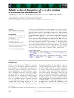
The proteolytic rate of skele tal muscle wa s measured as
net release of tyrosine for total protein and 3-MH for
myofibrillar protein. Com pared to the control group , the
rate of total protein proteolysis was significantly increased
in the LPS group ( 210.49 ±14.09 vs. 3 83.4 ± 12.72, P <
0.01). Although release of tyrosine was affected slightly in
the low-dose insulin group in sepsis rats (383.4 ± 12.72 vs.
361.3 ± 16.05, P = 0.26), when the infusion rate of insulin
was increased to 4.8 mU.kg
-1
.min
-1
, the net release of tyro-
sine was significantly decreased compared to the LPS and
low-dose insulin groups (298.21 ± 11.18 vs. 383.4 ± 12.72,
P < 0.01; 298.21 ± 11.18 vs. 361.3 ± 16.05, P < 0.01). Simi-
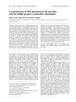
larly, compared to the control group, myofibrillar protein
breakdown in the LPS t reatment group was significantly
increased (1.98 ± 0.19 vs. 5.25 ± 0.29, P < 0.01). With
administration of 2.4 mU.kg
-1
.min
-1
insulin, the net release
of 3-MH was decreased in sepsis rats (5.25 ± 0.29 vs. 4.3 ±
0.27, P <0.01). When the infusion rate o f insulin was
increased (4.8 mU.kg
-1
.min
-1
), the inhibition effect on the
net release of 3-MH increased slightly compared to the
low-dose insulin group (4.3 ± 0.27 vs. 3.67 ± 0.14, P =
0.069) (Figure 1, 2).
Ubiquitin, E2-14KDa, and proteasome subunit C2
expression in gastrocnemius muscle
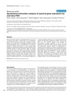
RT-PCR analysis indicated that Ub, E2-14KDa, and C2
mRNA in gas trocnemius muscle was upregulated signifi-
cantly after LPS injection. However, after infusion of insu-
lin, Ub mRNA levels in septic rats decreased gradually and
was dependent on the insulin infusion dos (Figure 3A).
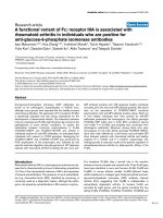
Although low-dose insulin had no notable influence on
E2-14KDa mRNA expression, when the insulin infusion
rate was increased to 4.8 mU.kg
-1
.min
-1
, E2-14KDa mRNA
expression wa s downregulated (Figure 4A). However,
Chen et al. Journal of Inflammation 2011, 8:13
/>Page 3 of 8
insulin infusion had no influence on C2 mRNA expression
in septic rats (Figure 5A).
Western blot analysis
Western blot analysis showed that LPS pretreatment
increasedUbconjugation,E2-14KDa,andtheprotea-
some subunit C2 proteins in gastrocnemius muscle, and
Ub conjugation was mainly to high-molecular weight
proteins. Although low-dose insulin infusion had no
influence on Ub conjugation, when the insulin infusion
was increased to 4.8 mU.kg
-1
.min
-1
, levels of proteins
Figure 1 Level of tyrosine in the medium or tissue samples
determined by HPLC. Data aremeans ± SE, and expressed as
nanomoles of tyrosine in medium per 2·h-1 g·muscle-1. Comparison
to control group, *P < 0.01; to LPS group, #P < 0.01; to low-dose
insulin group, $P < 0.01.
Figure 2 Level of 3-MH in medium or tissue samples
determined by HPLC. Data are means ± SE, and expressed as
nanomoles methylhistidine in medium per 2·h-1 g·muscle-1.
Comparison to control group, *P < 0.01; to LPS group, #P < 0.01.
B
A
Ubiquitin
£
£
-actin
H
igh-dose
Low-dose
LPS
Control
Ubiquitin
£
-actin
-180
-170
-140
-130
-100
High-dose
Low-dose
LPS
Control
Figure 3 A. mRNA for Ub in gastrocnemius muscles measured
by semiquantitative RT-PCR normalized to b-actin. Data are
means ± SE. Compared to control group, *P < 0.05; to LPS group,
#P < 0.05; to low-dose insulin group, $P < 0.05. B. Ubiquitinated
protein analysed by western blot with protein levels quantified by
densitometry and normalized to b-actin. Compared to control
group, *P < 0.05; to LPS group, #P < 0.05; to low-dose insulin
group, $P < 0.05.
Chen et al. Journal of Inflammation 2011, 8:13
/>Page 4 of 8
A
E2-14KDa
£
£
-actin
High-dose
Low-dose
LPS
Control
E2-14KDa
£
-actin
High-dose
Low-dose
LPS
Control
B
Figure 4 A. mRNA for E 2-14KDa in gastrocnemius muscle
measured by semiquantitative RT-PCR with normalization to
b-actin. Data are means ± SE. Compared to control group, *P <
0.05; to LPS group, #P < 0.05. B. E2-14KDa protein was by western
blot with protein levels quantified by densitometry and normalized
to b-actin. Compared to control group, *P < 0.05; to LPS group,
#P < 0.05; to low-dose insulin group, $P < 0.05.
C2
£
£
-actin
High-dose
Low-dose
LPS
Control
C2
£
-actin
High-dose
Low-dose
LPS
Control
A
B
Figure 5 A. mRNA for proteasome subunit C2 in gastrocnemius
muscle measured by semiquantitative RT-PCR with
normalization to b-actin. Data are means ± SE. Compared to
control group, *P < 0.05. B. Proteasome subunit C2 protein by
western blot with protein levels quantified by densitometry and
normalized to b-actin. Compared to control group, *P < 0.05; to LPS
group, #P < 0.05; to low-dose insulin group, $P < 0.05.
Chen et al. Journal of Inflammation 2011, 8:13
/>Page 5 of 8
conjugated to Ub in septic rats decreased compared to
the LPS and low-dose insulin groups (Figure 3B). Com-
paredtotheLPSgroup,nodifference was observed
after low-dose insulin infusion; however, when the insu-
lin infusion was 4.8 mU.kg
-1
.min
-1
, the concentr ation of
E2-14KDa protein decreased compared to the LPS and
low-dose insulin groups (Figure 4B). After insulin infu-
sion, the levels of the proteasome subunit C2 in septic
rats decreased gradually (Figure 5B).
Discussion
Severe injury, infection, and other critical illnesses are
associated with excessive loss o f body pro tein. Muscle
catabolism, resulting in muscl e wast ing and fatigue , is a
characteristic metabolic response to sepsis [1-3]. Sepsis-
induced muscle catabolism is mainly caused by increased
protein breakdown, in particular myofibrillar protein
breakdown, although reduced protein synthesis and
inhibited amino acid transport contribute to the meta-
bolic response. Muscle breakd own may impair recover y
in septic patients and increase the risk for ulmonary and
thrombo-embolic complications when respiratory mus-
cles and ambulation are affected [3-6]. Ub Ub A loss of
greater than 10% body protein contributes significantly
to morbidity and debility [20]. Methods to reduce the
catabolic response in skeletal muscle during sepsis, there-
fore, have great clinical significance.
Many studies have confirmed the i mportance of the
Ub-proteasome system for breaking down intracellular
proteins during pathophysiologic conditions, for example,
severe sepsis, burn, diabetes, or trauma [2,21-25]. In this
study, we used a classical rat LPS model of intraperito-
neal injection with 10 mg/kg LPS to mimic sepsis. After
induction of sepsis for 8 h, we found that expression of
the genes for Ub, E2-14KDa, and the 20S proteasome
subunit C2 were upregul ated signi ficantly. At the same
time, the concent ration of ubiquitinated proteins, E2-
14KDa, and C2, increased notably in the LPS group com-
pared to the control group. Molecular markers of skeletal
muscle proteolysis (tyrosine and 3-MH) increased
prominently.
Our results are consistent with other studies. For
example, Chai, et al. [25] suggested that after intraperi-
toneal injection of 10 mg/kg LPS, mRNA for Ub, E2-
14KDa, and C2 were upregulated significantly compared
to normal control rats, while therateoftotalprotein
breakdown and myofibrillar proteolysis increased. Van
Beneden et al. [26] similarly found that Ub and E2-
14KDa mRNA inc reased in rat tibialis anterior muscles
afte r LPS injection. Hobler et al. [27] suggested that the
Ub-proteasome system E214kDa increased 70% in EDL
in septic rats induced by cecal ligation and puncture.
Subsequently, they discovered a three- to four- fold
increase in mRNA levels for Ub and the 20s proteasome
in muscle tissue from septic patients, concomitant with
increased muscle levels of phenylalanine and 3-MH [8].
These data support our r esults that the Ub-proteasome
system was activated in skeletal muscle under septic
conditions.
The function of the Ub-proteasome system is indepen-
dent of the amount of protein consumed. Consequently,
simple nutritional supplementation would not be
expected to attenuate muscle catabolism [28]. Thus,
developing new therapeutic approaches for treating
muscle wasting is important, especially for h ypermeta-
bolism patients. Currently, several studies suggest that
insulin can inhibit the activation of the Ub-proteasome
system. For example, a low level of plasma insulin trig-
gers protein degradation in muscle through activation of
the Ub-proteasome pathway [13], while higher levels
downregulate the expression of th e 14-kDa E2 conjugat-
ing enzyme in vitro [14]. In vivo, a 6-hour hyperinsuli-
naemic euglycaemic clamp significantly reduced Ub
mRNA in fast twitch and m ixed skeletal muscle. Obser-
vations in hepatoma cells show that insulin regulates
anticatabolic activity by decreasing Ub mediated protea-
somal activity [17]. However, under sepsis conditions,
whether insulin also inhibits the expression of the Ub is
not currently known.
In our study, we hypothesized than continuous insulin
infusion would alleviate degradation of skeletal muscle
protein by inhibiting the Ub-proteasome system under
sepsis conditions. After intraperitoneal injection with
LPS, low-and high-dose insulin group anim als received a
continuous infusion of insulin at 2.4 mU.kg
-1
.min
-1
or
4.8 mU.kg
-1
.min
-1
, and blood glucose was controlled to
4.4-6.1 mmol/L. After 8 h, we found that mRNA for Ub,
and the protein concentra tion of the proteaso me su bunit
C2inthelow-doseinsulingroupweresignificantly
higher than in the LPS group. At the same time, 3-MH
was also reduced, but the concentration of tyrosine and
other mRNA and protein levels of the Ub system chan-
ged only slightly. When the infusion dose of insulin was
increased to 4.8 mU.kg
-1
.min
-1
,theUbmRNAlevelwas
furth er reduced compared to the low-dose insulin group.
Compared to the LPS group, E2-14KDa mRNA was
downregulated prominently. Although insulin infusion
had no influence on C2 mRNA expression, C2 protein
levels were significantly decreased and the extent of C2
protein decrease was proportional to the insulin dose.
The concentration of ubiquitinated proteins was also
downregulated. Because high-dose insulin infusion
reduced the activity of the Ub system, the release of tyro-
sine and 3-MH in EDL also decreased. The se findings, to
some extent, suggest that infusion of insulin alleviates
degradation of skeletal muscle protein by inhibiting the
Ub-proteasome system, and the effect is proportional to
the insulin infusion dose.
Chen et al. Journal of Inflammation 2011, 8:13
/>Page 6 of 8
Several possible mechanisms may result in insulin reg-
ulation of Ub-proteasome activity. Several animal
experiments and clinical evidence suggest that in dia-
betes, the PI3K-Akt pathway plays a key role in inhibit-
ing the activity of the Ub system [29-31]. However,
whether PI3K-Akt has the same effect under sepsis is
not yet known. Our preliminary experiments showed
that after administering LY294002, an inhibitor of the
PI3K-Akt pathway, the inhibiting effect of insulin was
clearly decreased. Insulin resistance is common in septic
conditions, resulting in a relative lack of in sulin in vivo
[29,32,33]. Hu et al. [14] found that insulin deficiency
activated the Ub-proteaso me system, resulting in cardiac
muscle protein catabolism in diabetes mellitus. They
also found that insulin resistance accelerates muscle
protein degradation by activation of the Ub-proteasome
[34]. Other studies suggested administration of insulin
significantly reduced Ub mRNA [14-16]. However in
our study, the relationship between insulin resistance
and activation of Ub system were not confirmed. Our
preliminary results show that insulin significantly inhib-
ited the release of inflammatory cytokines such as TNF-
a, IL-1 and IL-6 in septic patients [35]. These inflamma-
tory cytokines are key factors for activity of the Ub-pro-
teasome system [36]. Thus, insulin may inhibit the
activity of the Ub system by inhibiting inflammatory
cytokines. However, the correlation between insulin,
cytokines and the Ub system remains to be further
investigated. Another possibility is that insulin may inhi-
bit the proteasome through an associated protein, IDE.
The catalytic properties of the proteasome can vary
widely, depending on its association with regulatory pro-
teins [37]. Previous studies showed th at insulin inhibits
the proteasome in vitro and in cultured cells. Removal
of IDE from the extracts or introduction of a neutraliz-
ing antibody into cells results in a loss of insulin regula-
tion of the proteasome [38,39].
In conclusion, our results suggested that insulin admin-
istration to septic rats can inhibit the Ub-proteasome sys-
tem with an effect proportional to the insulin infusion
dose. These findings may provide a new therapeutic
strategy for hypercatabolism patients under septic cond i-
tions or other critical illnesses.
Acknowledgements
This study was supported by the National Natural Science Foundation (No.
30801086), the Natural Science Foundation of Jiangsu Province (No.
BK2007573), and the Research Fund for the Doctoral Program of Higher
Education of China (No. 200802841005)
Authors’ contributions
QC and TG participated in collection of data. NL and WY conceived and
designed this study. WL and JZ did the statistical analysis. QC and WY wrote
the first draft of the paper and JL commented on the draft. All other authors
provided comments and approved the final manuscript.
Competing interests
The authors declare that they have no competing interests.
Received: 30 August 2010 Accepted: 3 June 2011
Published: 3 June 2011
References
1. Hasselgren PO: Role of the ubiquitin-proteasome pathway in sepsis-
induced muscle catabolism. Mol Biol Rep 1999, 26(1-2):71-6.
2. Lin SY, Chen WY, Lee FY, Huang CJ, Sheu WH: Activation of ubiquitin-
proteasome pathway is involved in skeletal muscle wasting in a rat
model with biliary cirrhosis: potential role of TNF-alpha. Am J Physiol
Endocrinol Metab 2005, 288(3):E493-501.
3. Tiao G, Hobler S, Wang JJ, et al: Sepsis is associated with increased
mRNAs of the ubiquitin-proteasome proteolytic pathway in human
skeletal muscle. J Clin Invest 1997, 99(2):163-8.
4. Hasselgren PO, Fischer JE: The ubiquitin-proteasome pathway: review of a
novel intracellular mechanism of muscle protein breakdown during
sepsis and other catabolic conditions. Ann Surg 1997, 225(3):307-16.
5. Klaude M, Fredriksson K, Tjader I, et al: Proteasome proteolytic activity in
skeletal muscle is increased in patients with sepsis. Clin Sci (Lond) 2007,
112(9):499-506.
6. Minnaard R, Wagenmakers AJ, Combaret L, et al: Ubiquitin-proteasome-
dependent proteolytic activity remains elevated after zymosan-induced
sepsis in rats while muscle mass recovers. Int J Biochem Cell Biol 2005,
37(10):2217-25.
7. Dardevet D, Sornet C, Taillandier D, Savary I, Attaix D, Grizard J: Sensitivity and
protein turnover response to glucocorticoids are different in skeletal
muscle from adult and old rats. Lack of regulation of the ubiquitin-
proteasome proteolytic pathway in aging. JClinInvest1995, 96(5):2113-9.
8. Hobler SC, Wang JJ, Williams AB, et al: Sepsis is associated with increased
ubiquitinconjugating enzyme E214k mRNA in skeletal muscle. Am J
Physiol 1999, 276(2 Pt 2):R468-73.
9. Rajan VR, Mitch WE: Muscle wasting in chronic kidney disease: the role of
the ubiquitin proteasome system and its clinical impact. Pediatr Nephrol
2008, 23(4):527-35.
10. Solomon V, Madihally S, Yarmush M, Toner M: Insulin suppresses the
increased activities of lysosomal cathepsins and ubiquitin conjugation
system in burn-injured rats. J Surg Res 2000, 93(1):120-6.
11. Solomon V, Madihally S, Mitchell RN, Yarmush M, Toner M: Antiproteolytic
action of insulin in burn-injured rats. J Surg Res 2002, 105(2):234-42.
12. Van den Berghe G, Wilmer A, Hermans G, et al: Intensive insulin therapy in
the medical ICU. N Engl J Med 2006, 354(5):449-61.
13. Price SR, Bailey JL, Wang X, et al
: Muscle
wasting in insulinopenic rats
results from activation of the ATP-dependent, ubiquitin-proteasome
proteolytic pathway by a mechanism including gene transcription. J Clin
Invest 1996, 98(8):1703-8.
14. Mitch WE, Bailey JL, Wang X, Jurkovitz C, Newby D, Price SR: Evaluation of
signals activating ubiquitin-proteasome proteolysis in a model of muscle
wasting. Am J Physiol 1999, 276(5 Pt 1):C1132-8.
15. Hu J, Klein JD, Du J, Wang XH: Cardiac muscle protein catabolism in
diabetes mellitus: activation of the ubiquitin-proteasome system by
insulin deficiency. Endocrinology 2008, 149(11):5384-90.
16. Wang X, Hu Z, Hu J, Du J, Mitch WE: Insulin resistance accelerates muscle
protein degradation: Activation of the ubiquitin-proteasome pathway by
defects in muscle cell signaling. Endocrinology 2006, 147(9):4160-8.
17. Wing SS, Banville D: 14-kDa ubiquitin-conjugating enzyme: structure of
the rat gene and regulation upon fasting and by insulin. Am J Physiol
1994, 267(1 Pt 1):E39-48.
18. Sadiq F, Hazlerigg DG, Lomax MA: Amino acids and insulin act additively
to regulate components of the ubiquitin-proteasome pathway in C2C12
myotubes. BMC Mol Biol 2007, 8:23.
19. Sugita H, Kaneki M, Tokunaga E, et al: Inducible nitric oxide synthase plays
a role in LPS-induced hyperglycemia and insulin resistance. Am J Physiol
Endocrinol Metab 2002, 282(2):E386-94.
20. Ling PR, Lydon E, Qu Z, Frederich RC, Bistrian BR: Metabolic effects of
insulin and insulin-like growth factor-I in endotoxemic rats during total
parenteral nutrition feeding. Metabolism 2000, 49(5):611-5.
21. Tisdale MJ: The ubiquitin-proteasome pathway as a therapeutic target
for muscle wasting. J Support Oncol 2005, 3(3):209-17.
Chen et al. Journal of Inflammation 2011, 8:13
/>Page 7 of 8
22. Wing SS: Control of ubiquitination in skeletal muscle wasting. Int J
Biochem Cell Biol 2005, 37(10):2075-87.
23. Wray CJ, Mammen JM, Hershko DD, Hasselgren PO: Sepsis upregulates the
gene expression of multiple ubiquitin ligases in skeletal muscle. Int J
Biochem Cell Biol 2003, 35(5):698-705.
24. Breen HB, Espat NJ: The ubiquitin-proteasome proteolysis pathway:
potential target for disease intervention. JPEN J Parenter Enteral Nutr 2004,
28(4):272-7.
25. Chai J, Wu Y, Sheng ZZ: Role of ubiquitin-proteasome pathway in skeletal
muscle wasting in rats with endotoxemia. Crit Care Med 2003,
31(6):1802-7.
26. Olberding KE, Kelley ML, Butler RA, Van Beneden RJ: A HECT E3 ubiquitin-
protein ligase with sequence similarity to E6AP does not target p53 for
degradation in the softshell clam (Mya arenaria). Mutat Res 2004,
552(1-2):61-71.
27. Tiao G, Hobler S, Wang JJ, et al: Sepsis is associated with increased
mRNAs of the ubiquitin-proteasome proteolytic pathway in human
skeletal muscle. J Clin Invest 1997, 99(2):163-8.
28. Tisdale MJ: The ubiquitin-proteasome pathway as a therapeutic target
for muscle wasting. J Support Oncol 2005, 3(3):209-17.
29. Glass DJ: PI3 kinase regulation of skeletal muscle hypertrophy and
atrophy. Curr Top Microbiol Immunol 2010, 346:267-78.
30. Stitt TN, Drujan D, Clarke BA, et al: The IGF-1/PI3K/Akt pathway prevents
expression of muscle atrophy-induced ubiquitin ligases by inhibiting
FOXO transcription factors. Mol Cell 2004, 14(3):395-403.
31. Lee SW, Dai G, Hu Z, et al: Regulation of muscle protein degradation:
coordinated control of apoptotic and ubiquitin-proteasome systems by
phosphatidylinositol 3 kinase. J Am Soc Nephrol 2004, 15(6):1537-45.
32. Griesdale DE, de Souza RJ, van DRM, et al: Intensive insulin therapy and
mortality among critically ill patients: a meta-analysis including NICE-
SUGAR study data. CMAJ 2009, 180(8):821-7.
33. Yu WK, Li WQ, Li N, Li JS: Influence of acute hyperglycemia in human
sepsis on inflammatory cytokine and counterregulatory hormone
concentrations. World J Gastroenterol 2003, 9(8):1824-7.
34. Wang X, Hu Z, Hu J, Du J, Mitch WE: Insulin resistance accelerates muscle
protein degradation: Activation of the ubiquitin-proteasome pathway by
defects in muscle cell signaling. Endocrinology 2006, 147(9):4160-8.
35. Yu WK, Li WQ, Wang XD, et al: Influence and mechanism of a tight
control of blood glucose by intensive insulin therapy on human sepsis.
Zhonghua Wai Ke Za Zhi 2005, 43(1):29-32.
36. Llovera M, Garcia-Martinez C, Agell N, Lopez-Soriano FJ, Argiles JM: TNF can
directly induce the expression of ubiquitin-dependent proteolytic
system in rat soleus muscles. Biochem Biophys Res Commun 1997,
230(2):238-41.
37. DeMartino GN, Slaughter CA: Regulatory proteins of the proteasome.
Enzyme Protein 1993, 47(4-6):314-24.
38. Duckworth WC, Bennett RG, Hamel FG: A direct inhibitory effect of insulin
on a cytosolic proteolytic complex containing insulin-degrading enzyme
and multicatalytic proteinase. J Biol Chem 1994, 269(40):24575-80.
39. Hamel FG, Bennett RG, Harmon KS, Duckworth WC: Insulin inhibition of
proteasome activity in intact cells. Biochem Biophys Res Commun 1997,
234(3):671-4.
doi:10.1186/1476-9255-8-13
Cite this article as: Chen et al.: Insulin alleviates degradation of skeletal
muscle protein by inhibiting the ubiquitin-proteasome system in septic
rats. Journal of Inflammation 2011 8:13.
Submit your next manuscript to BioMed Central
and take full advantage of:
• Convenient online submission
• Thorough peer review
• No space constraints or color figure charges
• Immediate publication on acceptance
• Inclusion in PubMed, CAS, Scopus and Google Scholar
• Research which is freely available for redistribution
Submit your manuscript at
www.biomedcentral.com/submit
Chen et al. Journal of Inflammation 2011, 8:13
/>Page 8 of 8