Báo cáo y học: "Treatment of a femoral shaft fracture in a patient with congenital hip disease: a case repor" ppt
Bạn đang xem bản rút gọn của tài liệu. Xem và tải ngay bản đầy đủ của tài liệu tại đây (1.19 MB, 4 trang )
CAS E REP O R T Open Access
Treatment of a femoral shaft fracture in a patient
with congenital hip disease: a case report
George A Tsakotos, Stefanos D Koutsostathis
*
, George A Macheras
Abstract
Introduction: We present a rare case of two concomitant morbidities treated in one operation. To our knowledge,
this is the first report of its kind in the literature.
Case presentation: A 57-year-old Greek woman was admitted to the emergency department having sustained a
spiral mid-shaft femoral fracture. She also suffered from an ipsilateral hip congenital dysplasia with ankylosed hip
joint due to severe arthritis. She was treated with a total hip arthroplasty using a long stem performing as an
intramedullary nail.
Conclusion: We undertook a complex operative treatment of both co-morbidities in a one stage procedure with a
satisfactory clinical result.
Introduction
Femoral shaft fractures are usually high energy traumas,
with significant blood loss and pain. These injuries are
best treated by closed intramedullary nailing, which sta-
bilizes the fracture site and allows immediate mobiliza-
tion with full weight bearing. Congenital hip disease is
quite common in the adult Greek population. Its inci-
dence has been dramatically reduced as a result of early
screening, immediate diagnosis and treatment after
birth. Adults with congenital dysplasia usually present
with hip arthritis and restrictive pain between the fourth
and sixth decade of their life. Total hip arthroplasty in
such cases is a demanding and challenging operation.
Case presentation
A 57-year-old Greek housewife, who was 165 cm tall
and weighed 65 kg, was admitted to our hospital after a
closed injury of her right femur. She was a married
mother with one 18- year-old daughter who was a non-
smoker and who did not drink alcohol. She was suffer-
ing f rom an ipsilateral dysplastic hip [1]. As a child she
had undergone an unsuccessful operation for a non-
defined femoral osteotomy. She had no other significant
medical history and received no medication except pai n
kill ers. Her right leg was fixed in a flexed and internally
* Correspondence:
4th Orthopaedic Department, KAT Hospital, 2 Nikis str, 145 61 Kifissia,
Athens, Greece
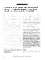
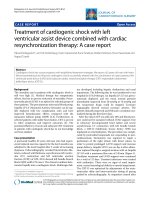
Figure 1 Initial post traumatic anteroposterior X-ray of the
femur. Hip dysplasia with severe arthritis is recognized. An oblique
mid shaft fracture is revealed
Tsakotos et al. Journal of Medical Case Reports 2010, 4:221
/>JOURNAL OF MEDICAL
CASE REPORTS
© 2010 Tsakotos et al; licensee BioMed Central Ltd. This is an Open Access article distributed under the terms of the Creative
Commons Attribution License ( which permits unrestricted use, distribution, and
reproductio n in any medium , provided the original work is properly cited.
rotated deformity. She had been walking with great diffi-
culty for more than 10 years, due to hip and knee stiff-
ness with concomitant severe hip arthritis.
She had fallen in her house while walking. On clinical
examination, the leg was in fixed flexion with adduction
and internal rotation. X-rays revealed an isolated spiral
mid-shaft fracture of the right femur (Figure 1): type 0
according to the Winquist-Hansen c lassification [2] or
32-A1 according to the AO-OTA classification [3].
We performed a total hip arthroplasty via a postero-
lateral incision, using a long cementless Wagner stem
[4] and a porous tantalum monoblock acetabular cup to
address both morbidities. The fixed deformity meant
that straight forward hip dislocation was impossible and,
therefore, the femoral neck had first to be osteotomised.
The cup was p laced in the anatomic position. Part of
the native head was used as a morselised autograft at
the true acetabular bed. The superolateral part of the
head was used as a structural graft and secured with
one screw. A cup was then inserted in a press fit man-
ner, basing the initial stability on the periphery of the
cup. After an additional small incision at the fracture
site, the fracture was initially reduced anatomically.
Reduction was secured with five cerclage wires and the
stem was inserted under direct vision. The operation
took 95 minutes. Tissues were sent for c ulture and his-
tological analysis: the results were negative for tumor or
infection, revealing that the fracture was not pathologi-
cal. The patient received three doses of prophylactic
antibiotic and was given low molecular weight heparin
for six weeks. There was no leg length discrepancy post-
operatively and no complications were recorded. She
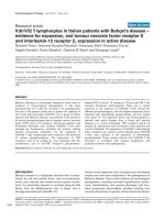
Figure 2 Pelvis anteroposterior X-ray at 3 months postoperatively. The cup has no sign of migration. The satisfactory healing process of
the morselised graft is seen at the acetabular bed. The structural autograft remains in its initial place held with one screw.
Tsakotos et al. Journal of Medical Case Reports 2010, 4:221
/>Page 2 of 4
was mobilized with partial weight bearing the second
postoperative day. Full weig ht bearing was allowed after
three weeks, due to the concomitant presence of acetab-
ular graft and diaphyseal cerclage wires. Three months
postoperatively, the fracture had healed, the cu p showed
no signs of migration (Figures 2,3,4,5), there was a nor-
mal hip range of motion and patient was walking and
free of symptoms.
Discussion
Femoral shaft fracture is usually caused by a high energy
trauma. In this case it is possible that trauma energy
was rotational and totally absorbed by the femoral shaft
due to the lack of motion at the dysplastic hip, causing
a low energy spiral fracture.
There was a debate about the best treatment for this
woman. The optimal treatment for femo ral mid-shaft
fractures is close-locked intramedullary nailing [5]. In
this case there was concern about the technical difficul-
ties of antegrade nailing due to the distorted anatomy
and the limited ability of intraoperative traction and
manipulation because of hip ankylosis in 15° of flexion
and as a result of previous surgery. Another o ption
would have been retrograde nailing or a compression
plate osteosynthesis. None of the above treatments
would have addressed the hip dysplasia and secondary
arthritis and stiffness which could have impeded proper
weight bearing and lead to the possible mechanical fail-
ure of the implants and/or an inability of the fracture to
unite. Additionally, it would have been necessary to per-
form a second operation, even with fracture healing,
which would have included material removal and total
hip arthroplasty to address the hip dysplasia.
We decided to perform a total hip a rthroplasty with a
long stem, in order to solve both the patient’ s problems
in one operation. The Wagner stem has been used for
many years in revision surgery. We applied a well known
technique that has been successful in treating peripros-
thetic fractures, combining a long stem with cerclage
wires. It was essential in this case to use secure open ana-
tomic reduction as it was not a simple femoral fracture
which could be treated by a closed intramedullary
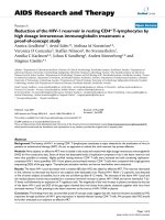
Figure 3 Anter oposterior X-ray at three postoperative months.
Fracture has healed.
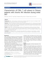
Figure 4 Lateral X-ray at three postoperative months.The
fracture has healed.
Tsakotos et al. Journal of Medical Case Reports 2010, 4:221
/>Page 3 of 4
nailing. The porous tantalum acetabular cup is a very
reliable material in dysplastic hip arthroplasties, where
acetabular bone stock is poor. It is strongly adheren t to
bone and, thus, offers excellent initial stability. It is also
highly osteoconductive and osteoinductive [6], properties
that are important for bone in-growth and long lasting
survivorship of the arthroplasty.
Conclusion
In this case an attempt was made to deal with two dif-
ferent and difficult co-morbidities in one operation. To
our knowledge, there has been no similar case re ported
in the literature. In orthopaedic surgery there is a variety
of impla nts and methods which, used correctly, can help
the surgeon to successfully treat high demanding
situations.
Consent
Written informed consent was obtained from the patient
for publication of this case report and the accompanying
images. A copy of the written consent is available for
review by the Editor-in-Chief of this journal.
Authors’ contributions
GM performed the operation and made the final review. GT analyzed the
data and wrote the manuscript. SK performed the follow-up, and reviewed
the manuscript. Both GT and SK participated in the operation. All authors
have read and approved the final manuscript.
Competing interests
The authors declare that they have no competing interests.
Received: 14 December 2009 Accepted: 22 July 2010
Published: 22 July 2010
References
1. Hartofilakidis G, Yiannakopoulos CK, Babis GC: The morphologic variations
of low and high hip dislocation. Clin Orthop Relat Res 2008, 466:820-824.
2. Johnson KD: From Femur: Trauma. Orthopaedic Knowledge Update: Trauma
Illinois: American Academy of Orthopaedic SurgeonsTornetta P III,
Baumgaertner M 1990, 3:514.
3. Rüedi TP, Buckley RE, Moran CG, (eds): AO Principles of Fracture Management
New York: Thieme, 2 2007, 767.
4. Fink B, Grossmann A, Schubring S, Schulz MS, Fuerst M: A modified
transfemoral approach using modular cementless revision stems. Clin
Orthop Relat Res 2007, 462:105-114.
5. Ricci WM, Gallagher B, Haidukewych GJ: Intramedullary nailing of femoral
shaft fractures: current concepts. J Am Acad Orthop Surg 2009, 17:296-305.
6. Gruen TA, Poggie RA, Lewallen DG, Hanssen AD, Lewis RJ, O’Keefe TJ,
Stulberg SD, Sutherland CJ: Radiographic Evaluation of a Monoblock
Acetabular Component. A Multicenter Study with 2- to 5-Year Results.
J Arthr 2005, 20:369-378.
doi:10.1186/1752-1947-4-221
Cite this article as: Tsakotos et al.: Treatment of a femoral shaft fracture
in a patient with congenital hip disease: a case report. Journal of Medical
Case Reports 2010 4:221.
Submit your next manuscript to BioMed Central
and take full advantage of:
• Convenient online submission
• Thorough peer review
• No space constraints or color figure charges
• Immediate publication on acceptance
• Inclusion in PubMed, CAS, Scopus and Google Scholar
• Research which is freely available for redistribution
Submit your manuscript at
www.biomedcentral.com/submit
Figure 5 Distal anteroposterior X-ray at three postoperative
months. The fracture has healed.
Tsakotos et al. Journal of Medical Case Reports 2010, 4:221
/>Page 4 of 4