Báo cáo y học: " Tropheryma whipplei tricuspid endocarditis: a case report and review of the literature" ppsx
Bạn đang xem bản rút gọn của tài liệu. Xem và tải ngay bản đầy đủ của tài liệu tại đây (571.25 KB, 4 trang )
CAS E REP O R T Open Access
Tropheryma whipplei tricuspid endocarditis:
a case report and review of the literature
Vincent Gabus
1*
, Zita Grenak-Degoumois
2
, Severin Jeanneret
1
, Riana Rakotoarimanana
2
, Gilbert Greub
3,4
,
Daniel Genné
2
Abstract
Introduction: The main clinical manifestations of Whipple’s disease are weight loss, arthropathy, diarrhea and
abdominal pain. Cardiac involvement is frequently described. However, endocarditis is rare and is not usually the
initial presentation of the disease. To the best of our knowledge, this is the first reported case of a patient with
Tropheryma whipplei tricuspid endocarditis without any other valve involved and not presenting signs of arthralgia
and abdominal involve ment.
Case presentation: We report a case of a 50-year-old Caucasian man with tricuspid endocarditis caused by
Tropheryma whipplei, showing signs of severe shock and an absence of other more classic clinical signs of
Whipple’s disease, such as arthralgia, abdominal pain and diarrhea. Tropheryma whipplei was documented by
polymerase chain reaction of the blood and pleural fluid. The infection was treated with a combined treatment of
doxycycline, hydroxychloroquine and sulfamethoxazole-trimethoprim for one year.
Conclusion: Tropheryma whipplei infectious endocarditis should always be considered when facing a blood-culture
negative endocarditis particularly in right-sided valves. Although not standardized yet, treatment of Tropheryma
whipplei endocarditis should probably include a bactericidal antibiotic (such as doxycycline) and should be given
over a prolonged period of time (a minimum of one year).
Introduction
The Gram positive bacillus Tropheryma whippelii was
first characterized by polymerase chain reaction (PCR)
in the early 1990s [1], and renamed Tropheryma whip -
plei in 2001 after its first culture and characterization
[2]. The main clinical manifestations of Whipple’sdis-
ease are weight loss (in 80 to 90% of reported cases),
arthropathy (70 to 90%), diarrhea (70 to 85%) and
abdominal pain (50 to 90%) [3]. Cardiac involvement is
reported in 17 to 55% of patients with classical Whip-
ple’s disease, pericarditis being the most frequent [4].
Endocarditis, however, is rare and 88% of cases occur in
patients with healthy valves without underlying disease
[5]. Endocarditis was the initial presentation of only a
few cases [6-10]. We report a case of a patient with tri-
cuspid endocarditis due to Tropheryma whipplei and
review all previously reported cases.
Case presentation
A 50-year-old Caucasian alcoholic man presented to the
emergency department with generalized weakness lasting
10 days and a history of weight loss. He had no other
complaints. His history was significant for excessive alco-
hol intake and cachexia. At the emergency department,
the patient was weak but alert, appeared ill and was very
pale. The clinical exam revealed: a temperature of 35.9°C,
blood pressure 60/38 mm Hg; a heart rate of 95 beats
per minute, a respiratory rate of 23 breaths per minute,
bilateral ankle edema, buccal candidiasis, and a faint sys-
tolic murmur. The neurological exam was normal, except
for psychomotor slowing and a fine tremor. Laboratory
results showed: hemoglobin 42 g/l, platelet count 23 G/l,
WBC 4.9 G/l (normally distributed), C-reacti ve protein
21 mg/l (N < 5), hypoalbuminaemia and cholestasis.
Other labor atory tests were norm al. A chest radiograph
showed cardiomegaly and pulmonary vascular redistribu-
tion with bilateral pleural fluid accumulation. Computed
tomography (CT) imaging excluded aortic dissection,
massive pulmonary embolism, pericardial fluid and
* Correspondence:
1
Department of Internal Medicine, Centre Hospitalier Universitaire Vaudois
and University of Lausanne, Switzerland
Full list of author information is available at the end of the article
Gabus et al . Journal of Medical Case Reports 2010, 4:245
/>JOURNAL OF MEDICAL
CASE REPORTS
© 2010 Gabus et al; licensee BioMed Central Ltd. This is an Open Access article dist ributed under th e terms of the Creative Commons
Attribution License (htt p://creativecommons.org/licenses/by/2.0), which permits unrestricted use, distribution, and reproduction in
any medium, pro vided the original work is properly cited.
retroperitoneal hematoma. After blood, urine and pleural
fluid had been collected for culture, he wa s empirically
treated intravenously with amoxicillin/clavulanate 2.2 g
four times a day and ciprofloxacin 200 mg twice a day
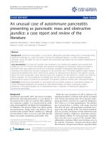
for presumed septic shock. A transthoracic echocardio-
graphy, that was performed because of the systolic mur-
mur and the hemodynamic instability, showed evidence
of tricuspid valvular involvement with several large vege-
tations of approximately 2.5 cm in diameter, severe valvu-
lar regurgitation, and a reduced ejec tion fraction (45%)
(Figure 1). As no pathogen could be isolated from blood
cultures after 60 hours of incubation, we considered all
agents of culture-negative endocarditis as possibl e etiol-
ogy. Investigations for HACEK microorganisms, and sero-
logic studies for Bartonella spp., Brucella spp.and
Coxiella burnetti were negative; PCR of the blood and
pleural fluid for Tropheryma whipplei was positi ve. The
PCR technique described by Meibach et al.in2003was
use d [11]. Having diagnos ed Tropheryma whipplei right
heart endocarditis, we switched the antibiotic regimen to
ceftriaxone 2 g once daily. Favorable clinical changes kept
him from requiring surgery, and he returned home after
25 days with a combined treatment of doxycycline 100
mg twice a day, hydroxychloroquine 200 mg and sulfa-
methoxazole-trimethoprim 160/800 mg three times a day
for a minimum of one year. The blood levels of doxycy-
cline and hydroxychloroquine were measured every other
mont h and doses adapted to therapeutic levels (doxycy-
cline: > 5 μg/ml, hydroxychloroquine 1 +/- 0.2 mg/l). At
a one-year follow-up he had completely recovered, gained
weight and all his laboratory values were back to normal.
A control echography performed after one year (Figure 2)
confirmed the treatment success.
Discussion
Right-sided endocarditis, which usually involves the tri-
cuspid valve, occurs predominantly in intravenous drug
users or is related to congenital defects, intracardiac
catheters, pacemakers or cardiac anomalies [12]. Physi-
cians often use the Duke criteria to diagnose endocardi-
tis, but in patients with blood culture-negative
endocarditis due to Tropheryma whipplei,twoofthe
criteria (fever and a history of valvulopathy) are gener-
ally absent, making them difficult to diagnose [4]. In
2001, Fenollar et al. reviewed the literature of Whipple’s
endocarditis based on valve histology [5]. According to
that study, patients with Whipple’s endoc arditis have no
previous heart disease and are most often afe brile, their
blood cultures are negative, and vegetation is observed
on an echocardiograph in 75% of cases. Fenollar et al
described 35 cases which came from a pathology series
without detailed clinical history. A tricuspid endocarditis
associated with involvement of other valves (mostly aor-
tic) is reported in 6% of cases [4]. To the best of our
knowledge, only one case of a patient diagnosed with
Tropheryma whipplei tricuspid endocarditis without any
other valve involved has b een completely reported [13].
It describes the case of a young female presenting with
migratory arthralgia, abdominal pain, diarrhea, and
weight loss of two years duration. Physical examination
revealed a systolic murmur on the left sternal margin.
The diagnosis of Whipple’ s disease was made on jejunal
biopsy by electron microscopy and transoesophageal
echocardiogram revealed a fixed vegetation on the tri-
cuspid valve. The patient wa s successfully treated with
penicillin G and streptomycin for 14 days, followed by
sulfamethoxazole-trimethoprim for one year [13]. No
surgery involving the valve was carried out.
Figure 1 Transthoracic echocardiography at time of diagnosis
showing large vegetations on the tricuspid valve caused by
Tropheryma whipplei. The tricuspid valve is indicated by an arrow.
VD; right ventricle: OD; right atrium.
Figure 2 Transthoracic echocardiography after one year of
treatment. No vegetation was found. The tricuspid valve is
indicated by an arrow. VD; right ventricle: OD; right atrium: VG; left
ventricle: OG; left atrium.
Gabus et al . Journal of Medical Case Reports 2010, 4:245
/>Page 2 of 4
Contrary to the patie nt described by Ferrari et al. who
presented symptoms (arthralgia and digestive involve-
ment) suggestive of Whipple’s disease [13], our patient
presented a Tropheryma whipplei endocardit is manifest-
ing as severe shock. Apart from weight loss, he didn’t
exhibit any o f the typical symptoms of Whipple’ sdis-
ease. He also did not have any risk factors for right-
sided endocarditis.
Diagnosis of Whipple’ s disease is suspected most of
the time on the basis of gastrointestinal symptoms and
is generally confirmed by intestinal biopsies. According
to recently published data it seems that the occurrence
of endocarditis due to Tropheryma whipplei, without
any of the classical features of Whipple’ s disease, is not
as rare as was previously thought [1 4]. As w e did not
suspect Whipple’s disease at the beginning, we did not
perform intestinal biopsies. No serology is yet avai lable.
PCR is especially useful for the diagnosis of Whipple
endocarditis and may be directly performed on blood
samples and pleural fluid, as we did, or on valvular sam-
ples [15]. PCR performed on blood allows a non-inva-
sive diagnosis and rapid results. However, cautious
interpretation of PCR results is needed since PCRs have
been positive in healthy patients, most likely as a result
of contamination [16]. Convers ely, sensiti vity of PCR on
bloo d sampl es may be impaired by the pre sence of PCR
inhibitors and by the relatively low amount of circulat-
ing DNA. For patients with concomitant gastrointestinal
involvement, diagnosis may a lso be made mor e easily
from a small bowel biopsy that will be positive on PAS-
staining. In the present case, the obvious vegetation on
cardiac ultrasound, the posit ive PCR on two different
samples (blood and pleural fluid), and the favorable
change in the condition with anti biotics makes the etio-
logical role of Tropheryma whipp lei in this right-sided
endocarditis absolutely clear.
Concerning treatment, our patient was initially treated
by ceftriaxone then with a combination of sulfamethoxa-
zole/trimethoprim, hydroxychloroquine and doxycycline
for one year. By analogy with what is known about Cox-
iella burnetii, the association of a lysotropic agent
(hydroxychloroquine) to doxycycline tends to reduce the
acidity of the vacuole in which Tropheryma whipplei is
located and thus improves the efficacy of doxycycline
inactive at lower pH [17,18]. Interestingly, between sul-
famethoxazole and tr imethoprim, only sulfametho xazole
is active and trimethoprim is absolutely not effective
against Tropheryma whipplei; thus, the association of
sulfamethoxazole and trimethoprim represents a
monotherapy.
Conclusion
In summary, Tropheryma whipplei infectious endocardi-
tis is a rare disease and tricuspid involvement is f ound
even less often. This diagnosi s shoul d always be consid-
ered when facing a blood-culture negative endocarditis
particularly in right-sided endocarditis without risk fac-
tors. Although not standardized yet, treatment o f
Tropheryma whipplei endocarditis should probably
include a bactericidal antibiotic (such as doxycycline)
andshouldbegivenforaprolongedperiodoftime
(a minimum of one year).
Competing interests
The authors declare that they have no competing interests.
Consent
Written informed consent was obtained from the patient for publication of
this case report and any accompanying images. A copy of the written
consent is available for review by the Editor-in-Chief of this journal.
Authors’ contributions
VG was responsible for writing the manuscript and reviewing the literature.
ZG, SJ and RR had significant roles in data gathering and were major
contributors to the content of the manuscript. VG, ZG, RR, GG and DG were
involved in patient management. GG and DG had a significant role in data
interpretation and provided significant revisions to the manuscript. All
authors read and approved the final manuscript.
Acknowledgements
We are indebted to Hans H. Siegrist and Thompson Kinge for their helpful
review of the manuscript and for their assistance with the preparation of the
manuscript. We are also indebted to Dr G. Bloemberg and to the Institute of
Medical Microbiology of the University of Zurich who performed the PCR of
Tropheryma whipplei. The authors had no financial support.
Author details
1
Department of Internal Medicine, Centre Hospitalier Universitaire Vaudois
and University of Lausanne, Switzerland.
2
Department of Internal Medicine,
Community Hospital, 2300 La Chaux-de-Fonds, Switzerland.
3
Infectious
disease service, Centre Hospitalier Universitaire Vaudois and University of
Lausanne, Switzerland.
4
Institute of Microbiology, Centre Hospitalier
Universitaire Vaudois and University of Lausanne, Switzerland.
Received: 19 September 2009 Accepted: 4 August 2010
Published: 4 August 2010
References
1. Relman DA, Schmidt TM, MacDermott RP, Falkow S: Identification of the
uncultured bacillus of Whipple’s disease. N Engl J Med 1992, 327:293-301.
2. La Scola B, Fenollar F, Fournier PE, Altwegg M, Mallet MN, Raoult D:
Description of Tropheryma whipplei gen. nov., sp. nov., the Whipple’s
disease bacillus. Int J Syst Evol Microbiol 2001, 51:1471-1479.
3. Dutly F, Altweg M: Whipple’s disease and “Tropheryma whippelii”. Clin
Microbiol Rev 2001, 14:561-583.
4. Fenollar F, Puéchal X, Raoult D: Whipple’s disease. New Eng J Med 2007,
356:55-66.
5. Fenollar F, Lepidi H, Raoult D: Whipple’s endocarditis: review of the
literature and comparisons with Q fever, Bartonella infection, and blood
culture-positive endocarditis. Clin Infect Dis 2001, 33:1309-1316.
6. Elkins C, Shuman TA, Pirolo JS: Cardiac Whipple’s disease without
digestive symptoms. Ann Thorac Surg 1999, 67:250-251.
7. Bostwick DG, Bensch KG, Burke JS, Billingham M E, Miller D C, Smith J C,
Keren D F: Whipple’s disease presenting as aortic insufficiency. N Engl J
Med 1981, 305:995-998.
8. Gubler JG, Kuster M, Bannwart F, Krause M, Vögelin HP, Garzoli G,
Altwegg M: Whipple endocarditis without overt gastrointestinal disease:
report of four cases. Ann Intern Med 1999, 131:112-116.
9. Geissdorfer W, Wittmann I, Seitz G, Cesnjevar R, Röllinghoff M, Schoerner C,
Bogdan C: A case of aortic valve disease associated with Tropheryma
Gabus et al . Journal of Medical Case Reports 2010, 4:245
/>Page 3 of 4
whippelii infection in the absence of other signs of Whipple’s disease.
Infection 2001, 29:44-47.
10. Smith MA: Whipple endocarditis without gastrointestinal disease. Ann
Intern Med 2000, 132:595.
11. Maibach RC, Altwegg M: Cloning and sequencing an unknown gene of
Tropheryma whipplei and development of two LightCycler PCR assays.
Diagn Microbiol Infect Dis 2003, 46:181-187.
12. Mesbahi R, Chaara A, Benomar M: Infectious endocarditis of the right
heart. A propos of 10 cases. Arch Mal Coeur Vaiss 1991, 84:355-359.
13. Ferrari MdLdA, Vilela EG, Faria LC, Couto CA, Salgado CJ, Leite VR, Brasileiro
Filho G, Bambirra EA, Mendes CM, Carvalho SdC, de Oliveira CA, da
Cunha AS: Whipple’s disease. Report of five cases with different clinical
features. Rev Inst Med Trop Sao Paulo 2001, 43:45-50.
14. Escher R, Roth S, Droz S, Egli K, Altwegg M, Täuber MG: Endocarditis due
to Tropheryma whipplei: rapid detection, limited genetic diversity, and
long-term clinical outcome in a local experience. Clin Microbiol Infect
2009.
15. Muller C, Stain C, Burghuber O: Tropheryma whippelii in peripheral blood
mononuclear cells and cells of pleural effusion. Lancet 1993, 341:701.
16. Ehrbar HU, Bauerfeind P, Dutly F, Koelz HR, Altwegg M: PCR-positive tests
for Tropheryma whippelii in patients without Whipple’s disease. Lancet
1999, 353:2214.
17. Boulos A, Rolain JM, Raoult D: Antibiotic susceptibility of Tropheryma
whipplei in MRC5 cells. Antimicrob Agents Chemother 2004, 48:747-752.
18. Ghigo E, Capo C, Aurouze M, Tung CH, Gorvel JP, Raoult D, Mege JL:
Survival of Tropheryma whipplei, the agent of Whipple’s disease,
requires phagosome acidification. Infect Immun 2002, 70:1501-1506.
doi:10.1186/1752-1947-4-245
Cite this article as: Gabus et al.: Tropheryma whipplei tricuspid
endocarditis: a case report and review of the literature. Journal of
Medical Case Reports 2010 4:245.
Submit your next manuscript to BioMed Central
and take full advantage of:
• Convenient online submission
• Thorough peer review
• No space constraints or color figure charges
• Immediate publication on acceptance
• Inclusion in PubMed, CAS, Scopus and Google Scholar
• Research which is freely available for redistribution
Submit your manuscript at
www.biomedcentral.com/submit
Gabus et al . Journal of Medical Case Reports 2010, 4:245
/>Page 4 of 4