Báo cáo khoa học: "Post-traumatic soft tissue tumors: Case report and review of the literature a propos a Post-traumatic paraspinal desmoid tumor" doc
Bạn đang xem bản rút gọn của tài liệu. Xem và tải ngay bản đầy đủ của tài liệu tại đây (445.3 KB, 4 trang )
BioMed Central
Page 1 of 4
(page number not for citation purposes)
World Journal of Surgical Oncology
Open Access
Case report
Post-traumatic soft tissue tumors: Case report and review of the
literature a propos a Post-traumatic paraspinal desmoid tumor
Sarit Cohen
1
, Dean Ad-El
1
, Ofer Benjaminov
2
and Haim Gutman*
3
Address:
1
Department of Plastic Surgery, Rabin Medical Center, Beilinson Campus, Petah Tiqwa; and Sackler Faculty of Medicine, Tel Aviv
University, Tel Aviv, Israel,
2
Department of Diagnostic Imaging, Rabin Medical Center, Beilinson Campus, Petah Tiqwa; and Sackler Faculty of
Medicine, Tel Aviv University, Tel Aviv, Israel and
3
Department of Surgery, Rabin Medical Center, Beilinson Campus, Petah Tiqwa; and Sackler
Faculty of Medicine, Tel Aviv University, Tel Aviv, Israel
Email: Sarit Cohen - ; Dean Ad-El - ; Ofer Benjaminov - ;
Haim Gutman* -
* Corresponding author
Abstract
Background: Antecedent trauma has been implicated in the causation of soft tissue tumors.
Several criteria have been established to define a cause-and-effect relationship. We postulate
possible mechanisms in the genesis of soft tissue tumors following antecedent traumatic injury.
Case presentation: We present a 27-year-old woman with a paraspinal desmoid tumor,
diagnosed 3-years following a motor vehicle accident. Literature is reviewed.
Conclusion: Soft tissue tumors arising at the site of previous trauma may be desmoids,
pseudolipomas or rarely, other soft tissue growths. The cause-and-effect issue of desmoid or other
soft tissue tumors goes beyond their diagnosis and treatment. Surgeons should be acquainted with
this diagnostic entity as it may also involve questions of longer follow-up and compensation and
disability privileges.
Background
The etiology of most soft tissue tumors is unknown. Our
search of the English literature revealed a few case reports
of soft tissue tumors developing at the site of a previous
traumatic injury [1-17]. Desmoid tumors, lipoma and
lymphoma were among the tumors reportedly associated
with such injuries.
We describe a young woman with a left paraspinal
desmoid tumor at the site of a recent trauma, possibly
associated with a cause-and-effect mechanism. We hope
this study will shed more light on this phenomenon.
Case presentation
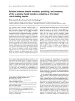
A 27-year-old woman presented with a large subcutaneous
mass in the upper back (Figure 1) of 8 months' duration.
Family history and past medical history were unremarka-
ble. The patient reported that she had been involved in a
motor vehicle accident 3 years previously, in which she
sustained a brain concussion, fracture of the right lamina
of the C-6 vertebra, and comminuted fractures of the left
radius, ulna and femur.
Physical examination revealed a firm mass measuring 15
× 10 cm, adherent to its surroundings, with no apparent
pathological vasculature or satellite lesions. Cytological
Published: 29 February 2008
World Journal of Surgical Oncology 2008, 6:28 doi:10.1186/1477-7819-6-28
Received: 19 June 2007
Accepted: 29 February 2008
This article is available from: />© 2008 Cohen et al; licensee BioMed Central Ltd.
This is an Open Access article distributed under the terms of the Creative Commons Attribution License ( />),
which permits unrestricted use, distribution, and reproduction in any medium, provided the original work is properly cited.
World Journal of Surgical Oncology 2008, 6:28 />Page 2 of 4
(page number not for citation purposes)
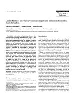
examination was inconclusive. Magnetic resonance imag-
ing (MRI) demonstrated a solid space-occupying lesion
measuring 12 × 4.8 × 7.6 cm, located in the left paraspinal
region beneath the trapezium muscle (asterisk), com-
pressing the paraspinal muscles medially (Figure 2). The
tumor has a heterogeneous appearance on T
2
weighted
images and enhanced with the injection of contrast mate-
rial, demonstrating its vascularity. Findings on core needle
biopsy were compatible with desmoid tumor. Colonos-
copy revealed no abnormalities.
Owing to the large size of the tumor and its close proxim-
ity to the spine, the initial treatment consisted of
tamoxifen 20 mg twice daily and indomethacin 250 mg
q8h. The treatment was well tolerated. However, after 4
months, neither subjective nor objective changes in tumor
consistency or size were noted. The tamoxifen dosage was
therefore doubled. Computerized tomography (CT) scan,
4 months later demonstrated tumor growth. There was no
evidence of infiltration of adjacent bony structures or pul-
monary metastases. The patient was offered surgery.
The tumor was surgically excised. It measured 9 × 12 × 22
cm and weighed 1970 grams. It was relatively well circum-
scribed, with a fibrous consistency, and no areas of hem-
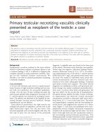
orrhage or necrosis. Microscopic study revealed relatively
low (up to 2–3/10HPF) mitotic activity (Figure 3, 4). The
surgical margins were clear. At present, 24 months post-
operatively, the patient is tumor-free.
Discussion
Desmoid tumor is a benign, locally aggressive neoplasm
that arises from fascial or musculoaponeurotic tissue. It
has a tendency to infiltrate surrounding tissue. The term
'desmoid', derived from the Greek "desmos" which means
tendon-like was first employed by Müller [12] in 1838.
Desmoid tumors account for 0.03% of all neoplasms
[13,14], and 3.0% of all soft tissue tumors [15,16].
Patients with familial adenomatous polyposis (FAP) have
a 1000-fold increased risk of developing desmoid tumors
Large subcutaneous mass in the left paraspinal regionFigure 1
Large subcutaneous mass in the left paraspinal region.
MRI of the tumor: T1W pre-(A) and post-(B) gadolinium injection, T2W (C) and T1W post gadolinium, sagittal view (D)Figure 2
MRI of the tumor: T1W pre-(A) and post-(B) gadolinium
injection, T2W (C) and T1W post gadolinium, sagittal view
(D). The tumor (arrows) has a heterogenous appearance on
T2W images and enhances with the injection of contrast
material, demonstrating its vascularity. It is located beneath
the trapezius muscle (asterisk) which is atrophic. The parasp-
inal muscle is compressed medially.
Histopathologic specimen demonstrating spindle cell prolifer-ation without significant atypia or pleomorphism (HE × 40)Figure 3
Histopathologic specimen demonstrating spindle cell prolifer-
ation without significant atypia or pleomorphism (HE × 40).
World Journal of Surgical Oncology 2008, 6:28 />Page 3 of 4
(page number not for citation purposes)
compared to the general population. The abdomen is the
most common site of the tumors in this patient group,
many times following a surgical insult.
The reported female: male ratio for sporadic desmoid
tumors is 5:2 [17]; most women are affected during or
after pregnancy. Reitamo et al., [13] found that 80% of
desmoid tumors occur in females, 50% of them in the
third to fifth decade of life. The female predominance is
less prominent in patients with FAP [18,19].
Recently, It was found that virtually all desmoid tumors
have somatic [beta]-catenin or adenomatous polyposis
coli (APC) gene mutation leading to intranuclear accu-
mulation of [beta]-catenin [20]. The expression of
nuclear [beta]-catenin may play a role in the differential
diagnosis of desmoid tumors from a host of fibroblastic
and myofibroblastic lesions as well as from smooth mus-
cle neoplasms [20]. The treatment of desmoid tumors is
usually surgical. Local recurrences may occur even after
clear margin resection. Distant metastases are extremely
rare.
The pathogenesis of desmoid tumor may involve genetic
abnormalities, sex hormones, and trauma [17], includ-
ing surgical trauma, especially in patients with FAP [19].
One study found that 10–30% of all sporadic abdominal
wall desmoid tumors occurred following surgical inter-
vention. Half these tumors developed within 4 years of
surgery [17].
Gebhart et al., [3] reported a case of desmoid tumor aris-
ing at the site of a total hip replacement. Desmoid tumors
developing around silicone implants have also been
described [13]. Skhiri et al., [1] reported a case of cervical
desmoid following placement of an internal jugular cath-
eter, and Wiel Marin et al., [2] described a thoracic
desmoid tumor at the site of a previous rib fracture.
Traumatic injury has been implicated as a causative factor
in the genesis of other soft tissues as well. Radhi et al., [6]
reported 3 cases of diffuse centroblastic lymphoma at a
site of previous surgery with metallic implants. Two of
them were preceded by atypical lymphoid infiltrate.
In 1969, Brooke and MacGregor [21] suggested that
lipoma may be secondary to trauma because of the pro-
lapse of normal deep adipose tissue through a tear in the
overlying Scarpa's fascia, namely, "pseudolipoma". Pseu-
dolipoma consists of normal adipose tissue in an abnor-
mal location, and is not considered a true lipoma because
it is not encapsulated. Meggit and Wilson [22] reported 12
cases of post-traumatic so-called lipoma. They speculated
that the tumors were the consequence of a rupture in the
septa that normally surround adipose tissue. A later report
by Herbert and DeGeus [23] described a young girl with
an abdominal wall lipoma due to pressure from tightly fit-
ting briefs. They demonstrated an anatomical defect in the
Scarpa's fascia at the level of a perforating vessel with fat
herniating through it.
The largest series of 24 pseudolipomas was reported by
Rozner and Isaacs [24] in 1977, wherein scar contracture
following a shearing fascial injury was the etiological
mechanism. Penoff [25] described 3 cases of traumatic
lipoma of the hip, although he found no anatomic confir-
mation of an injury to Scarpa's fascia.
In 1988, Dodenhoff [26] described a "saddle-bag deform-
ity" of the right hip secondary to trauma. Post-traumatic
lipoma was also reported by Elsahy [27] (5 cases) and
David et al., [8] (10 cases). Signorini and Campiglio [9]
described 9 cases of subcutaneous lipoma that appeared
within a few months of a blunt trauma. They proposed
that the differentiation of mesenchymal precursors
(preadipocytes) to mature adipocytes – a process triggered
by the trauma – could lead to the formation of subcutane-
ous lipoma.
Warren [28] listed several criteria defining a post-trau-
matic neoplasm: (a) prior integrity of the tumor site; (b)
injury severe enough to initiate reparative proliferation of
cells; (c) reasonable latent period; and (d) tumor compat-
ible with the scar tissue and anatomic location of the
injury. Ewing [29] suggested slightly different criteria to
establish a cause/effect relationship: (a) authenticity and
severity of the injury; (b) previous integrity of the
wounded part; (c) tumor originating within the boundary
of the injury; (d) histologic variety of tumor compatible
with underlying scar tissue; and (e) proper latent period.
Photomicrograph at high power magnification (HE × 100)Figure 4
Photomicrograph at high power magnification (HE × 100).
World Journal of Surgical Oncology 2008, 6:28 />Page 4 of 4
(page number not for citation purposes)
In our case, the wounded part (upper back) was previ-
ously tumor-free, the authenticity of the trauma was con-
firmed by MRI, the tumor originated within the boundary
of the injury, and the latency period was reasonable. Fur-
thermore, the desmoid histology was compatible with a
scar or other reparative process. Thus, the tumor met the
criteria of both Warren [28] and Ewing [29] for post-trau-
matic neoplasm.
Conclusion
The cause-and-effect issue of desmoid or other soft tis-
sue tumors goes beyond their diagnosis and treatment.
It may also involve questions of longer follow-up and
compensation and disability privileges.
Pseudolipomas are not real neoplasia, but they seem to
account for the reports of the so-called post-traumatic
lipomas. The post-injury local reparatory mechanisms
better explain the creation of desmoid tumors, which, in
these rare cases, seem to have lost control of cell growth,
giving rise to a soft tissue tumor. The rarity of desmoid
tumor, its specific biology, the well-documented associ-
ation between abdominal wall desmoids and preg-
nancy, and even the tendency of surgery to induce new
desmoid tumors in patients with FAP support the
notion that trauma/tissue injury is a likely cause of at
least, some of these tumors, including the one described
here.
Abbreviations
CT-computerized tomography; FAP-familial adenoma-
tous polyposis; MRI-magnetic resonance imaging
Competing interests
The author(s) declare that they have no competing inter-
ests.
Authors' contributions
CS participated in drafting the manuscript, interpretation
of data and conceptual design, AD conceived the study
and participated in drafting the manuscript, BO carried
out the imaging analysis and interpretation of data, GH
carried out the surgical procedure, conceptual design, par-
ticipated in drafting the manuscript and revised it criti-
cally for important intellectual content.
All authors read and approved the final manuscript.
Acknowledgements
Written consent was obtained from the patient for publication of this case
report.
References
1. Skhiri H, Zellama D, Ameur Frih M, Moussa A, Gmar Bouraoui S,
Achour A, Ben Dhia N, Zakhama A, Elmay M: Desmoid cervical
tumor following the placing of an internal jugular catheter.
Presse Med 2004, 33:95-97. (French)
2. Wiel Marin A, Romagnoli A, Carlucci I, Veneziani A, Mercuri M, Des-
tito C: Thoracic desmoid tumors: a rare evolution of rib frac-
ture. Etiopathogenesis and therapeutic considerations. G
Chir 1995, 16:341-344.
3. Gebhart M, Fourmarier M, Heymans O, Alexiou J, Yengue P, De Saint-
Aubain N: Development of a desmoid tumor at the site of a
total hip replacement. Acta Orthop Belg 1999, 65:230-234.
4. Pereyo NG, Heimer WL 2: Extraabdominal desmoid tumor. J
Am Acad Dermatol 1996, 34(2 Pt 2):352-356.
5. Flores RAR: Abdominal desmoid tumors and the surgeon. Rev
Gastroenterol Mex 1995, 60:207-210.
6. Radhi JM, Ibrahiem K, al-Tweigeri T: Soft tissue malignant lym-
phoma at sites of previous surgery. J Clin Pathol 1998,
51:629-632.
7. Delpla PA, Rouge D, Durroux R, Rouquette I, Arbus L: Soft tissue
tumors following traumatic injury: two observations of inter-
est for the medicolegal causality. Am J Forensic Med Pathol 1998,
19:152-156.
8. David LR, DeFranzo A, Marks M, Argenta LC: Posttraumatic pseu-
dolipoma. J Trauma 1996, 40:396-400.
9. Signorini M, Campiglio GL: Posttraumatic lipomas: where do
they really come from? Plast Reconstr Surg 1998, 101:699-705.
10. Copcu E, Sivrioglu NS: Posttraumatic lipoma: analysis of 10
cases and explanation of possible mechanisms. Dermatol Surg
2003, 29:215-220.
11. Bashara ME, Jules KT, Potter GK: Dermatofibrosarcoma protu-
berans: 4 years after local trauma. J Foot Surg 1992, 31:160-165.
12. Müller J: Veber den Feinern Bau und die Formen der Krankhaftlichen
Geschwulste
Berlin: G Reimer; 1838:80.
13. Reitamo JJ, Hayry P, Nykyri E, Saxen E: The desmoid tumor. I.
Incidence, sex-, age- and anatomical distribution in the Finn-
ish population. Am J Clin Pathol 1982, 77:665-673.
14. Suit HD: Radiation dose and response of desmoid tumors. Int
J Radiat Oncol Biol Phys 1990, 19:225-227.
15. Taylor LJ: Musculoaponeurotic fibromatosis. A report of 28
cases and review of the literature. Clin Orthop Relat Res 1987,
224:294-302.
16. Nuyttens JJ, Rust PF, Thomas CR Jr, Turrisi AT 3rd: Surgery versus
radiation therapy for patients with aggressive fibromatosis
or desmoid tumors: A comparative review of 22 articles.
Cancer 2000, 88:1517-1523.
17. Kulaylat MN, Karakousis CP, Keaney CM, McCorvey D, Bem J, Abrus
JL Sr: Desmoid tumor: a pleomorphic lesion. Eur J Surg Oncol
1999, 25:487-497.
18. Shields CJ, Winter DC, Kirwan WO, Redmond HP: Desmoid
tumors. Eur J Surg Oncol 2001, 27:701-706.
19. Gurbuz AK, Giardiello FM, Petersen GM, Krush AJ, Offerhaus GJ,
Booker SV, Kerr MC, Hamilton SR: Desmoid tumors in familial
adenomatous polyposis. Gut 1994, 35:377-381.
20. Bhattacharya B, Dilworth HP, Iacobuzio-Donahue C, Ricci F, Weber
K, Furlong MA, Fisher C, Montgomery E: Nuclear [beta]-catenin
expression distinguishes deep fibromatosis from other
benign and malignant fibroblastic and myofibroblastic
lesions. Am J Surg Pathol 2005, 29:653-659.
21. Brooke RI, MacGregor AJ: Traumatic pseudolipoma of the buc-
cal mucosa. Oral Surg Oral Med Oral Pathol 1969, 28:223-225.
22. Meggitt BF, Wilson JN: The battered buttock syndrome: fat
fractures: a report on a group of traumatic lipomata. Br J Surg
1972, 59:165-169.
23. Herbert DC, DeGeus J: Post-traumatic lipomas of the abdomi-
nal wall. Br J Plast Surg 1975, 28:
303-306.
24. Rozner L, Isaacs GW: The traumatic pseudolipoma. Aust N Z J
Surg 1977, 47:779-782.
25. Penoff JH: Traumatic lipomas/pseudolipomas. J Trauma 1982,
22:63-65.
26. Dodenhoff TT: Trauma induced saddle-bag: case report. Lipo-
plasty Newsletter 1988, 5:55-57.
27. Elsahy NI: Post-traumatic fatty deformities. Eur J Plast Surg 1989,
12:208-211.
28. Warren S: Minimal criteria required to improve causation of
traumatic or occupational neoplasms. Ann Surg 1943, 117:585.
29. Ewing J: Buckley lecture: Modern attitude toward traumatic
cancer. Arch Pathol 1935, 19:690.