Báo cáo khoa hoc:" Biodegradable Nanoparticles are Excellent Vehicle for Site Directed in-vivo Delivery of Drugs and Vaccines" pptx
Bạn đang xem bản rút gọn của tài liệu. Xem và tải ngay bản đầy đủ của tài liệu tại đây (413.88 KB, 34 trang )
This Provisional PDF corresponds to the article as it appeared upon acceptance. Fully formatted
PDF and full text (HTML) versions will be made available soon.
Biodegradable Nanoparticles are Excellent Vehicle for Site Directed in-vivo
Delivery of Drugs and Vaccines
Journal of Nanobiotechnology 2011, 9:55 doi:10.1186/1477-3155-9-55
Anil Mahapatro ()
Dinesh K Singh ()
ISSN 1477-3155
Article type Review
Submission date 27 September 2011
Acceptance date 28 November 2011
Publication date 28 November 2011
Article URL />This peer-reviewed article was published immediately upon acceptance. It can be downloaded,
printed and distributed freely for any purposes (see copyright notice below).
Articles in JN are listed in PubMed and archived at PubMed Central.
For information about publishing your research in JN or any BioMed Central journal, go to
/>For information about other BioMed Central publications go to
/>Journal of Nanobiotechnology
© 2011 Mahapatro and Singh ; licensee BioMed Central Ltd.
This is an open access article distributed under the terms of the Creative Commons Attribution License ( />which permits unrestricted use, distribution, and reproduction in any medium, provided the original work is properly cited.
1
Biodegradable Nanoparticles are Excellent
Vehicle for Site Directed in-vivo Delivery of
Drugs and Vaccines
Anil Mahapatro
1
and Dinesh K. Singh
2
*
1
Bioengineering Program & Department of Industrial and Manufacturing Engineering,
Wichita State University, 1845 Fairmount Street, Wichita, KS 67260, USA
2
Department of Life Sciences, Winston- Salem State University, 601 S MLK Jr. Drive
Winston Salem, NC 27110, USA
Corresponding Author details:
*
Corresponding author.
Dr. Dinesh K. Singh, 217 WBA Science Bldg. Winston- Salem State University,
601 MLK Jr. Drive, Winston Salem, NC 27110
Phone: 336-750-8616 Office), 336-750- 8775, 8776, 8942, 8943 (Lab)
Fax: 336-750-3094
Email addresses:
A.M.:
D.K.S.:
2
ABSTRACT: Biodegradable nanoparticles (NPs) are gaining increased attention for their ability
to serve as a viable carrier for site specific delivery of vaccines, genes, drugs and other
biomolecules in the body. They offer enhanced biocompatibility, superior drug/vaccine
encapsulation, and convenient release profiles for a number of drugs, vaccines and biomolecules
to be used in a variety of applications in the field of medicine. In this manuscript, the methods of
preparation of biodegradable NPs, different factors affecting optimal drug encapsulation, factors
affecting drug release rates, various surface modifications of nanoparticles to enhance in-vivo
circulation, distribution and multimodal functionalities along with the specific applications such
as tumor targeting, oral delivery, and delivery of these particles to the central nervous system
have been reviewed.
KEYWORDS: Biodegradable, nanoparticles, polyesters, vaccine delivery, drug delivery, gene
delivery
3
REVIEW
Nanotechnology, although not a new concept, has gained significant momentum in recent years.
Due to the recent advances in material science and nano-engineering in the last decade, the
nanoparticles have become very attractive for their applications in the fields of biology and
medicine. Nanostructured materials are materials with sizes in the 1-100nm range, which
demonstrate unique properties and functions due to their “size effect”[1]. Since most biologically
active macromolecules and agents such as viruses, membranes and protein complexes are natural
nanostructures [2], it is assumed that nano-sized structures will be capable of enhanced
interaction with cell membrane and proteins. The size and structure of nanoparticles also makes
it easier for these materials to be integrated in to a number of biomedical devices. Within past
few years, rapid developments have been made to use nanomaterials in a wide variety of
applications in various fields of medicine such as cardiovascular and orthopedic. In medicine,
nanomaterials have been used in specific applications such as tissue engineered scaffolds and
devices, site specific drug delivery systems, cancer therapy and clinical bioanalytical diagnostics
and therapeutics [3-5]. In recent years significant efforts have been made to use nanotechnology
for the purpose of drug and vaccine delivery. The nanoparticles offer a suitable means to deliver
small molecular weight drugs as well as macromolecules such as proteins, peptides or genes in
the body using various routes of administration. The nano-sized materials provide a mechanism
for local or site specific targeted delivery of macromolecules to the tissue/organ of interest, in-
vivo. The newer developments in material science and nanoengineering are currently being
leveraged to formulate therapeutic agents in biocompatible nanocomposites such as
nanoparticles, nanocapsules, micellar systems and conjugates. In this manuscript, we have
reviewed preparation of polymer based biodegradable nanoparticles and their applications in the
field of medicine.
Polymer-based nanoparticles are submicron-sized polymeric colloidal particles in which a
therapeutic agent of interest can be embedded or encapsulated within their polymeric matrix or
adsorbed or conjugated onto the surface [6]. These nanoparticles serve as an excellent vehicle for
delivery of a number of biomolecules, drugs, genes and vaccines to the site of interest in-vivo.
During the 1980’s and 1990’s several drug delivery systems were developed to improve the
efficiency of drugs and minimize toxic side effects [7]. The early nanoparticles (NPs) and
microparticles were mainly formulated from poly-alkyl-cyanoacrylate. The initial enthusiasm for
the use of microparticles in medicine was later on dampened due to the size of the
4
microparticles. There is a size limit for the particles to be able to cross the intestinal mucosal
barrier of the gastrointestinal (GI) tract after the drug has been delivered orally. Most often,
macroparticles could not cross mucosal barrier due to their bigger sizes resulting in failed
delivery of drugs. Nanoparticles on the other hand have an advantage over microparticles due
their nano-sizes. They are also better suited for intravenous (i.v.) delivery [8] compared to
microparticles. Nanoparticles, however, had a different set of problems of their own. They had a
very short circulating life span within the body after intravenous administration. The
nanoparticles administered intravenously were rapidly cleared from the body by phagocytic cells.
The therapeutic effect of drugs delivered via nanoparticles was thus minimized and could not be
sustained. In recent years the problem of phagocytic removal of nanoparticles has been solved
by surface modification of nanoparticles [7]. The surface modification protected nanoparticles
from being phagocytosed and removed from the blood vascular system after intravenous
injections. Now, a wide variety of biomolecules, vaccines and drugs can be delivered into the
body using nanoparticulate carriers and a number of routes of delivery. NPs can be used to safely
and reliably deliver hydrophilic drugs, hydrophobic drugs, proteins, vaccines, and other
biological macromolecules in the body. They can be specifically designed for targeted drug
delivery to the brain, arterial walls, lungs, tumor cells, liver, and spleen. They can also be
designed for long-term systemic circulation within the body. In addition, nanoparticles tagged
with imaging agents offer additional opportunities to exploit optical imaging or MRI in cancer
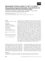
diagnosis and guided hyperthermia therapy [9]. Figure 1 illustrates the possibility of using a
multimodal approach and integrated systems that combine differing properties such as tumor
targeting, cancer therapy and imaging in an-all-in one system [9]. Numerous techniques now
exist for synthesizing different set of nanoparticles based on the type of drugs used, and the
targeted organ and delivery mechanism selected. Depending upon the protocol of choice, the
parameters can be tailored to create the best possible characteristics for the nanoparticles. In this
manuscript we have reviewed a number of biodegradable nanoparticles currently in use, and the
techniques of their preparation. We will also discuss advances in surface modifications, drug
encapsulation and specific end applications of various types of NPs.
PREPARATION OF NANOPARTICLES
Biodegradable nanoparticles can be prepared from a variety of materials such as proteins,
polysaccharides and synthetic biodegradable polymers. The selection of the base polymer is
based on various designs and end application criteria. It depends on many factors such as 1) size
5
of the desired nanoparticles, 2) properties of the drug (aqueous solubility, stability, etc.) to be
encapsulated in the polymer, 3) surface characteristics and functionality, 4) degree of
biodegradability and biocompatibility, and 5) drug release profile of the final product.
Depending upon selection of desired criteria for the preparation of the nanoparticles, the methods
can be classified as following 1) dispersion of preformed polymers, 2) polymerization of
monomers and 3) ionic gelation method for hydrophilic polymers. The general advantages and
disadvantages of individual methods are summarized in Table 1 [10].
Dispersion of preformed polymers: This is the most commonly used technique to prepare
biodegradable nanoparticles from poly-lactic acid (PLA); poly -D- L-glycolide (PLG); poly-D-
L-lactide-co-glycolide (PLGA) and poly-cyanoacrylate (PCA). This technique can be used in
several ways as described below.
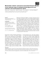
(a) Solvent evaporation method: In this technique the polymer is dissolved in an organic
solvent such as dichloromethane, chloroform or ethyl acetate. The drug is dissolved or dispersed
in the preformed polymer solution followed by emulsification of the mixture to form an oil/water
(o/w) emulsion using an appropriate surfactant / emulsifying agents. Most commonly used
surfactant/emulsifying agents for this purpose are gelatin and polyvinyl alcohol. After formation
of a stable emulsion the organic solvent is evaporated by increasing the temperature or pressure
along with continuous stirring of the solution. Figure 2 shows a schematic representation of this
method [10]. Process parameters such as stabilizer and polymer concentration and stirring speed
have a great influence on the particle size of the NPs formed [8, 11].
(b) Spontaneous emulsification / solvent diffusion method: This is a modified solvent
diffusion method where a water-miscible solvent such as acetone or methanol along with a
water-insoluble organic solvent such as dichloromethane or chloroform are used as an oil phase
[12]. Due to the spontaneous diffusion of solvents, an interfacial turbulence is created between
the two phases leading to the formation of smaller particles. As the concentration of water-
soluble solvent increases, smaller particle sizes of NPs can be achieved [10, 12].
(c) Nanoprecipitation method: Typically, this method is used for hydrophobic drug
entrapment, but it has been adapted for hydrophilic drugs as well. Polymers and drugs are
dissolved in a polar, water-miscible solvent such as acetone, acetonitrile, ethanol, or methanol.
The solution is then poured in a controlled manner (i.e. drop-by-drop addition) into an aqueous
solution with surfactant. Nanoparticles are formed instantaneously by rapid solvent diffusion.
Finally, the solvent is removed under reduced pressure [13].
6
(d) Salting out method: In this method, the polymer is dissolved in the organic phase,
which should be water-miscible, like acetone or tetrahydrofuran (THF). The organic phase is
emulsified in an aqueous phase, under strong mechanical shear stress. The aqueous phase
contains the emulsifier and a high concentration of salts which are not soluble in the organic
phase. Typically, the salts used are 60% w/w of magnesium chloride hexahydrate [14] or
magnesium acetate tetrahydrate in 1:3 polymer to salt ratio [15] . Contrary to the emulsion
diffusion method, there is no diffusion of the solvent due to the presence of salts. The fast
addition of pure water to the o/w emulsion under mild stirring reduces the ionic strength and
leads to the migration of the water-soluble organic solvent to the aqueous phase inducing
nanosphere formation. The final step is purification of nanoparticles by cross flow filtration or
centrifugation to remove the salting out agent [14, 15].
Polymerization Methods: NPs are prepared from monomers that are polymerized to form NPs
in an aqueous solution. Vaccines or drugs/therapeutic agents are incorporated in the NPs either
by dissolving the drug in the polymerization medium or by adsorption/attachment of the drug
onto the polymerized and fully formed NPs. The NP suspension is then purified by removing
stabilizers. The surfactants may be recycled for subsequent polymerization. This technique of
NPs preparation has been reported for making polybutylcyanoacrylate or poly-alkyl-
cyanoacrylate NPs [16, 17]. The concentration of surfactant and the stabilizer determines the
final size of the NPs formed [18].
Ionic gelation method for hydrophilic polymers: Some of the natural macromolecules have
been used to prepare NPs. These polymers include gelatin, alginate, chitosan and agarose. They
are hydrophilic natural polymers and have been used to synthesize biodegradable NPs by the
ionic gelation method. This involves the transition of materials from liquid to gel due to ionic
interaction at room temperature. An example of preparation of gelatin NPs includes hardening of
the droplets of emulsified gelatin solution into gelatin NPs. The gelatin emulsion droplets are
cooled below the gelation point in an ice bath leading to gelation of the droplets [19] into gelatin
NPs. Alginate NPs are reported to be produced by drop-by-drop extrusion of the sodium alginate
solution into the calcium chloride solution [20]. Sodium alginate is a water-soluble polymer that
gels in the presence of multivalent cations such as calcium [21]. Chitosan NPs are prepared by
spontaneous formation of complexes between chitosan and polyanions or by the gelation of a
chitosan solution dispersed in an oil emulsion [22].
7
BIODEGRADABLE NANOPARTICLES
Biodegradable nanoparticles have been used for site-specific delivery of drugs, vaccines and
various other biomolecules. A few of the most extensively used biodegradable polymer matrices
for preparation of nanoparticles are:
Poly-D-L- lactide-co-glycolide (PLGA): Poly-D-L- lactide-co-glycolide (PLGA) is one of the
most successfully used biodegradable polymers. It undergoes hydrolysis in the body to produce
biodegradable metabolite monomers such as lactic acid and glycolic acid. Figure 3 depicts the
schematic representation of the chemical structure of PLGA. Since lactic acid and glycolic acids
are normally found in the body and participate in a number of physiological and biochemical
pathways, there is very minimal systemic toxicity associated with the use of PLGA for the drug
delivery or biomaterial applications. PLGA NPs have been mostly prepared by the
emulsification-diffusion, the solvent evaporation and the nanoprecipitation methods [23]. PLGA
nanoparticles have been used to develop protein and peptide based nanomedicines, nano-
vaccines, and genes containing nanoparticles for in-vivo delivery systems [23, 24].
Polylactic acid (PLA): PLA (Figure 4) is a biocompatible and biodegradable polymer which is
broken down to monomeric units of lactic acid in the body. Lactic acid is a natural
intermediate/by product of anaerobic respiration, which is converted into glucose by the liver
during the Cori cycle. Glucose then is used as an energy source in the body. The use of PLA
nanoparticles is therefore safe and devoid of any major toxicity. PLA nanoparticles have been
mostly prepared by the solvent evaporation, solvent displacement, salting out and solvent
diffusion methods [10, 25]. The salting out procedure is based on the separation of a water-
miscible solvent from aqueous solution by adding a salting out agent like magnesium chloride or
calcium chloride. The main advantage of the salting out procedure is that it minimizes stress to
protein encapsulants [23].
Poly-ε-caprolactone (PCL): poly-ε-caprolactone (Figure 5) is degraded by hydrolysis of its
ester linkages under the normal physiological conditions in the human body and has minimal or
no toxicity. Therefore, PCL has grabbed the attention of researchers as a candidate of choice for
use in drug delivery and long-term implantable devices. PCL’s slower rate of degradation
compared to polylactides has made it better candidate for making long-term implantable devices.
8
PCL nanoparticles have been prepared mostly by nanoprecipitation, solvent displacement and
solvent evaporation [23, 26, 27].
Chitosan: Chitosan (Figure 6) is a modified natural carbohydrate polymer prepared by the
partial N-deacetylation of the crustacean-derived natural biopolymer chitin. There are at least
four methods reported for the preparation of chitosan nanoparticles. The four methods are
ionotropic gelation, microemulsion, emulsification solvent diffusion and polyelectrolyte complex
formation [23, 28, 29].
Gelatin: Gelatin (Figure 7) is extensively used in food and medical products and is a nontoxic
alternative. Gelatin NPs are very efficient in delivery and controlled release of the drugs. They
are nontoxic, biodegradable, bioactive and inexpensive. Gelatin is a poly-ampholyte consisting
of both cationic and anionic groups along with a hydrophilic group. It is known that the
mechanical properties such as swelling behavior and thermal properties of gelatin NPs depend
significantly on the degree of cross-linking between cationic and anionic groups. These
properties of gelatin can be manipulated to prepare desired type of NPs from gelatin. Gelatin
nanoparticles can be prepared by the desolvation/coacervation or emulsion methods [23, 30, 31].
Poly-alkyl-cyano-acrylates (PAC): The biodegradable as well as biocompatible poly-
alkylcyanoacrylates (Figure 8) are degraded by enzyme esterases found in the body. On
degradation they produce some toxic products that may stimulate or damage the central nervous
system. Thus this polymer is not authorized for application in humans. PAC nanoparticles are
prepared mostly by emulsion polymerization, interfacial polymerization and nanoprecipitation
[10, 23].
SURFACE MODIFICATION
One of the problems faced in the use of nanoparticles via the intravenous route was their speedy
removal by the phagocytic cells (macrophages) in the body. Macrophages are powerful
phagocytic cells and are the important constituent of mononuclear phagocytic system (MPS).
The mononuclear phagocytic system (MPS) is one of the body’s innate defenses. MPS filters and
eliminates any injected particulate matter including NPs from the blood stream if they are
recogniozed as foreign body. Unless the injected nanoparticles are modified in a way to escape
recognition as foreign particles, they will be phagocytosed and removed from the circulation.
9
This necessitated modification of the surface of nanoparticles in order for them to escape MPS
recognition and subsequent clearance. Surface modification of the NPs therefore plays a critical
role in their successful applications in-vivo [32]. Once NPs are surface modified with
biomolecules found normally in the body, they will be able to circulate within the blood vascular
system for longer period of time. This increases the probability of nanoparticles reaching their
target rapidally and safely when compared to non- modified NPs. Smaller particles (<100 nm)
circulating in blood vascular system with a hydrophilic surface have the greatest ability to evade
the MPS [33, 34]. Several methods have been developed for surface modification of the NPs.
The most preferred method of surface modification is the adsorption or grafting of poly-ethylene
glycol (PEG) to the surface of nanoparticles. Addition of PEG and PEG-containing copolymers
to the surface of nanoparticles results in an increase in the blood circulation half-life of the
particles. The exact mechanisms by which PEG prolonged circulation time of the surface
modified NPs are still not well understood. It is generally thought that the increased residency of
the nanoparticles in blood is mainly due to prevention of opsonization of nanoparticles by a
certain serum or plasma proteins (opsonins). It is believed that PEG causes steric repulsion by
creating hydrated barriers on nanoparticle surfaces that prevents coating of PEG modified NPs
by serum opsonins.
Studies have shown that the degree to which proteins (opsonins) adsorb onto particle surface
can be minimized by increasing the PEG density on the particle surface. Increasing the molecular
weight of the PEG chains [33] has also been shown to minimize opsonization of nanoparticles
and improve retention in the circulation. For example, Leroux et al. [35] showed that an increase
in PEG molecular weight was associated with less interaction with the MPS, and longer systemic
circulation of PLGA nanoparticles. PEG has been shown to impart stability on PLA particles
submerged in simulated gastric fluid (SGF). Tobio et al. [36] showed that after 4 hours in SGF,
9% of PLA nanoparticles converted to lactic acid versus 3% conversion for PEG-PLA particles
[36]. PEG is also believed to facilitate mucoadhesion and consequent transport through the
Peyer’s patches of the GALT (gut associated lymphoid tissue) [37]. In addition, PEG may
benefit nanoparticle’s interaction with blood constituents. Thus, the presence of PEG on the
nanoparticles imparts additional functionality during the use of polymeric NPs.
Apart from PEG, there are other hydrophilic polymers such as poloxamers, polysorbate 80,
TPGS, polysorbate 20, polysaccharides like dextran and different type of copolymers that can be
used to efficiently coat conventional nanoparticles to add number of variations in the surface
properties of NPs [38, 39]. These coatings provide a dynamic cloud of hydrophilic and neutral
10
chains at the particle surface, which repels plasma proteins. Surface modification by TPGS
increases the adhesion of nanoparticles to tumor cell’s surfaces. It also provides safer
environments to the encapsulated proteins. IgG coating on the surface of nanoparticles increases
the immunoresponse to the encapsulated proteins within the nanoparticles. Hydrophilic
polymers can be applied at the surface of NPs by adsorption of surfactants or by use of block
copolymers or branched copolymers [38-40].
DRUG LOADING AND ENCAPSULATION
One of the most desired qualities of a successful nanoparticle is its high loading capacity for
the drugs. The high loading ability of NPs reduces the amount of the polymer carrier required for
vaccine/drug delivery in the body. The loading of drugs/vaccine into/onto nanoparticles is
achieved by two methods: 1) by incorporating the drug at the time of nanoparticle production or
2) by adsorbing the drug after the formation of nanoparticles. Adsorption of drugs is achieved by
incubating the NPs in a concentrated drug solution [8]. These two methods provide number of
ways by which the drug is adsorbed / attached to the NPs. The encapsulation of the drug in the
polymer, dispersion of the drug in the polymer, adsorption of the drug onto the surface of the
nanoparticles and chemical binding of the drug to the polymer can be accomplished using
incorporation/adsorption techniques. The amount of drugs bound to NPs and the type of
interaction –between drugs and nanoparticles depend on the chemical structure of the drug,
chemical structure of the polymer and the conditions of drug loading [41]. The amount of bound
drug can be determined by subtracting the drug content in the supernatant from the primary
amount of drug present in the suspension.
The drug release mechanisms are an equally important consideration during drug polymer
formulation. It will influence the effectiveness of the proposed application and successful
sustained drug delivery. In general, the drug release rate depends on solubility of the drug,
desorption of the surface bound/adsorbed drug, drug diffusion through the polymer matrix, NP
matrix erosion/degradation and combination of the erosion diffusion process [23]. For
manipulation of the drug release, a good understanding of the mechanisms of drug release is
needed which would involve knowledge of the solubility, diffusion and biodegradation of the
matrix. One way to modify the drug release profile is by adopting appropriate polymer matrices.
Drug release kinetics also depend upon size of the NPs and the loading efficiency of the vaccine
or drug. The vaccine or drug loading efficiency will determine the initial burst and the sustained
release rate of nanoencapsulated drug molecule. Larger particles have a smaller initial burst
11
release than smaller particles. In the case of nanospheres, where the vaccine/drug is uniformly
distributed, the release occurs by diffusion or erosion of the matrix under sink conditions. If the
diffusion of a vaccine/drug is faster than the matrix erosion, the release mechanism is
predominately through a diffusion process. The rapid initial release or burst of vaccine/drug seen
in release profiles is mainly attributed to weakly bound or adsorbed vaccine/drug on to the
surface [7, 42].
SPECIFIC APPLICATIONS OF BIODEGRADABLE NPs
Tumor Targeting: The rationale of using nanoparticles for tumor targeting is based on 1) NP’s
ability to deliver the requisite dose load of drug in the vicinity of the tumor due to the enhanced
permeability and retention effect or active targeting by ligands on the surface of NPs and 2) NP’s
ability to reduce the drug exposure to healthy tissues by limiting drug distribution to the target
organ. Active tumor targeting of NPs may be achieved with either direct targeting or the
pretargeting method. In direct targeting method NPs are covalently coupled with the ligands. The
ligand coupled NPs are received by the tumor cells expressing a homologous receptor on their
surfaces. The specific ligand-receptor binding ensures that the NPs carrying drugs will get
attached specifically to the tumor cells. This will facilitate delivery of drugs only to the cells
(tumor cells) expressing receptor and not the normal healthy cells. In the pretargeting approach,
the therapeutic molecule is not coupled with the ligand and is administered after an appropriate
delay time following the administration of the targeting ligand. Nobs et al. [43] explored both -
approaches to target PLA NPs to tumor cells. In the direct approach, NPs with mAbs exposed on
their surface were incubated with the two tumor cells, while in the pretargeting protocol, tumor
cells were pretargeted with biotinylated MABs prior to the administration of avidin-labelled NPs
[43].
Verdun et al. [44] in an elegant experiment demonstrated positive effects of using poly-
isohexylcyanoacrylate-nanospheres in the delivery of doxorubicin in mice. The doxorubicin
incorporated into poly (isohexylcyanoacrylate) nanopsheres and delivered in mice showed higher
concentrations of doxorubicin in the liver, spleen and the lungs than in mice treated with only
free doxorubicin[44]. Studies show that the drug distribution pattern in the body is greatly
influenced by selected drug’s molecular weight, polymeric composition (type, hydrophobicity
and biodegradation profile) of nanoparticles, localization of drug in the nanospheres, and drug
incorporation techniques such as adsorption or incorporation.[45].
12
Extensive efforts have been devoted to achieving “active targeting” of nanoparticles in order
to deliver drugs to the right targets. The molecular recognition processes such as ligand-receptor
specificity or antigen-antibody interaction plays important role in such targeting. Considering
that folate receptors are over expressed on the surface of some human malignant cells and that
cell adhesion molecules such as selectins and integrins are involved in metastatic events,
nanoparticles bearing specific ligands such as folate may be used to target ovarian carcinoma
while specific peptides or carbohydrates may be used to target integrins and selectins [46].
Oyewumi et al. [47] demonstrated that the benefits of folate ligand coating were to facilitate
internalization and retention of Gd-nanoparticles in the tumor cells/tissues [47]. Targeting with
small ligands appears more likely to succeed since they are easier to handle and manufacture.
Furthermore, it could be advantageous to use active targeting ligands in combination with the
long-circulating nanoparticles to maximize the likelihood of active targeting of nanoparticles.
Nanoparticles for Oral delivery: In recent years, significant research has been done using
nanoparticles as oral drug delivery vehicles. Oral delivery of drugs using nanoparticles has been
shown to be far superior to the delivery of free drugs in terms of bioavialability, residence time,
and biodistribution [48]. Advances in biotechnology and biochemistry have led to the discovery
of a large number of bioactive molecules and vaccines based on peptides and proteins.
Development of suitable carriers remains a challenge due to the fact that bioavailability of these
molecules is limited by the epithelial barriers of the gastrointestinal tract. The drugs may also be
susceptible to gastrointestinal degradation by digestive enzymes. The advantage of using
polymeric nanoparticles is to allow encapsulation of bioactive molecules and protect them
against enzymatic and hydrolytic degradation. For instance, it has been found that insulin-loaded
nanoparticles have preserved insulin activity and produced blood glucose reduction in diabetic
rats for up to 14 days following the oral administration [49].
Another study showed that an antifungal drug encapsulated in particles of less than 300 nm
in diameter was detected in the lungs, liver, and spleen of mice seven days post oral
administration. The oral-free formulations on the other hand were cleared within 3 hours post
administration [48]. For this application, the major interest lies in lymphatic uptake of the
nanoparticles by the Peyer’s patches in the GALT (gut associated lymphoid tissue). There have
been many reports as to the optimum size for Peyer’s patch uptake ranging from less than 1 µm
to 5 µm [50,51]. However, it has also been shown that microparticles remain in the Peyer’s
Patches while nanoparticles are disseminated systemically [52, 53]
13
Nanoparticles can be engineered not only for oral absorption, but can also be used to
deliver a drug directly to the source for gastrointestinal uptake, thereby protecting the drug from
low pH and enzymes in the stomach. The pH-sensitive nanoparticles made from a
poly(methylacrylic acid and methacyrlate) copolymer can increase the oral bioavailability of
drugs like cyclosporine-A by releasing their load at a specific pH within the gastrointestinal tract.
The pH sensitivity allows this to happen as close as possible to the drug’s absorption window
through the Peyer’s patches [54].
Nanoparticles for vaccine/gene delivery: Polynucleotide vaccines/DNA vaccines/plasmid
vaccines work by delivering genes encoding relevant antigens to host cells where they are
expressed, producing the antigenic protein within the vicinity of professional antigen presenting
cells to initiate immune response. Such vaccines produce both humoral and cell-mediated
immunity because intracellular production of protein, as opposed to extracellular deposition,
stimulates both arms of the immune system [55]. The key ingredient of polynucleotide vaccines,
DNA, can be produced cheaply and has much better storage and handling properties than the
ingredients of the majority of protein-based vaccines. Hence, polynucleotide vaccines/DNA
vaccines are set to supersede many conventional vaccines particularly for immunotherapy.
However, there are several issues related to the delivery of polynucleotides which limit their
application. These issues include efficient delivery of the polynucleotide to the target cell
population, its localization to the nucleus of these cells, and ensuring that the integrity of the
polynucleotides is maintained during delivery to the target site [2]. Nanoparticles loaded with
plasmid DNA could also serve as an efficient sustained release gene delivery system due to their
rapid escape from the degradative endo-lysosomal compartment to the cytoplasmic compartment
[56]. Hedley et al. [57] reported that following their intracellular uptake and endolysosomal
escape, nanoparticles could release DNA at a sustained rate resulting in continuous gene
expression. This gene delivery strategy could be applied to facilitate bone healing by using
PLGA nanoparticles containing therapeutic genes such as bone morphogenic protein.
Nanoparticles for drug delivery into the brain: The blood-brain barrier (BBB) is the most
important factor limiting the development of new drugs for the central nervous system [58]. The
BBB is characterized by relatively impermeable endothelial cells with tight junctions, enzymatic
activity and active efflux transport systems. It effectively prevents the passage of water-soluble
molecules from the blood circulation into the CNS, and consequently only permits selective
14
transport of molecules that are essential for brain function [59]. Strategies for nanoparticle
targeting to the brain rely on nanoparticle’s interaction with the specific receptor-mediated
transport systems in the BBB. For example, polysorbate 80/LDL, transferrin receptor binding
antibody (such as OX26), lactoferrin, cell penetrating peptides and melanotransferrin have been
shown to be capable of delivery of a self non transportable drug into the brain via the chimeric
construct that can undergo receptor-mediated transcytosis [60-63]. It has been reported that
poly(butylcyanoacrylate) nanoparticles were able to deliver hexapeptide dalargin, doxorubicin
and other agents into the brain which is significant because of the great difficulty for drugs to
cross the BBB [62]. Despite some reported success with polysorbate 80 coated NPs, this system
does have many shortcomings including desorption of polysorbate coating, rapid NP degradation
and toxicity caused by presence of high concentration of polysorbate 80 [64]. OX26 MAbs (anti-
transferrin receptor MAbs), the most studied BBB targeting antibody, have been used to enhance
the BBB penetration of lipsosomes [65].
Another study by Kreuter et. al. [66] demonstrates the delivery of several drugs successfully
through the blood brain barrier using polysorbate 80 coated PACA nanoparticles [66]. It is
thought that after administration of the polysorbate 80-coated particles, apolipoprotein E (ApoE)
adsorbs onto the surface. The ApoE protein mimics low density lipoprotein (LDL) causing the
particles to be transported across the blood brain barrier via the LDL receptors. The effects of
polysorbate-80 on transport through the blood brain barrier were confirmed by Sun et al. with
PLA nanoparticles [67]. Nanoparticles were also functionalized with thiamine surface ligands.
These particles, with an average diameter of 67 nm, were able to associate with the blood brain
barrier thiamine transporters and thereby increase the unidirectional transfer coefficient for the
particles into the brain [68].
CONCLUSION
In summary, NPs are a potentially viable vaccine and drug delivery system capable of delivering
a multitude of therapeutic agents and biomolecules at the targeted sites in the body. To optimize
NPs as a delivery system, greater understanding of the different mechanisms of biological
interactions and particle engineering is still required. However, biodegradable NPs appear to be a
promising drug delivery carrier system because of their versatile formulation, sustained release
properties, sub cellular size and biocompatibility with various cells and tissue in the body.
15
LIST OF ABBREVIATIONS USED
Nanoparticles: NPs, PLA: Poly-lactic acid, PLG: poly (D, L-glycolide), PLGA: Poly (D, L-
lactide-co-glycolide, PCA: Poly-cyanoacrylate, THF: Tetrahydrofuran, PCL: Poly-ε-
caprolactone, PAC: Poly-alkyl-cyano-acrylate, MPS: Mononuclear Phagocytic System, PEG:
Poly-ethylene glycol, SGF: Simulated gastric fluid, GALT: Gut-associated lymphoid system,
TPGS: Tocopheryl polyethylene glycol 1000 succinate, mABs: Monoclonal antibodies, BBB:
Blood-brain barrier, ApoE: Apolipoprotein E, LDL: low density lipoprotein,
16
COMPETING INTERESTS
The authors declare that they have no competing interests.
AUTHOR’S CONTRIBUTIONS
Both authors have read and approved the final manuscript. AM participated in conceptualization
and preparation of this manuscript. He contributed in preparation of nanoparticles, biodegradable
nanoparticles and surface modification of nanoparticles sections of this manuscript. DKS
participated in specific application of biodegradable NPs section. DKS also participated in the
conceptualization of the manuscript, writing, editing and revision of this report. His lab provided
materials and resources used in this study.
AUTHORS INFORMATION
AM: is an assistant professor of Bioengineering in the Department of Industrial and
Manufacturing Engineering at Wichita State University, Wichita, KS. AM’s lab is working on
development and application of biodegradable implants and drug delivery systems. AM’s lab is
developing biodegradable nanoparticles for gene and drug delivery and is also participating in a
collaborative project with DKS’ lab on the use of biodegradable nanoparticles in delivering a
HIV- DNA vaccine within the cervical and vaginal mucosa.
DKS: is an associate professor of microbiology at the Winston Salem State University. DKS’ lab
is working on development of a DNA vaccine for HIV/AIDS. His other research interest
involves prevention of HIV-1 transmission at the cervical/vaginal mucosal surfaces, use of
nanoparticles in preventing transmission of HIV at the mucosal surfaces. DKS is also
participating in a collaborative project with AMS’ lab on the use of biodegradable nanoparticles
in delivering a HIV- DNA vaccine within the cervical and vaginal mucosa. His current research
is funded by two NIH grants.
17
ACKNOWLEDGEMENTS
The work described was supported by Award Number P20MD002303 from the National Center
on Minority Health and Health Disparities, and SC3GM084802 from National Institute of
General Medical Sciences of NIH to DKS. The content is solely the responsibility of the authors
and does not necessarily represent the official views of the National Center on Minority Health
and Health Disparities or NIGMS or the National Institutes of Health. This research is a project
supported by Winston-Salem State University’s Center of Excellence for the Elimination of
Health Disparities.
18
REFERENCES
1. Xu T, Zhang N, Nichols HL, Shi D, Wen X: Modification of nanostructured materials
for biomedical applications. Materials Science and Engineering: C 2007, 27(3):579-
594.
2. Mohanraj VJ, Chem Y: Nanoparticles - A Review. Tropical Journal of Pharmaceutical
Research 2006, 5(1):561-573.
3. Liu Y, Miyoshi H, Nakamura M: Nanomedicine for drug delivery and imaging: A
promising avenue for cancer therapy and diagnosis using targeted functional
nanoparticles. International Journal of Cancer 2007, 120(12):2527-2537.
4. van Vlerken LE, Amiji MM: Multi-functional polymeric nanoparticles for tumour-
targeted drug delivery. Expert Opinion on Drug Delivery 2006, 3(2):205-216.
5. Vasir JK, Labhasetwar V: Biodegradable nanoparticles for cytosolic delivery of
therapeutics. Advanced Drug Delivery Reviews 2007, 59(8):718-728.
6. Labhasetwar V, Song C, Levy RJ: Nanoparticle drug delivery system for restenosis.
Advanced Drug Delivery Reviews 1997, 24(1):63-85.
7. Hans ML, Lowman AM: Biodegradable nanoparticles for drug delivery and
targeting. Current Opinion in Solid State and Materials Science 2002, 6(4):319-327.
8. Soppimath KS, Aminabhavi TM, Kulkarni AR, Rudzinski WE: Biodegradable
polymeric nanoparticles as drug delivery devices. Journal of Controlled Release 2001,
70(1-2):1-20.
9. Park K, Lee S, Kang E, Kim K, Choi K, Kwon IC: New Generation of Multifunctional
Nanoparticles for Cancer Imaging and Therapy. Advanced Functional Materials
2009, 19(10):1553-1566.
10. Pinto Reis C, Neufeld RJ, Ribeiro AJ, Veiga F: Nanoencapsulation I. Methods for
preparation of drug-loaded polymeric nanoparticles. Nanomedicine: Nanotechnology,
Biology and Medicine 2006, 2(1):8-21.
11. Scholes PD, Coombes AGA, Illum L, Daviz SS, Vert M, Davies MC: The preparation
of sub-200 nm poly(lactide-co-glycolide) microspheres for site-specific drug delivery.
Journal of Controlled Release 1993, 25(1-2):145-153.
12. Niwa T, Takeuchi H, Hino T, Kunou N, Kawashima Y: Preparations of biodegradable
nanospheres of water-soluble and insoluble drugs with D,L-lactide/glycolide
copolymer by a novel spontaneous emulsification solvent diffusion method, and the
drug release behavior. Journal of Controlled Release 1993, 25(1-2):89-98.
19
13. Govender T, Stolnik S, Garnett MC, Illum L, Davis SS: PLGA nanoparticles prepared
by nanoprecipitation: drug loading and release studies of a water soluble drug.
Journal of Controlled Release 1999, 57(2):171-185.
14. Zweers MLT, Engbers GHM, Grijpma DW, Feijen J: In vitro degradation of
nanoparticles prepared from polymers based on DL-lactide, glycolide and
poly(ethylene oxide). Journal of Controlled Release 2004, 100(3):347-356.
15. Eley JG, Pujari VD, McLane J: Poly (Lactide-co-Glycolide) Nanoparticles Containing
Coumarin-6 for Suppository Delivery: In Vitro Release Profile and In Vivo Tissue
Distribution. 2004, 11(4):255-261.
16. Zhang Q, Shen Z, Nagai T: Prolonged hypoglycemic effect of insulin-loaded
polybutylcyanoacrylate nanoparticles after pulmonary administration to normal
rats. International Journal of Pharmaceutics 2001, 218(1-2):75-80.
17. Boudad H, Legrand P, Lebas G, Cheron M, Duchêne D, Ponchel G: Combined
hydroxypropyl-[beta]-cyclodextrin and poly(alkylcyanoacrylate) nanoparticles
intended for oral administration of saquinavir. International Journal of
Pharmaceutics 2001, 218(1-2):113-124.
18. Puglisi G, Fresta M, Giammona G, Ventura CA: Influence of the preparation
conditions on poly(ethylcyanoacrylate) nanocapsule formation. International Journal
of Pharmaceutics 1995, 125(2):283-287.
19. Toshio Y, Mitsuru H, Shozo M, Hitoshi S: Specific delivery of mitomycin c to the
liver, spleen and lung: Nano- and m1crospherical carriers of gelatin. International
Journal of Pharmaceutics 1981, 8(2):131-141.
20. Kwok KK, Groves M, Burgess D: Production of 5–15 µm Diameter Alginate-
Polylysine Microcapsules by an Air-Atomization Technique. Pharmaceutical
Research 1991, 8(3):341-344.
21. Aslani P, Kennedy RA: Studies on diffusion in alginate gels. I. Effect of cross-linking
with calcium or zinc ions on diffusion of acetaminophen. Journal of Controlled
Release 1996, 42(1):75-82.
22. Calvo P, Remuñan-López C, Vila-Jato JL, Alonso MJ: Chitosan and Chitosan/Ethylene
Oxide-Propylene Oxide Block Copolymer Nanoparticles as Novel Carriers for
Proteins and Vaccines. Pharmaceutical Research 1997, 14(10):1431-1436.
23. Kumari A, Yadav SK, Yadav SC: Biodegradable polymeric nanoparticles based drug
delivery systems. Colloids and Surfaces B: Biointerfaces 2010, 75(1):1-18.
20
24. Carrasquillo KG, Stanley AM, Aponte-Carro JC, De Jésus P, Costantino HR, Bosques
CJ, Griebenow K: Non-aqueous encapsulation of excipient-stabilized spray-freeze
dried BSA into poly(lactide-co-glycolide) microspheres results in release of native
protein. Journal of Controlled Release 2001, 76(3):199-208.
25. Fessi H, Puisieux F, Devissaguet JP, Ammoury N, Benita S: Nanocapsule formation by
interfacial polymer deposition following solvent displacement. International Journal
of Pharmaceutics 1989, 55(1):R1-R4.
26. Choi C, Chae SY, Nah J-W: Thermosensitive poly(N-isopropylacrylamide)-b-
poly([epsilon]-caprolactone) nanoparticles for efficient drug delivery system.
Polymer 2006, 47(13):4571-4580.
27. Kim SY, Lee YM: Taxol-loaded block copolymer nanospheres composed of methoxy
poly(ethylene glycol) and poly(caprolactone) as novel anticancer drug carriers.
Biomaterials 2001, 22(13):1697-1704.
28. Sinha VR, Singla AK, Wadhawan S, Kaushik R, Kumria R, Bansal K, Dhawan S:
Chitosan microspheres as a potential carrier for drugs. International Journal of
Pharmaceutics 2004, 274(1-2):1-33.
29. Gan Q, Wang T: Chitosan nanoparticle as protein delivery carrier Systematic
examination of fabrication conditions for efficient loading and release. Colloids and
Surfaces B: Biointerfaces 2007, 59(1):24-34.
30. Zillies JC, Zwiorek K, Hoffmann F, Vollmar A, Anchordoquy TJ, Winter G, Coester C:
Formulation development of freeze-dried oligonucleotide-loaded gelatin
nanoparticles. European Journal of Pharmaceutics and Biopharmaceutics 2008,
70(2):514-521.
31. Ofokansi K, Winter G, Fricker G, Coester C: Matrix-loaded biodegradable gelatin
nanoparticles as new approach to improve drug loading and delivery. European
Journal of Pharmaceutics and Biopharmaceutics, 76(1):1-9.
32. Kim D, El-Shall H, Dennis D, Morey T: Interaction of PLGA nanoparticles with
human blood constituents. Colloids and Surfaces B: Biointerfaces 2005, 40(2):83-91.
33. Gref R, Lück M, Quellec P, Marchand M, Dellacherie E, Harnisch S, Blunk T, Müller
RH: 'Stealth' corona-core nanoparticles surface modified by polyethylene glycol
(PEG): influences of the corona (PEG chain length and surface density) and of the
core composition on phagocytic uptake and plasma protein adsorption. Colloids and
Surfaces B: Biointerfaces 2000, 18(3-4):301-313.
21
34. Gref R, Couvreur P, Barratt G, Mysiakine E: Surface-engineered nanoparticles for
multiple ligand coupling. Biomaterials 2003, 24(24):4529-4537.
35. Leroux J-C, Allémann E, De Jaeghere F, Doelker E, Gurny R: Biodegradable
nanoparticles From sustained release formulations to improved site specific drug
delivery. Journal of Controlled Release 1996, 39(2-3):339-350.
36. Tobío M, Sánchez A, Vila A, Soriano I, Evora C, Vila-Jato JL, Alonso MJ: The role of
PEG on the stability in digestive fluids and in vivo fate of PEG-PLA nanoparticles
following oral administration. Colloids and Surfaces B: Biointerfaces 2000, 18(3-
4):315-323.
37. Vila A, Sánchez A, TobIo M, Calvo P, Alonso MJ: Design of biodegradable particles
for protein delivery. Journal of Controlled Release 2002, 78(1-3):15-24.
38. Torchilin VP, Trubetskoy VS: Which polymers can make nanoparticulate drug
carriers long-circulating? Advanced Drug Delivery Reviews 1995, 16(2-3):141-155.
39. Stolnik S, Illum L, Davis SS: Long circulating microparticulate drug carriers.
Advanced Drug Delivery Reviews 1995, 16(2-3):195-214.
40. Storm G, Belliot SO, Daemen T, Lasic DD: Surface modification of nanoparticles to
oppose uptake by the mononuclear phagocyte system. Advanced Drug Delivery
Reviews 1995, 17(1):31-48.
41. Shenoy DB, Amiji MM: Poly(ethylene oxide)-modified poly(caprolactone)
nanoparticles for targeted delivery of tamoxifen in breast cancer. International
Journal of Pharmaceutics 2005, 293(1-2):261-270.
42. Mahapatro A, Johnson DM, Patel DN, Feldman MD, Ayon AA, Agrawal CM: Drug
Delivery from Therapeutic Self-Assembled Monolayers (T-SAMs) on 316L Stainless
Steel Current Topics in Medicinal Chemistry 2008, 8(4):281-289.
43. Nobs L, Buchegger F, Gurny R, Allémann E: Biodegradable Nanoparticles for
Direct or Two-Step Tumor Immunotargeting. Bioconjugate Chemistry 2005,
17(1):139-145.
44. Verdun C, Brasseur F, Vranckx H, Couvreur P, Roland M: Tissue distribution of
doxorubicin associated with polyisohexylcyanoacrylate nanoparticles. Cancer
Chemotherapy and Pharmacology 1990, 26(1):13-18.
45. Couvreur P, Kante B, Lenaerts V, Scailteur V, Roland M, Speiser P: Tissue distribution
of antitumor drugs associated with polyalkylcyanoacrylate nanoparticles. Journal of
Pharmaceutical Sciences 1980, 69(2):199-202.
22
46. Stella B, Arpicco S, Peracchia MT, Desmaële D, Hoebeke J, Renoir M, D'Angelo J,
Cattel L, Couvreur P: Design of folic acid-conjugated nanoparticles for drug
targeting. Journal of Pharmaceutical Sciences 2000, 89(11):1452-1464.
47. Oyewumi MO, Yokel RA, Jay M, Coakley T, Mumper RJ: Comparison of cell uptake,
biodistribution and tumor retention of folate-coated and PEG-coated gadolinium
nanoparticles in tumor-bearing mice. Journal of Controlled Release 2004, 95(3):613-
626.
48. Pandey R, Ahmad Z, Sharma S, Khuller GK: Nano-encapsulation of azole antifungals:
Potential applications to improve oral drug delivery. International Journal of
Pharmaceutics 2005, 301(1-2):268-276.
49. Damgé C, Michel C, Aprahamian M, Couvreur P, Devissaguet JP: Nanocapsules as
carriers for oral peptide delivery. Journal of Controlled Release 1990, 13(2-3):233-
239.
50. Lemoine D, Préat V: Polymeric nanoparticles as delivery system for influenza virus
glycoproteins. Journal of Controlled Release 1998, 54(1):15-27.
51. Torché A-M, Jouan H, Le Corre P, Albina E, Primault R, Jestin A, Le Verge R: Ex vivo
and in situ PLGA microspheres uptake by pig ileal Peyer's patch segment.
International Journal of Pharmaceutics 2000, 201(1):15-27.
52. Jenkins PG, Howard KA, Blackball NW, Thomas NW, Davis SS, O'Hagan DT:
Microparticulate absorption from the rat intestine. Journal of Controlled Release
1994, 29(3):339-350.
53. Eldridge JH, Hammond CJ, Meulbroek JA, Staas JK, Gilley RM, Tice TR: Controlled
vaccine release in the gut-associated lymphoid tissues. I. Orally administered
biodegradable microspheres target the peyer's patches. Journal of Controlled Release
1990, 11(1-3):205-214.
54. Dai J, Nagai T, Wang X, Zhang T, Meng M, Zhang Q: pH-sensitive nanoparticles for
improving the oral bioavailability of cyclosporine A. International Journal of
Pharmaceutics 2004, 280(1-2):229-240.
55. Gurunathan S, Wu C-Y, Freidag BL, Seder RA: DNA vaccines: a key for inducing
long-term cellular immunity. Current Opinion in Immunology 2000, 12(4):442-447.
56. Panyam J, Zhou W-Z, Prabha S, Sahoo SK, Labhasetwar V: Rapid endo-lysosomal
escape of poly(DL-lactide-co-glycolide) nanoparticles: implications for drug and
gene delivery. The FASEB Journal 2002, 16(10):1217-1226.
23
57. Hedley ML, Curley J, Urban R: Microspheres containing plasmid-encoded antigens
elicit cytotoxic T-cell responses. Nat Med 1998, 4(3):365-368.
58. Chakraborty C, Sarkar B, Hsu C, Wen Z, Lin C, Shieh P: Future prospects of
nanoparticles on brain targeted drug delivery. Journal of Neuro-Oncology 2009,
93(2):285-286.
59. Chen Y, Dalwadi G, Benson HAE: Drug Delivery Across the Blood-Brain Barrier
Current Drug Delivery 2004, 1(4):361-376.
60. Gabathuler R, Arthur G, Kennard M, Chen Q, Tsai S, Yang J, Schoorl W, Vitalis TZ,
Jefferies WA: Development of a potential protein vector (NeuroTrans) to deliver
drugs across the blood-brain barrier. International Congress Series 2005, 1277:171-
184.
61. Ji B, Maeda J, Higuchi M, Inoue K, Akita H, Harashima H, Suhara T: Pharmacokinetics
and brain uptake of lactoferrin in rats. Life Sciences 2006, 78(8):851-855.
62. Pardridge WM: Drug and gene targeting to the brain with molecular trojan horses.
Nat Rev Drug Discov 2002, 1(2):131-139.
63. Scherrmann J-M, Temsamani J: The use of Pep: Trans vectors for the delivery of
drugs into the central nervous system. International Congress Series 2005, 1277:199-
211.
64. Olivier J-C: Drug Transport to Brain with Targeted Nanoparticles. NeuroRx : the
journal of the American Society for Experimental NeuroTherapeutics 2005, 2(1):108-
119.
65. Pardridge WM: Drug and gene targeting to the brain via blood-brain barrier
receptor-mediated transport systems. International Congress Series 2005, 1277:49-62.
66. Kreuter J: Nanoparticulate systems for brain delivery of drugs. Advanced Drug
Delivery Reviews 2001, 47(1):65-81.
67. Sun W, Xie C, Wang H, Hu Y: Specific role of polysorbate 80 coating on the targeting
of nanoparticles to the brain. Biomaterials 2004, 25(15):3065-3071.
68. Lockman PR, Oyewumi MO, Koziara JM, Roder KE, Mumper RJ, Allen DD: Brain
uptake of thiamine-coated nanoparticles. Journal of Controlled Release 2003,
93(3):271-282.
24
Figure legends
Figure 1: Multifunctional nanoparticles. Multifunctional nanoparticles can combine a specific
targeting agent (usually with an antibody or peptide) with nanoparticles for imaging (such as
quantum dots or magnetic nanoparticles), a cell-penetrating agent (e.g., the polyArg peptide
TAT), a stimulus-selective element for drug release, a stabilizing polymer to ensure
biocompatibility polyethylene glycol most frequently), and the therapeutic compound.
Development of novel strategies for controlled released of drugs will provide nanoparticles with
the capability to deliver two or more therapeutic agents. Adapted from ref [9] Copyright 2009
Wiley interscience.
Figure 2: Schematic representation of the emulsification-evaporation technique. Adapted from
ref [10] Copyright 2006 Elsevier
Figure 3: Structure of PLGA. The suffixes x and y represent the number of lactic and
glycolic acid respectively.
Figure 4: Chemical structure of poly lactic acid (PLA)
Figure 5: Chemical structure of Poly-ε-caprolactone (PCL)
Figure 6: Chemical structure of chitosan
Figure 7: Chemical structure of Gelatin
Figure 8: Chemical structure of Poly-alkyl-cyano-acrylates