Báo cáo y học: "Dexamethasone inhibits IL-9 production by human T cells" doc
Bạn đang xem bản rút gọn của tài liệu. Xem và tải ngay bản đầy đủ của tài liệu tại đây (441.09 KB, 9 trang )
BioMed Central
Page 1 of 9
(page number not for citation purposes)
Journal of Inflammation
Open Access
Research
Dexamethasone inhibits IL-9 production by human T cells
Lauren E Holz
†1,2
, Kristoffer P Jakobsen
†1
, Jacques Van Snick
3
,
Francoise Cormont
3
and William A Sewell*
1,2,4
Address:
1
Garvan Institute of Medical Research, 384 Victoria St, Darlinghurst, NSW 2010, Australia,
2
Centre for Immunology, St. Vincent's
Hospital, University of NSW, NSW 2052, Australia,
3
Ludwig Institute of Cancer Research, Brussels Branch and the Experimental Medicine Unit,
Universite de Louvain, B-1200 Brussels, Belgium and
4
St Vincent's Clinical School, University of NSW, NSW 2052, Australia
Email: Lauren E Holz - ; Kristoffer P Jakobsen - ; Jacques Van
Snick - ; Francoise Cormont - ; William A Sewell* -
* Corresponding author †Equal contributors
Abstract
Background: Interleukin 9 (IL-9) is produced by activated CD4+ T cells. Its effects include
stimulation of mucus production, enhanced mast cell proliferation, enhanced eosinophil function,
and IgE production. These effects are consistent with a role in allergic diseases. Glucocorticoids
have potent anti-inflammatory effects, including suppression of cytokine synthesis, and are widely
used in the treatment of allergic conditions.
Methods: We examined the effect of the glucocorticoid dexamethasone (Dex) on IL-9 mRNA
expression and protein secretion with real-time RT-PCR and ELISA. Peripheral blood mononuclear
cells (PBMC) were prepared from human volunteers and activated with OKT3. CD4+ T cells were
purified from PBMC and activated with OKT3 plus PMA.
Results: IL-9 mRNA abundance and protein secretion were both markedly reduced following
treatment of activated PBMC with Dex. mRNA levels were reduced to 0.7% of control values and
protein secretion was reduced to 2.8% of controls. In CD4+ T cells, Dex reduced protein secretion
to a similar extent. The IC
50
value of Dex on mRNA expression was 4 nM.
Conclusion: These results indicate that IL-9 production is very markedly inhibited by Dex. The
findings raise the possibility that the beneficial effects of glucocorticoids in the treatment of allergic
diseases are in part mediated by inhibition of IL-9 production.
Background
CD4+ T cells of the T helper 2 (Th2) type have been impli-
cated as major contributors to the pathology of allergic
asthma [1]. Th2 cells produce the cytokines IL-4, IL-5, IL-
9 and IL-13. IL-9, which was first identified as a T cell
growth factor [2], has multiple effects consistent with a
role in allergic inflammation. IL-9 acts on the pulmonary
epithelium to induce production of mucus [3] and chem-
okines [4]. It enhances eosinophil function via induction
of the IL-5 receptor [5]. IL-9 induces immunoglobulin
synthesis of all isotypes, especially IgE [6]. Mast cell num-
bers are elevated in the lung by IL-9 [7].
There is evidence in clinical studies for an association
between IL-9 and allergic asthma. In bronchial biopsies,
the cells expressing IL-9, which were predominantly T
cells, were increased in patients with allergic asthma, and
this was associated with bronchial hyper-reactivity [8,9].
Published: 20 April 2005
Journal of Inflammation 2005, 2:3 doi:10.1186/1476-9255-2-3
Received: 03 December 2004
Accepted: 20 April 2005
This article is available from: />© 2005 Holz et al; licensee BioMed Central Ltd.
This is an Open Access article distributed under the terms of the Creative Commons Attribution License ( />),
which permits unrestricted use, distribution, and reproduction in any medium, provided the original work is properly cited.
Journal of Inflammation 2005, 2:3 />Page 2 of 9
(page number not for citation purposes)
An association between IL-9 expressing cells and eosi-
nophilia has also been described [10]. In allergic asthma
patients, IL-9 in the bronchoalveolar fluid was increased
after segmental allergen challenge [11]. Among a range of
cytokines produced by in vitro stimulated PBMC, IL-9 was
found to have the best correlation with allergic reactivity
as measured by skin prick tests [12].
Several animal studies have investigated the role of IL-9 in
allergic asthma. In transgenic mice with elevated pulmo-
nary expression of IL-9, there was increased influx of
inflammatory cells to the lungs, increased mucus produc-
tion and increased mast cell numbers [13]. In two separate
mouse model studies of allergen-induced asthma, admin-
istration of neutralising anti-IL-9 antibodies reduced eosi-
nophilia, BHR, airway damage and IgE [14,15]. In a
model of parasitic infection with a Th2 response, IL-9
knockout mice displayed markedly reduced goblet cell
hyperplasia and mastocytosis [16]. However, in a model
of allergic asthma, airway hyperreactivity, eosinophilia
and goblet cell hyperplasia were not impaired in IL-9
knock-out mice [17]. Despite the findings in knock-out
mice, overall the evidence from animal models is consist-
ent with clinical evidence that IL-9 may have a role in
allergic asthma.
Glucocorticoids (GC) are a major component of the treat-
ment of asthma and other allergic disorders. GC bind to
cytoplasmic glucocorticoid receptors (GR) and GC/GR
complexes translocate to the cell nucleus where they stim-
ulate or inhibit the transcription of a large number of
genes. The anti-inflammatory effects of GC have been
associated with inhibition of transcription of numerous
cytokines [18]. GC markedly reduce gene transcription of
the Th2 cytokines IL-4 [19], IL-5 [20] and IL-13 [21] as
well as inhibiting the production of many other cytokines
including IL-2 [22], GM-CSF [23] and interferon gamma
(IFN-γ) [24]. By contrast, GC induce the expression of cer-
tain cytokines, including IL-10 [25], IL-1 receptor antago-
nist (IL-1Ra) [26] and transforming growth factor-beta
[27], and GC do not affect expression of M-CSF [23].
Given the extensive evidence indicating IL-9 may be a can-
didate cytokine in the pathogenesis of allergic diseases,
further research into the regulation of IL-9 production is
warranted. Because glucocorticoids are effective in the
treatment of allergic diseases, it is important to under-
stand their effects on genes that are potentially relevant to
the pathogenesis of these diseases. Therefore we have
investigated the effect of the synthetic glucocorticoid dex-
amethasone (Dex) on IL-9 production.
Methods
Cell culture
Peripheral blood was donated by healthy volunteers from
the Garvan Institute of Medical Research and the Centre
for Immunology. The procedures were approved by the
Human Research Ethics Committee, St Vincent's Hospital,
Sydney and are in compliance with the Helsinki Declara-
tion. Peripheral blood mononuclear cells (PBMC) were
isolated by ficoll-based density centrifugation. Cells were
resuspended in complete medium consisting of RPMI
1640 medium (JRH Biosciences, Lenexa, KS, USA) supple-
mented with 10% v/v heat-inactivated foetal bovine
serum (FBS) (CSL Ltd, Parkville, Australia), 2 mM L-
glutamine, 20 mM HEPES buffer, 100 U/mL penicillin
and 100 µg/mL streptomycin (all from Invitrogen,
Carlsbad, CA, USA). Cell counts and viabilities were deter-
mined by trypan blue exclusion in a haemocytometer.
Viability was always greater than 95%.
PBMC were adjusted to 1 × 10
6
cells/mL and were incu-
bated at 37°C in 5% CO
2
for the activation period. PBMC
were treated with 100 ng/mL OKT3 (diluted in PBS) or
with a corresponding volume of PBS. OKT3, a kind gift of
Janssen-Cilag, Sydney, Australia, causes T cell activation
by binding to the T-cell specific surface molecule CD3.
Cells were treated with Dex (Sigma, Castle Hill, Australia)
or with a corresponding volume of PBS. Dex was diluted
in PBS and added immediately after OKT3.
In some experiments, CD4+ T cells were purified by incu-
bating PBMC in complete medium for 90 minutes at
37°C and 5% CO
2
to deplete adherent cells. The non-
adherent cells were then centrifuged and resuspended in
MACS Buffer (0.5% FBS and 2 mM EDTA in PBS) and
MACS human CD4
+
micro beads (Miltenyi Biotec,
Auburn, California, USA) according to the manufacturer's
instructions. After incubation, the cells were washed and
CD4
+
cells were then isolated by a MACS LS Column
placed in a MACS Separator according to the manufac-
turer's instructions (Miltenyi).
Small aliquots of the CD4
+
cells were analysed by flow
cytometry. Cells were stained with anti-CD3 FITC and
anti-CD4 PE antibodies and analysed on a FACSCalibur
using CellQuest software (all BD Biosciences, San Jose,
CA). At least 98% of the cells expressed CD3 and CD4.
CD4
+
cells were cultured as above except that they were
stimulated with a combination of 8 ng/mL PMA (Sigma)
and plate-bound OKT3. OKT3 was bound to 12-well
plates by addition of 10 µg/mL of OKT3 in PBS at 4°C
overnight. The antibody solution was removed immedi-
ately prior to addition of the cells.
RT-PCR
After culture for 24 hours, cells were centrifuged at 440 g
for 5 min. Total RNA was extracted by Trizol (Invitrogen)
according to the manufacturer's instructions. RNA was
Journal of Inflammation 2005, 2:3 />Page 3 of 9
(page number not for citation purposes)
dissolved in DEPC-treated water and stored at -70°C until
required. RNA concentration was determined by spectro-
photometry. 2 µg of total RNA was heated to 65°C for 5
min, cooled for 2–3 min on ice, and reverse transcribed by
avian myeloblastosis virus reverse transcriptase (AMV-
RT), with 1 µM oligo (dT)
15
primer (Roche, Castle Hill,
Australia), 20U AMV-RT enzyme (Roche), 1 mM dNTP
(Roche), AMV-RT buffer (50 mM Tris-HCl, 8 mM MgCl
2
,
30 mM KCl, 1 mM dithiothreitol) (Roche) and DEPC-
water in a 20 µL volume at 42°C for 1 hour. Tubes were
heated to 65°C for 5 min and stored at -20°C until
required.
For IL-9 PCR, in a 20 µL reaction mixture, 1 µL cDNA was
amplified by Platinum Quantitative PCR Supermix UDG
(1.5 U Platinum Taq Polymerase, 20 mM Tris-HCl, 50
mM KCl, 3 mM MgCl
2
, 200 µM dGTP, 200 µM dATP, 200
µM dCTP, 200 µM dUTP, 1U Uracil DNA glycosylase
(UDG)) (Invitrogen), Milli-Q water, 0.4 µM of the for-
ward and reverse primers and Taqman probe (Geneworks,
Rundle Mall, Adelaide, Australia) with sequences
5'CCTGGACATCAACTTCCTCATC3',
5'CATGGCTGTTCACAGGAAAA3' and 5'FAM-CTCT-
GACAACTGCACCAGA-TAMRA3', respectively. PCR was
performed with a Rotorgene 3000 real-time PCR machine
(Corbett Research, Mortlake, Sydney, Australia). No tem-
plate controls (NTC) with water instead of cDNA were
included in all experiments. The reaction conditions for
the IL-9 real-time PCR were 95°C for 3 min followed by
40 cycles of 95°C for 15 sec then 60°C for 60 sec. Forward
and reverse primers were designed to bind to different
exons so that any genomic DNA amplification could be
distinguished from cDNA.
The PCR amplification efficiency was determined in every
experiment by serial four-fold dilutions of the activated
sample containing no Dex. These diluted samples and all
the undiluted samples were analysed again in duplicate by
real-time PCR under the same conditions. The amplifica-
tion efficiency was determined by plotting the mean
threshold cycle (Ct) value of the diluted samples against
the log of the dilution. IL-9 amplification efficiencies
ranged from 1.63 to 1.99. The actual amplification effi-
ciencies were then used to determine the ratios of samples
treated with and without Dex.
A β-actin PCR was also performed on each sample. 1 µL
cDNA was amplified in 25 µL in PCR buffer (10 mM Tris-
HCl, 1.5 mM MgCl
2
, 50 mM KCl) (Roche), 0.25 mM
dNTP (Roche), 1 X SybGr (Molecular Probes, Eugene, OR,
USA), 0.75 U Taq polymerase (Roche), 2 mM MgCl
2
and
0.32 µM of forward and reverse β-actin primer
(Geneworks) with sequences
5'CCAACTGGGACGACATG3' and
5'CAGGGATAGCACAGCCT3' respectively [20]. Samples
were amplified by 94°C for 2 min followed by 30 cycles
of 94°C for 15 sec, 56°C for 20 sec and 72°C for 20 sec.
To confirm the identity of PCR products, all products were
size-fractionated by agarose gel electrophoresis, and prod-
ucts with apparent mobility consistent with the expected
size (277 bp for IL-9 and 203 bp for β-actin) were
detected.
ELISA assays
ELISA assays were used to determine the IL-4, IL-9 and
IFN-γ concentration in the culture supernatants. The IL-9
reagents (capture antibody, standard, and detection anti-
body) have been described previously [28], whereas the
IL-4 and IFN-γ kits were purchased from BD Biosciences.
384 well flat bottom MAXISorp plates (Nunc, Roskilde,
Denmark) were used. In the IL-9 ELISA the capture anti-
body, mh9a4, was diluted in a coating buffer (20 mM gly-
cine, 30 mM NaCl, pH 9.2) at a concentration of 5 µg/mL.
After overnight incubation at 4°C and washing with
0.05% Tween-20 in PBS, the plate was blocked with the
assay diluent, 1% (w/v) BSA in PBS, incubated at 37°C for
at least 2 hours and washed again. Before a final overnight
incubation at 4°C the samples and standards were pre-
pared in assay diluent, and loaded into the wells in tripli-
cate. The standards were prepared in two-fold dilutions
from 500 pg/mL to 3.9 pg/mL. After washing, detection
antibody mh9a3-biotin was added in a 1:2000 dilution
for 2 hours at 37°C. The plates were washed and strepta-
vidin-horseradish peroxidase conjugate was added (Dako-
Cytomation, Glostrup, Denmark) 1:500 in assay diluent,
and plates were incubated at room temperature for 30
min. The IL-4 and IFN-γ ELISA assays were performed
according to the manufacturer's instructions. The lower
limits of detection were 3.9–15.6 pg/mL for IL-9, 7.8 pg/
mL for IL-4 and 3.9 pg/mL for IFN-γ. When results with
and without Dex were presented as percentages, if a sam-
ple was undetectable in the ELISA, the lower limit of
detection of the assay was used in the calculation.
All assays were washed, loaded with TMB Substrate solu-
tion (BD Biosciences) in a 1:1 mixture of TMB substrate A
and B, and incubated at room temperature in the dark for
30–45 minutes before the reaction was stopped with 2 M
H
2
SO
4
. Absorbance was measured by a Spectra Image
reader using X-read Plus software (both Tecan, Maenne-
dorf, Switzerland).
Statistics
Samples were compared with the Wilcoxon signed rank
test (Statview Software 5.0, Abacus Concepts, Berkeley,
California, USA). A p value of <0.05 was considered
significant.
Journal of Inflammation 2005, 2:3 />Page 4 of 9
(page number not for citation purposes)
Results
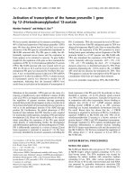
Dexamethasone reduces IL-9 mRNA abundance
In preliminary experiments, real-time RT-PCR revealed
that OKT3 was a highly effective stimulus of IL-9 expres-
sion in PBMC, as previously reported [2]. IL-9 mRNA was
induced from 4 to 48 h after activation (Fig. 1A), and 24
h was chosen as a suitable time for detection of mRNA in
subsequent experiments. The effect of Dex on IL-9 mRNA
abundance in PBMC was examined in 13 healthy individ-
uals by real-time RT-PCR. PBMC were cultured with
OKT3, with or without 10
-6
M Dex. In all samples treated
with OKT3 without Dex, IL-9 mRNA expression was read-
ily detected. Addition of Dex to cultures stimulated by
OKT3 was followed by a marked reduction in IL-9 mRNA
abundance. All samples treated with Dex had much
higher Ct values than those without Dex (Table 1). Statis-
tical analysis revealed a highly significant effect of Dex (p
< 0.01). All RT-PCR products were subjected to gel electro-
phoresis and the results were consistent with the real-time
data. In the samples activated with OKT3, a single strong
band was detected with apparent mobility consistent with
the predicted fragment size of 277 bp (Fig. 1B). After treat-
ment with OKT3 and Dex, a very faint band of the same
mobility was detected, and no bands were detected in the
unactivated samples. Except for some very low molecular
size material, there was no evidence of any other band
apart from the 277 bp band. The cDNA samples were also
assessed for the housekeeping gene β-actin by real-time
RT-PCR, and Dex had no significant effect. In activated
cells, Ct values for β-actin were 15.5 ± 2.7 (SD) for sam-
ples given Dex, compared with 16.5 ± 4.6 for samples not
given Dex. The findings with β-actin indicate that Dex did
not cause a generalized reduction of gene expression.
The relative change in IL-9 mRNA expression produced by
Dex was ascertained by calculating the difference in Ct val-
ues between the activated and activated + Dex samples
(Table 1). This difference was then corrected for the
amplification efficiency of samples from each individual
PBMC donor. Amplification efficiency was determined by
serial dilution of each of the samples activated and not
treated with Dex. The percentage of IL-9 transcription in
the Dex-treated samples compared to controls ranged
from 0.03% to 3.57% with a mean of 0.67% and a
median of 0.20%.
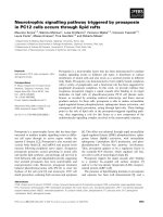
Concentration-response studies
The effectiveness of Dex was assessed by comparing IL-9
transcription in samples not treated with Dex to samples
treated with 10
-6
M to 10
-11
M Dex. Mean Ct values of
duplicate samples were determined, and in each individ-
ual the mean Ct value of the sample not treated with Dex
was given a figure of 100%. PCR was then performed on
serial dilutions of the samples not treated with Dex to cor-
rect for amplification efficiency as described in the Meth-
ods. Dex inhibited IL-9 transcription in PBMC activated
with OKT3 in a concentration dependent manner in four
different individuals. The average percentage value for
each Dex concentration is plotted in Figure 2. 10
-7
M Dex
was almost as inhibitory as 10
-6
M Dex, and 10
-8
M Dex
reduced IL-9 transcript abundance to 20% of control lev-
els. At lower concentrations of Dex, transcription
increased towards control levels. In 2 of 4 experiments,
the samples treated with 10
-10
M Dex had a higher level of
transcription than control samples, contributing to the
slightly higher average IL-9 expression level at 10
-10
M Dex
compared with no Dex (Fig. 2). The concentration of Dex
Table 1: Effect of Dex on IL-9 mRNA in 13 different individuals.
Expt Ct no Dex Ct with Dex Amplification Efficiency % IL-9 in Dex vs no Dex
1 22.6 34.3 1.74 0.15
2 21.1 32.1 1.76 0.20
3 24.0 35.0 1.91 0.08
4 21.6 30.8 1.85 0.34
5 27.0 35.8 1.70 0.91
6 23.1 34.6 1.79 0.12
7 20.9 37.8 1.85 0.03
8 24.5 33.7 1.85 0.34
9 22.0 30.3 1.63 1.72
10 24.5 30.2 1.79 3.57
11 19.7 32.2 1.70 0.13
12 24.0 30.7 1.99 1.00
13 23.2 34.6 1.79 0.13
PBMC were activated with or without 10
-6
M Dex, and Ct values for IL-9 were determined. The amplification efficiencies were measured for each
sample, and were applied to the Ct differences between the Dex and no Dex samples to determine the proportion of IL-9 in samples treated with
and without Dex.
Journal of Inflammation 2005, 2:3 />Page 5 of 9
(page number not for citation purposes)
that inhibited 50% of IL-9 transcription in activated
PBMC, the IC
50
, was calculated to be 10
-8.4
M or 4 nM.
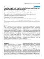
Dex inhibits IL-9 protein secretion
After activation with OKT3, IL-9 secretion was readily
detected by sandwich ELISA. Supernatants were harvested
at various times after activation, and IL-9 was measured in
triplicate. The amount of IL-9 after 72 h of culture was
defined as 100%. IL-9 was not detected at 0 h. At 24 h, the
IL-9 level was 16 ± 1 % (mean ± SD) and at 48 h it was 85
± 3 %. Thus IL-9 levels had almost peaked by 48 h, and
supernatants were harvested at this time in subsequent
experiments. PBMC from 11 different donors were treated
with or without OKT3 and with or without 10
-6
M Dex
throughout the culture period. In all samples stimulated
with OKT3, there were high levels of IL-9 secretion in the
absence of Dex, and IL-9 concentrations were in the range
of 207–2,526 pg/mL. IL-9 secretion was very markedly
reduced after treatment with Dex (p < 0.005). In the Dex-
treated samples, secretion was only 2.8 % ± 2.5% (SD) of
control values (Fig. 3). In 8 of the 11 samples treated with
OKT3 and Dex, the IL-9 concentration was below the
lower limit of detection of the assay. In most of the cul-
tures not treated with OKT3, IL-9 could not be detected. It
was detected at very low levels in 3 samples in the absence
of Dex and in 1 sample in the presence of Dex.
In six of the 11 samples, the culture supernatants were
also tested for IFN-γ and IL-4. In activated cells treated
with Dex, the IFN-γ and IL-4 concentrations were always
above the lower detection limit of the assays. Dex signifi-
cantly reduced the concentrations of both cytokines (p <
0.05 in both cases). The effect of Dex on IFN-γ secretion
was similar to that on IL-9. Activated cells treated with
Dex secreted 2.4 ± 2.1 % as much IFN-γ as control acti-
vated cells. By contrast, Dex had substantially less inhibi-
tory effect on IL-4 secretion. The Dex-treated cells secreted
IL-9 RT-PCRFigure 1
IL-9 RT-PCR. A. Time course. PBMC were incubated for
various times with OKT3, RNA was extracted, IL-9 real time
RT-PCR was performed, and the mean threshold cycle (Ct)
was determined. The data shown are from an experiment on
one representative individual. The values are means of dupli-
cate determinations. B. Gel electrophoresis. PBMC were
incubated for 24 h with or without OKT3 and with or with-
out Dex (10-6 M). RNA was extracted and IL-9 RT-PCR per-
formed for 40 cycles. For each condition, duplicate PCRs
were performed on cDNA from one representative individ-
ual. Products were analysed in a 2% agarose gel. The left lane
contains HaeIII cut ΦX174 molecular size markers (Roche);
the arrow indicates the position of the 281/271 bp markers.
- Dex + Dex
-OKT3 +OKT3
- Dex + Dex
Time after activation (h)
Ct value
20
25
30
35
010203040
50
A
B
Concentration-response effect of Dex on IL-9 mRNA in acti-vated PBMCFigure 2
Concentration-response effect of Dex on IL-9 mRNA
in activated PBMC. Cells were incubated with OKT3 and
the stated concentration of Dex. 24 hours later, RNA was
extracted and real time RT-PCR for IL-9 was performed.
Data were corrected for amplification efficiency as described
in Methods. Each sample was measured in duplicate. The
results are expressed as the % of the response in cells not
treated with Dex. The data are the mean ± SEM of four dif-
ferent individuals.
Dexamethasone (-log
10
M)
% response w ithout Dex
Nil 11 10 9 8 7 6
0
20
40
60
120
140
80
100
Journal of Inflammation 2005, 2:3 />Page 6 of 9
(page number not for citation purposes)
31.4 ± 14.1 (SD) % as much IL-4 compared with control
activated cells.
CD4+ T cells were purified from 7 individuals, to deter-
mine whether Dex was acting directly on these cells. In
cells not activated with OKT3, IL-9 was detected at very
low levels in 4 samples without Dex and in 2 samples in
the presence of Dex. Activated cells not treated with Dex
secreted IL-9 in the range 222–1,939 pg/mL. As with
PBMC, Dex markedly inhibited IL-9 secretion in activated
cells (Fig. 4) (p < 0.02). Samples treated with 10
-6
M Dex
secreted only 2.9 % ± 2.5% (SD) as much IL-9 as control
samples. In activated cells treated with Dex, IL-9 was
below the detection limit of the assay in 3 of 7 cultures in
these experiments.
Discussion
The study demonstrates that Dex is an efficient pharma-
ceutical agent for inhibition of IL-9 production. In
activated PBMC, Dex reduced IL-9 secretion to a mean of
2.8% of control levels, whereas in the case of mRNA, the
corresponding value was 0.7 %. The difference between
these 2 percentage values may have arisen because in the
real-time PCR analysis, it was always possible to deter-
mine a value for IL-9 mRNA in the Dex-treated samples,
whereas in the ELISA assay, the corresponding samples
were usually undetectable. In the latter samples, the lower
limit of detection of the assay was used to calculate per-
centages, which may have over-estimated the IL-9 concen-
tration in the Dex-treated samples.
To determine if the inhibitory effect was specific for helper
T cells, experiments were also carried out with purified
CD4
+
cells. These populations contained at least 98%
CD3+CD4+ cells, making it very likely that the observed
effects directly involve helper T cells. The data indicate
that CD4+ T cells produce substantial amounts of IL-9,
although the possibility that other cells in PBMC also
produce IL-9 has not been excluded. Dex markedly
reduced IL-9 secretion in CD4
+
T cells, and the data are
most consistent with a direct effect of Dex on CD4+ T
cells.
Dex was found to inhibit the synthesis of IL-9 mRNA in
PBMC in a concentration dependent manner. Marked
inhibition of IL-9 transcription was observed with Dex
concentrations as low as 10
-8
M, and Dex had an IC
50
value of 4 nM. Similar Dex concentration response curves
have been observed with IL-2 [22] and IL-5 [20] expres-
sion in T cells, as well as IL-4 and IL-5 in mast cells [29].
ICAM-1 expression [30] as well as prostaglandin synthesis
and release in alveolar tissue [31] have also been found to
have similar responses to a range of concentrations of
Dex. IC
50
values for Dex have been obtained for ICAM-1
expression of <1 nM [30], COX activity of 1–10 nM [31],
IL-11 expression of 1 nM [32] and IL-5 expression in T
cells of 1 nM [20]. In mast cells, Dex had an IC
50
value of
1.6 nM on IL-5 expression indicating that the sensitivity of
T cells and mast cells to Dex is similar for Th2 cytokines
[29]. These findings, taken together, suggest that Dex may
be inhibiting similar pathways involved in regulation of
Effect of Dex on IL-9 secretion by PBMCFigure 3
Effect of Dex on IL-9 secretion by PBMC. Cells from 11
different individuals were treated with OKT3 and with or
without 10
-6
M Dex. Culture supernatants were harvested 48
hours later and measured for IL-9 by sandwich ELISA. Data
represent the mean ± SD of triplicate determinations.
0
500
1000
1234567891011
Experiment
IL-9 concentration (pg/mL)
2500
3000
OKT3
OKT3 + Dex
Effect of Dex on IL-9 secretion by CD4+ T cellsFigure 4
Effect of Dex on IL-9 secretion by CD4+ T cells. Cells
from 7 different individuals were treated with OKT3 and
with or without 10
-6
M Dex. Culture supernatants were har-
vested 48 hours later and measured for IL-9 by sandwich
ELISA. Data represent the mean ± SD of triplicate
determinations.
0
500
1000
1500
2000
2500
1234567
Experiment
IL-9 concentration (pg/mL)
OKT3
OKT3 + Dex
Journal of Inflammation 2005, 2:3 />Page 7 of 9
(page number not for citation purposes)
expression of a variety of different genes in T cells and
mast cells.
Glucocorticoids can mediate effects on transcription in
two ways. After translocation of the GC/GR to the nucleus,
the GR can bind directly to glucocorticoid response ele-
ment (GRE) sequences in the promoter regions of target
genes. The expression of many genes is stimulated in this
fashion. However there is limited evidence for GRE
involved in inhibition of gene expression. Alternatively,
GC act indirectly by GR binding to transcription factors so
as to prevent them from interacting with DNA. Previous
studies have found that the two mechanisms are mediated
by different concentrations of Dex. The inhibitory effect of
Dex on collagenase expression was found to be mediated
by interaction between GC/GR and the transcription fac-
tor AP-1 [33]. In the absence of GC, AP-1 binds to the pro-
moter of the collagenase gene to stimulate transcription,
whereas in the presence of GC, binding between GC/GR
and AP-1 prevents the latter from associating with DNA,
so that transcription is inhibited. Half maximal repression
of collagenase expression was reached with 1.5 nM Dex,
whereas half-maximal induction of gene expression via
GRE binding required 10 nM or greater [33]. We found
Dex to have an IC
50
value of 4 nM, consistent with an indi-
rect effect via interference with transcription factor(s).
Among possible transcription factors, NF-AT is a likely
candidate. In the case of the IL-5 promoter, we observed
that Dex inhibited binding to the NF-AT site but not to the
GATA-3 site [34]. The IL-9 promoter contains binding
sequences for NF-AT [35], and the transcription of other
cytokines including IL-2 [36] and IL-4 [37] involves NF-
AT. IL-4, IL-5 and IL-9 all reside within the Th2 gene clus-
ter on human chromosome 5 [38] raising the possibility
that they may have similar regulatory mechanisms. Other
factors which may be involved include AP-1, NF-κB and
CREB, which have DNA binding sites in the IL-9 promoter
[35] and which can be inhibited by glucocorticoids
[36,39,40].
Expression of IL-9 by T cells may depend on the effects of
other cytokines produced after activation [41]. This is con-
sistent with the delayed induction of IL-9 mRNA, which
did not peak until 24 h after activation (Fig. 1A). It is
therefore possible that the effect of Dex on IL-9 produc-
tion may be a consequence of its inhibitory effect on
cytokines produced earlier after T cell activation. Dex
inhibited the production of the key Th1 cytokine IFN-γ to
a similar extent to IL-9 (Table 2). In other experiments on
PBMC, we observed that 10
-6
M Dex reduced the secretion
of IL-5 to 0.8 % of control PHA activated cells, and that of
IL-13 to 6.2 % of controls (n = 6 for IL-5 and IL-13) (M.
Irvine & W. A. Sewell, unpublished observations). How-
ever, not all Th2 cytokines are as markedly inhibited by
Dex, because IL-4 was only inhibited to 31% of control
levels (Table 2). The relative resistance of IL-4 to the
inhibitory effects of Dex may explain an unexpected effect
of Dex in enhancing the development of Th2 cells [42];
these findings could be explained by more efficient sup-
pression by Dex of IFN-γ than IL-4, leaving sufficient IL-4
to favour differentiation of T cells into Th2 cells.
Conclusion
IL-9 mRNA expression and protein secretion were very
markedly inhibited by Dex. The findings suggest that the
beneficial effects of glucocorticoids in the treatment of
allergic diseases may, in part, be mediated by inhibition of
IL-9 production. Glucocorticoids are a mainstay in the
treatment of allergic asthma and other allergic diseases,
but their usefulness is limited by side effects. Drugs that
inhibit effector cytokines, but lack the side effects of glu-
cocorticoids, would potentially be very useful in the
treatment of allergy. Our findings suggest that, when such
novel drugs are evaluated, their effects on IL-9 should be
taken into consideration.
Competing interests
The author(s) declare that they have no competing
interests.
Table 2: Effect of Dex on IFN-γ, IL-4 and IL-9 secretion.
Cytokine OKT3 range OKT3 plus Dex range % cytokine in Dex vs no Dex
IFN-γ (ng/mL) 11–56 0.15–1.1 2.4 ± 2.1
IL-4 (pg/mL) 23–81 12–22 31 ± 14
§ IL-9 (pg/mL) 234–781 * undetectable 4.3 ± 2.9
PBMC from 6 different individuals were activated with OKT3 and treated with or without 10
-6
M Dex. Cytokine concentration was measured in
triplicate. For each individual, the % cytokine secretion in Dex versus no Dex was determined, and the Table shows the mean ± SD of these values.
§ The IL-9 data are for these 6 individuals only; the results are not significantly different from the results for all 11 individuals shown in Fig. 3. * For
IL-9, all the Dex treated samples were below the lower limit of detection of the assay which was 7.8–15.8 pg/mL. The latter figures were used to
calculate the % cytokine figure.
Journal of Inflammation 2005, 2:3 />Page 8 of 9
(page number not for citation purposes)
Authors' contributions
LEH performed the RT-PCR experiments. KPJ performed
the ELISA experiments. LEH and KPJ drafted the manu-
script. JvS prepared the anti-IL-9 antibodies and revised
the manuscript. FC prepared the anti-IL-9 antibodies.
WAS conceived of the project, supervised its design and
coordination, and revised the manuscript. All authors
read and approved the final manuscript.
Acknowledgements
The work was supported by a grant from the St Vincent's Hospital
Research Committee.
References
1. Robinson DS, Hamid Q, Ying S, Tsicopoulos A, Barkans J, Bentley AM,
Corrigan C, Durham SR, Kay AB: Predominant TH2-like bron-
choalveolar T-lymphocyte population in atopic asthma. N
Engl J Med 1992, 326:298-304.
2. Renauld JC, Goethals A, Houssiau F, Merz H, Van Roost E, Van Snick
J: Human P40/IL-9. Expression in activated CD4+ T cells,
genomic organization, and comparison with the mouse
gene. J Immunol 1990, 144:4235-4241.
3. Longphre M, Li D, Gallup M, Drori E, Ordonez CL, Redman T, Wen-
zel S, Bice DE, Fahy JV, Basbaum C: Allergen-induced IL-9 directly
stimulates mucin transcription in respiratory epithelial cells.
J Clin Invest 1999, 104:1375-1382.
4. Dong Q, Louahed J, Vink A, Sullivan CD, Messler CJ, Zhou Y, Haczku
A, Huaux F, Arras M, Holroyd KJ, Renauld JC, Levitt RC, Nicolaides
NC: IL-9 induces chemokine expression in lung epithelial
cells and baseline airway eosinophilia in transgenic mice. Eur
J Immunol 1999, 29:2130-2139.
5. Gounni AS, Gregory B, Nutku E, Aris F, Latifa K, Minshall E, North J,
Tavernier J, Levit R, Nicolaides N, Robinson D, Hamid Q: Inter-
leukin-9 enhances interleukin-5 receptor expression, differ-
entiation, and survival of human eosinophils. Blood 2000,
96:2163-2171.
6. Dugas B, Renauld JC, Pene J, Bonnefoy JY, Peti-Frere C, Braquet P,
Bousquet J, Van Snick J, Mencia-Huerta JM: Interleukin-9 potenti-
ates the interleukin-4-induced immunoglobulin (IgG, IgM
and IgE) production by normal human B lymphocytes. Eur J
Immunol 1993, 23:1687-1692.
7. Godfraind C, Louahed J, Faulkner H, Vink A, Warnier G, Grencis R,
Renauld JC: Intraepithelial infiltration by mast cells with both
connective tissue-type and mucosal-type characteristics in
gut, trachea, and kidneys of IL-9 transgenic mice. J Immunol
1998, 160:3989-3996.
8. Shimbara A, Christodoulopoulos P, Soussi-Gounni A, Olivenstein R,
Nakamura Y, Levitt RC, Nicolaides NC, Holroyd KJ, Tsicopoulos A,
Lafitte JJ, Wallaert B, Hamid QA: IL-9 and its receptor in allergic
and nonallergic lung disease: increased expression in
asthma. J Allergy Clin Immunol 2000, 105:108-115.
9. Tsicopoulos A, Shimbara A, de Nadai P, Aldewachi O, Lamblin C, Las-
salle P, Walls AF, Senechal S, Levitt RC, Darras J, Hamid Q, Wallaert
B: Involvement of IL-9 in the bronchial phenotype of patients
with nasal polyposis. J Allergy Clin Immunol 2004, 113:462-469.
10. Ying S, Meng Q, Kay AB, Robinson DS: Elevated expression of
interleukin-9 mRNA in the bronchial mucosa of atopic asth-
matics and allergen-induced cutaneous late-phase reaction:
relationships to eosinophils, mast cells and T lymphocytes.
Clin Exp Allergy 2002, 32:866-871.
11. Erpenbeck VJ, Hohlfeld JM, Volkmann B, Hagenberg A, Geldmacher
H, Braun A, Krug N: Segmental allergen challenge in patients
with atopic asthma leads to increased IL-9 expression in
bronchoalveolar lavage fluid lymphocytes. J Allergy Clin Immunol
2003, 111:1319-1327.
12. Macaubas C, Sly PD, Burton P, Tiller K, Yabuhara A, Holt BJ, Smalla-
combe TB, Kendall G, Jenmalm MC, Holt PG: Regulation of T-
helper cell responses to inhalant allergen during early
childhood. Clin Exp Allergy 1999, 29:1223-1231.
13. Temann UA, Ray P, Flavell RA: Pulmonary overexpression of IL-
9 induces Th2 cytokine expression, leading to immune
pathology. J Clin Invest 2002, 109:29-39.
14. Kung TT, Luo B, Crawley Y, Garlisi CG, Devito K, Minnicozzi M, Egan
RW, Kreutner W, Chapman RW: Effect of anti-mIL-9 antibody
on the development of pulmonary inflammation and airway
hyperresponsiveness in allergic mice. Am J Respir Cell Mol Biol
2001, 25:600-605.
15. Cheng G, Arima M, Honda K, Hirata H, Eda F, Yoshida N, Fukushima
F, Ishii Y, Fukuda T: Anti-interleukin-9 antibody treatment
inhibits airway inflammation and hyperreactivity in mouse
asthma model. Am J Respir Crit Care Med 2002, 166:409-416.
16. Townsend MJ, Fallon PG, Matthews DJ, Smith P, Jolin HE, McKenzie
AN: IL-9-deficient mice establish fundamental roles for IL-9
in pulmonary mastocytosis and goblet cell hyperplasia but
not T cell development. Immunity 2000, 13:573-583.
17. McMillan SJ, Bishop B, Townsend MJ, McKenzie AN, Lloyd CM: The
absence of interleukin 9 does not affect the development of
allergen-induced pulmonary inflammation nor airway
hyperreactivity. J Exp Med 2002, 195:51-57.
18. Umland SP, Schleimer RP, Johnston SL: Review of the molecular
and cellular mechanisms of action of glucocorticoids for use
in asthma. Pulm Pharmacol Ther 2002, 15:35-50.
19. Robinson D, Hamid Q, Ying S, Bentley A, Assoufi B, Durham S, Kay
AB: Prednisolone treatment in asthma is associated with
modulation of bronchoalveolar lavage cell interleukin-4,
interleukin-5, and interferon-gamma cytokine gene
expression. Am Rev Respir Dis 1993, 148:401-406.
20. Rolfe FG, Hughes JM, Armour CL, Sewell WA: Inhibition of inter-
leukin-5 gene expression by dexamethasone. Immunology 1992,
77:494-499.
21. Naseer T, Minshall EM, Leung DY, Laberge S, Ernst P, Martin RJ,
Hamid Q: Expression of IL-12 and IL-13 mRNA in asthma and
their modulation in response to steroid therapy. Am J Respir
Crit Care Med 1997, 155:845-851.
22. Boumpas DT, Anastassiou ED, Older SA, Tsokos GC, Nelson DL,
Balow JE: Dexamethasone inhibits human interleukin 2 but
not interleukin 2 receptor gene expression in vitro at the
level of nuclear transcription. J Clin Invest 1991, 87:1739-1747.
23. Tobler A, Meier R, Seitz M, Dewald B, Baggiolini M, Fey MF: Gluco-
corticoids downregulate gene expression of GM-CSF, NAP-
1/IL-8, and IL-6, but not of M-CSF in human fibroblasts. Blood
1992, 79:45-51.
24. Umland SP, Nahrebne DK, Razac S, Beavis A, Pennline KJ, Egan RW,
Billah MM: The inhibitory effects of topically active glucocorti-
coids on IL-4, IL-5, and interferon-gamma production by cul-
tured primary CD4+ T cells. J Allergy Clin Immunol 1997,
100:511-519.
25. John M, Lim S, Seybold J, Jose P, Robichaud A, O'Connor B, Barnes PJ,
Chung KF: Inhaled corticosteroids increase interleukin-10 but
reduce macrophage inflammatory protein-1alpha, granulo-
cyte-macrophage colony-stimulating factor, and interferon-
gamma release from alveolar macrophages in asthma. Am J
Respir Crit Care Med 1998, 157:256-262.
26. Levine SJ, Benfield T, Shelhamer JH: Corticosteroids induce intra-
cellular interleukin-1 receptor antagonist type I expression
by a human airway epithelial cell line. Am J Respir Cell Mol Biol
1996, 15:245-251.
27. Batuman OA, Ferrero A, Cupp C, Jimenez SA, Khalili K: Differential
regulation of transforming growth factor beta-1 gene
expression by glucocorticoids in human T and glial cells. J
Immunol 1995, 155:4397-4405.
28. Jenmalm MC, Van Snick J, Cormont F, Salman B: Allergen-induced
Th1 and Th2 cytokine secretion in relation to specific aller-
gen sensitization and atopic symptoms in children. Clin Exp
Allergy 2001, 31:1528-1535.
29. Sewell WA, Scurr LL, Orphanides H, Kinder S, Ludowyke RI: Induc-
tion of interleukin-4 and interleukin-5 expression in mast
cells is inhibited by glucocorticoids. Clin Diagn Lab Immunol 1998,
5:18-23.
30. Cronstein BN, Kimmel SC, Levin RI, Martiniuk F, Weissmann G: A
mechanism for the antiinflammatory effects of corticoster-
oids: the glucocorticoid receptor regulates leukocyte adhe-
sion to endothelial cells and expression of endothelial-
leukocyte adhesion molecule 1 and intercellular adhesion
molecule 1. Proc Natl Acad Sci U S A 1992, 89:9991-9995.
31. Newton R, Seybold J, Kuitert LM, Bergmann M, Barnes PJ: Repres-
sion of cyclooxygenase-2 and prostaglandin E2 release by
dexamethasone occurs by transcriptional and post-tran-
Publish with BioMed Central and every
scientist can read your work free of charge
"BioMed Central will be the most significant development for
disseminating the results of biomedical research in our lifetime."
Sir Paul Nurse, Cancer Research UK
Your research papers will be:
available free of charge to the entire biomedical community
peer reviewed and published immediately upon acceptance
cited in PubMed and archived on PubMed Central
yours — you keep the copyright
Submit your manuscript here:
/>BioMedcentral
Journal of Inflammation 2005, 2:3 />Page 9 of 9
(page number not for citation purposes)
scriptional mechanisms involving loss of polyadenylated
mRNA. J Biol Chem 1998, 273:32312-32321.
32. Wang J, Zhu Z, Nolfo R, Elias JA: Dexamethasone regulation of
lung epithelial cell and fibroblast interleukin-11 production.
Am J Physiol 1999, 276:L175-185.
33. Jonat C, Rahmsdorf HJ, Park KK, Cato AC, Gebel S, Ponta H, Herrlich
P: Antitumor promotion and antiinflammation: down-modu-
lation of AP-1 (Fos/Jun) activity by glucocorticoid hormone.
Cell 1990, 62:1189-1204.
34. Quan A, McCall MN, Sewell WA: Dexamethasone inhibits the
binding of nuclear factors to the IL-5 promoter in human
CD4 T cells. J Allergy Clin Immunol 2001, 108:340-348.
35. Zhu YX, Kang LY, Luo W, Li CC, Yang L, Yang YC: Multiple tran-
scription factors are required for activation of human inter-
leukin 9 gene in T cells. J Biol Chem 1996, 271:15815-15822.
36. Paliogianni F, Raptis A, Ahuja SS, Najjar SM, Boumpas DT: Negative
transcriptional regulation of human interleukin 2 (IL-2) gene
by glucocorticoids through interference with nuclear tran-
scription factors AP-1 and NF-AT. J Clin Invest 1993,
91:1481-1489.
37. Chen R, Burke TF, Cumberland JE, Brummet M, Beck LA, Casolaro V,
Georas SN: Glucocorticoids inhibit calcium- and calcineurin-
dependent activation of the human IL-4 promoter. J Immunol
2000, 164:825-832.
38. Renauld JC: New insights into the role of cytokines in asthma.
J Clin Pathol 2001, 54:577-589.
39. Ray A, Prefontaine KE: Physical association and functional
antagonism between the p65 subunit of transcription factor
NF-kappa B and the glucocorticoid receptor. Proc Natl Acad Sci
U S A 1994, 91:752-756.
40. Imai E, Miner JN, Mitchell JA, Yamamoto KR, Granner DK: Gluco-
corticoid receptor-cAMP response element-binding protein
interaction and the response of the phosphoenolpyruvate
carboxykinase gene to glucocorticoids. J Biol Chem 1993,
268:5353-5356.
41. Houssiau FA, Schandene L, Stevens M, Cambiaso C, Goldman M, van
Snick J, Renauld JC: A cascade of cytokines is responsible for IL-
9 expression in human T cells. Involvement of IL-2, IL-4, and
IL-10. J Immunol 1995, 154:2624-2630.
42. Ramirez F: Glucocorticoids induce a Th2 response in vitro. Dev
Immunol 1998, 6:233-243.