Báo cáo y học: "Mesenchymal stem cells avoid allogeneic rejection" pptx
Bạn đang xem bản rút gọn của tài liệu. Xem và tải ngay bản đầy đủ của tài liệu tại đây (806.96 KB, 11 trang )
BioMed Central
Page 1 of 11
(page number not for citation purposes)
Journal of Inflammation
Open Access
Review
Mesenchymal stem cells avoid allogeneic rejection
Jennifer M Ryan
1
, Frank P Barry
2
, J Mary Murphy
2
and Bernard P Mahon*
1
Address:
1
Institute of Immunology, National University of Ireland, Maynooth, Co. Kildare Ireland and
2
Regenerative Medicine Institute (REMEDI),
National Centre for Biomedical Engineering Science, National University of Ireland, Galway, Ireland
Email: Jennifer M Ryan - ; Frank P Barry - ; J Mary Murphy - ;
Bernard P Mahon* -
* Corresponding author
Abstract
Adult bone marrow derived mesenchymal stem cells offer the potential to open a new frontier in
medicine. Regenerative medicine aims to replace effete cells in a broad range of conditions
associated with damaged cartilage, bone, muscle, tendon and ligament. However the normal
process of immune rejection of mismatched allogeneic tissue would appear to prevent the
realisation of such ambitions. In fact mesenchymal stem cells avoid allogeneic rejection in humans
and in animal models. These finding are supported by in vitro co-culture studies. Three broad
mechanisms contribute to this effect. Firstly, mesenchymal stem cells are hypoimmunogenic, often
lacking MHC-II and costimulatory molecule expression. Secondly, these stem cells prevent T cell
responses indirectly through modulation of dendritic cells and directly by disrupting NK as well as
CD8+ and CD4+ T cell function. Thirdly, mesenchymal stem cells induce a suppressive local
microenvironment through the production of prostaglandins and interleukin-10 as well as by the
expression of indoleamine 2,3,-dioxygenase, which depletes the local milieu of tryptophan.
Comparison is made to maternal tolerance of the fetal allograft, and contrasted with the immune
evasion mechanisms of tumor cells. Mesenchymal stem cells are a highly regulated self-renewing
population of cells with potent mechanisms to avoid allogeneic rejection.
Review
Introduction: What are Stem Cells?
The term "stem cell" can be applied to a remarkably
diverse group of cells. These cells, regardless of their
source, share two characteristic properties. Firstly, they
have the capacity for prolonged or unlimited self-renewal
under controlled conditions, and secondly they retain the
potential to differentiate into a variety of more specialized
cell types [1,2]. The stem cells that arise during the first
days of mammalian embryonic development are pluripo-
tent and are referred to as embryonic stem (ES) cells.
These are usually derived from the inner cell mass of the
pre-implantation embryo, at the blastocyst stage[3]. How-
ever stem cells are not confined to tissues of early develop-
ment, but can also be found at various sites in the adult
mammal. Adult stem cells are more differentiated then ES
cells but can still give rise to specialized lineages[1,2]. The
best-described populations to date are the hematopoietic
stem cells (HSC) of the bone marrow that can generate
various blood cells[4]. However the bone marrow also
contains a population of mesenchymal stem cells (MSC)
[1,2]. These cells, first characterized by Friedenstein and
colleagues more than thirty years ago, are multipotent
cells capable of differentiating into several lineages
including; cartilage, bone, muscle, tendon, ligament and
adipose tissue[2,5,6]. In their undifferentiated state, MSC
Published: 26 July 2005
Journal of Inflammation 2005, 2:8 doi:10.1186/1476-9255-2-8
Received: 01 April 2005
Accepted: 26 July 2005
This article is available from: />© 2005 Ryan et al; licensee BioMed Central Ltd.
This is an Open Access article distributed under the terms of the Creative Commons Attribution License ( />),
which permits unrestricted use, distribution, and reproduction in any medium, provided the original work is properly cited.
Journal of Inflammation 2005, 2:8 />Page 2 of 11
(page number not for citation purposes)
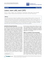
are spindle-shaped and resemble fibroblasts[5,6] (Fig 1).
There are no cell surface markers that specifically and
uniquely identify MSC, and their characterization in the
literature lacks consistency. The diversity of characteristics
associated with MSC can be explained by differences in
tissue origin, isolation methods and culture conditions
between laboratories, in addition there appear to be
strain-to-strain differences in murine derived MSC[2,7-9].
Whilst there is an obvious need for standardization
between research groups, some consensus can be found
among the conflicting data. In broad terms, MSC
expanded in vitro do not express the hematopoietic or
endothelial surface markers CD11b, CD14, CD31, CD34
or CD45 but stain positive for CD29, CD44, CD73,
CD105, CD106 and CD166 [2,5,10]. The non-embryonic
source of this population, the reduced likelihood of neo-
plasia, and the more limited differentiation potential,
have made these cells attractive candidates for application
in cell based therapies usually termed "regenerative med-
icine"[2]. There is one confounding influence on this
approach; whilst self derived MSC pose few immunologi-
cal problems, in practice regenerative medicine is likely to
rely on mismatched (allogeneic) cells to repair or replace
damaged tissue. Normally, allogeneic cells are deleted by
host immune responses. The major surprise to Immunol-
ogists working in this field have been findings that suggest
that MSC do not obey the normal "rules" of allogeneic
rejection. This review will survey recent data, which con-
vincingly indicate the mechanisms by which MSC escape
the normal process of alloantigen recognition.
MSC evade allorejection
The major limit to solid organ graft survival is T cell recog-
nition by the recipient of alloantigen (dominated by, but
not confined to MHC/HLA antigens)[11]. There are two
mechanisms mediating this powerful rejection response;
"direct" recognition, involving recognition by recipient
CD8+ or CD4+ T cells of donor MHC class I and class II
molecules; and "indirect" mechanisms involving recogni-
tion of peptides from the allogeneic tissue[11]. Recipient
antigen presenting cells (APC) such as dendritic cells
(DC) process alloantigen into peptides and present these
to naive T cells on self-MHC molecules [12]. However
there are notable exceptions to these allorejection proc-
esses; the fetal allograft evades rejection by the mother
through a complex series of actions (reviewed in[13]),
similarly tissue which has limited lymphatic drainage is
less prone to allorejection[14]. Interestingly tumor cells,
whilst not allogeneic, are in many cases both "altered-self"
and immunogenic but often actively modulate immune
responsiveness to evade immune surveillance[15]. Thus
mechanisms of tumor evasion of the immune system may
provide insight into how allogeneic MSC are tolerated by
the mismatched host.
There is supporting evidence for the use of allogeneic MSC
from both in vitro and in vivo studies that show MSC
avoid normal alloresponses. A small number of in- vivo
studies suggest that MSC play a role in enabling alloanti-
gen tolerance. Koc et al, showed no evidence of alloreac-
tive T cells and no incidence of graft v host disease when
allogeneic MSC were infused into patients with Hurler's
syndrome or metachromatic leukodystrophy[16]. In a
previous study by the same group, autologous culture-
expanded MSC were infused to breast cancer patients to
investigate whether MSC would enhance the engraftment
of peripheral blood stem cells after myeloablative therapy
[17]. Results showed rapid hematopoietic recovery and no
signs of toxicity from MSC infusion[17]. Horwitz and col-
leagues, reported that donor MSC contributed to bone
remodelling after allogeneic stem cell transplantation in
three children with osteogenesis imperfecta (OI)[18], a
rare genetic disorder of type I collagen. This is supported
by data from Bartholomew et al who showed that in-vivo
administration of allogeneic MSC prolonged 3rd party
skin graft survival in animal models[19]. Furthermore,
Saito et al, demonstrated that MSC undergoing differenti-
ation to a cardiac phenotype were tolerated in a xenoge-
neic environment, retaining their ability to be recruited to
the injured myocardium[20]. More recent work by Aggar-
wal and Pittenger supported the feasibility of MSC-trans-
plantation showing that MSC altered the phenotypes of
specific immune cell subtypes thereby creating a
tolerogenic environment[21]. These reports suggest that
transplantation of MSC could be beneficial in patients
with various disorders requiring tissue regeneration, and
Human mesenchymal stem cells (MSC) are spindle shaped, fibroblast-like cellsFigure 1
Human mesenchymal stem cells (MSC) are spindle shaped,
fibroblast-like cells. Original magnification × 100, phase-con-
trast light microscopy, scale bar represents 50 µm.
Journal of Inflammation 2005, 2:8 />Page 3 of 11
(page number not for citation purposes)
provide evidence supporting the tolerance of allogeneic
MSC by recipients.
Data supporting the contention that MSC avoid alloge-
neic responses has also come from a large body of in vitro
experiments, usually involving co-culture or mixed lym-
phocyte reactions (MLR). Evidence from these studies
indicate that the use of mismatched MSC does not pro-
voke a proliferative T cell response in allogeneic MLR, thus
suggesting an immunosuppressive role for MSC[19,22-
26]. Le Blanc et al, showed that MSC failed to elicit prolif-
eration of allogeneic lymphocytes[27]. Additionally, they
demonstrated that MSC remained immunosuppressive
even after IFN-γ stimulation[27]. Evidence from Krampera
et al confirms these findings, they showed that murine
MSC lack MHC class II and inhibited T cell prolifera-
tion[25]. Tse et al, also showed that human MSC fail to
elicit allogeneic T cell response in a MLR even when MHC
class II was upregulated[28]. Consistent with these stud-
ies, Bartholomew et al showed that allogeneic baboon
MSC suppressed the proliferative activity of lymphocytes
in vitro and prolonged graft survival[19]. These findings
support the view that MSC can be transplanted between
MHC-incompatible individuals. Although these data
show that successful use of allogeneic MSC in regenerative
therapy is possible, such approaches are unlikely to be
broadly acceptable until it is understood why MSC are not
rejected. This question has been the subject of intense
recent study and three candidate mechanisms are emerg-
ing. MSC appear to evade allogeneic rejection by a) being
hypoimmunogenic; b) modulating T cell phenotype and
c) creating an immunosuppressive local milieu. These
mechanisms are inter-related and will involve cell contact
dependent and independent interactions. The challenge
facing the field is to unravel the contribution of these
diverse interactions.
MSC are hypoimmunogenic
There is controversy surrounding the cell surface expres-
sion of MHC alloantigens by MSC. Although conflicting
evidence exists, most studies describe human MSC as
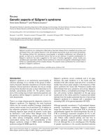
MHC class I positive and MHC class II negative (Fig 2).
The data conflicting with these findings may represent dif-
ferent stem cell lineages or be the result of the recently
described process of cell-cell transfer [29-31]. The expres-
sion of MHC class I by MSC is important because expres-
sion protects MSC from certain NK cell mechanisms of
deletion. For instance, a major function of NK and NK-
like cells is to kill tumor cells that have downregulated
class I [32]. HLA-G is an MHC-like protein that is known
to protect the fetal allograft against NK mediated rejec-
tion[33,34]. This protein has been shown to bind to the
two major inhibitory NK receptors, KIR1 and KIR2, and to
inhibit NK killing [35-37]. However no studies of HLA-G
expression by MSC have been reported to date.
As MHC class II proteins are potent alloantigens, the
expression by MSC is another important factor. Again
there is some controversy over expression, which may be
explained by the diversity of models described above.
However there are widespread observations that under
non-inflammatory conditions, human MSC are MHC-II
Human MSC cultured according to [106, 107] are A) MHC-I positive (HLA-A,B,C, antibody W6/32-FITC), B) MHC class II negative (HLA-DR, antibody LN-3-PE); C) CD14 negative (antibody MEM-18-FITC), D) CD86 negative (antibody IT2.2-PE); and E) CD40L/ CD154 (antibody 24-31-FITC), F) CD95L (FasL) negative (antibody NOK-1-PE)Figure 2
Human MSC cultured according to [106, 107] are A) MHC-I
positive (HLA-A,B,C, antibody W6/32-FITC), B) MHC class II
negative (HLA-DR, antibody LN-3-PE); C) CD14 negative
(antibody MEM-18-FITC), D) CD86 negative (antibody IT2.2-
PE); and E) CD40L/ CD154 (antibody 24-31-FITC), F)
CD95L (FasL) negative (antibody NOK-1-PE). Isotype
matched control antibody labelling are shown as unshaded
plots, FITC conjugates are shown in blue, PE conjugates
shown in pink. Flow cytometry performed according to
methods previously described [108-110].
Journal of Inflammation 2005, 2:8 />Page 4 of 11
(page number not for citation purposes)
negative, supporting a role for MSC as having reduced
immunogenicity through the control of alloantigen
expression [38-40]. The absence of MHC class II gives
MSC the potential to escape recognition by alloreactive
CD4+ T cells. In addition to being MHC II negative, MSC
do not appear to express the co-stimulatory molecules
CD40, CD40L, CD80 or CD86 required for effector T cell
induction[28,39]. The absence of co-stimulatory mole-
cules is a significant observation. It implies that any resid-
ual engagement of the T cell receptor on Th cells would
result in anergy and contribute to tolerance rather than
allogeneic responses. Although this is a comforting sce-
nario, based largely on in vitro studies, it cannot fully
explain the evasion of alloreactivity demonstrated by
MSC. Experiments involving allogeneic co-cultures or
MLR have demonstrated that both cell-cell contact and
action by soluble factors contribute to the immunomod-
ulatory function of MSC[25,41-43]. Thus it is likely that
evasion of alloreactivity is a result of both MSC hypoim-
munogenicity, modulation of T cell immune induction
and the creation of a suppressive milieu around MSC.
Although the mechanisms governing the suppressive
effect are not fully understood, several studies have given
indicators to the processes involved.
MSC interfere with DC maturation and function
Dendritic cells (DC) are the most influential APC, playing
a key role in directing cellular and humoral immune
responses against self and non-self antigens [44]. DC con-
tribute to the establishment of tolerance, especially in the
periphery[45]. Immature DC are not fully differentiated
to carry out their known roles as inducers of immu-
nity[45]. Despite this, immature DC circulate through tis-
sues and the lymph system, capturing self and non-self
antigens[45]. Immature DC that are loaded with antigen
can silence T cells by deletion or by expanding regulatory
T cell populations[45,46]. It has long been believed that
this process contributes to graft survival during transplan-
tation [14]. The capacity of DC to induce peripheral toler-
ance is a potential mechanism by which MSC could
manipulate immunity in order to escape T cell recogni-
tion. Thus MSC could prevent normal allogeneic
responses either through modulation of DC function or
by direct effects on T cells. Indications from different stud-
ies encourage this hypothesis. Zhang et al [24] provides
evidence that MSC interfere with DC maturation. Co-cul-
ture experiments showed that MSC down-regulate CD1a,
CD40, CD80, CD86, and HLA-DR expression during DC
maturation[24]. This is also shown by Beyth et al. [42],
who suggest that human MSC converted APC into an
inhibitory or suppressor phenotype via cell-to-cell con-
tact, thus locking DC into a semi-mature state and thereby
inducing peripheral tolerance. Their findings also show
reduced IFN-γ, IL-12 and TNF-α in human MSC/mono-
cyte co-culture [42]. Similarly Jiang et al reported that
MSC maintain DC in an immature state[26] and show
that MSC inhibit up regulation of IL-12p70 [26]. These
results suggest that MSC mediate allogeneic tolerance by
directing APC towards a suppressor or inhibitory pheno-
type that results in an attenuated or regulatory T cell
response.
MSC modulate CD4+ T cell responses
Evidence has emerged that MSC interact directly with T
cells to suppress alloreativity[25]. Krampera et al showed
that MSC impair T cell contact with APC in a non-cognate
but transient fashion[25]. This supported work from Bar-
tholomew et al showing that the addition of IL-2 to MLR/
MSC co-cultures reduced MSC suppression and restored T
cell proliferation[19]. Taken together, these results
strongly support a role for either a direct (T cell pheno-
type) or indirect (DC phenotype) mechanism of immune
modulation directed by MSC.
MSC modulation of CD4+ T cell responses is more exten-
sive than the straightforward effect described above. The
regular process of antigen specific CD4+ T cell induction
requires antigen capture and processing by DC (or other
amenable cells), followed by a process of maturation and
trafficking to local lymph nodes[14,47-49]. There is evi-
dence that MSC prevent normal allogeneic responses by
directing CD4+ T cells to a suppressive or counter-regula-
tory phenotype[46,50]. Di Nicola et al, showed that MSC
strongly suppressed CD4+ (and CD8+) T cells in
MLR[43], findings supported by Tse et al, who showed
that MSC suppress the proliferation of T-cell subsets[28].
Studies of T cell differentiation have shown that in the
presence of human MSC, Th1 cell secretion of IFN-γ
dropped by 50% compared to cultures without MSC.
Conversely, effector T cells undergoing Th2 differentiation
when co-cultured with human MSC showed a significant
increase in IL-4 production compared to controls[21].
These findings suggest that MSC exert a counter regula-
tory, anti-inflammatory role by directing cytokine-medi-
ated immunity[21].
A strategy of regulation and deletion of specific T cells is
an effective control against unwanted immune respon-
siveness especially after transplantion[51]. Consequently,
enormous interest has focused on the possibility of Treg
cells as a marker for T cell tolerance during transplanta-
tion. Treg can act directly on other T cells or indirectly
through APC[46]. Aggrawal et al, demonstrated that
CD4+ CD25+ T reg populations increased significantly in
MLR when MSC were present compared to controls[21].
However, data exists showing that human MSC-mediated
inhibition is not suppressed by removing T reg cells from
co-cultures [25,42]. Nevertheless a role for these cells can
not be excluded, it is possible that an incomplete replica-
tion of the suppressive microenvironment in vitro or
Journal of Inflammation 2005, 2:8 />Page 5 of 11
(page number not for citation purposes)
indeed the diversity of Treg cell populations mean that
these studies do not fully explore the potential role of sup-
pressive or regulatory T cells in promoting MSC tolerance.
MSC influence control over cell division cycle pathways in
cells of immunological relevance. Glennie et al have
shown that T cells stimulated in co-cultures with MSC
exhibit an extensive inhibition of cyclin D2 and upregula-
tion of the cyclin dependent kinase inhibitor p27
kip1
[52].
As T cell inhibition could not be reversed, these cells were
not interpreted as anergic in the classical sense. The
authors suggest that MSC are most likely inducing the
alternative condition of divisional arrest anergy in T cells,
an occurrence usually associated with CTLA-4 signal-
ling[53]. In addition, removal of MSC from the system
only restored IFN-γ production but not T cell prolifera-
tion[52]. This suggests that MSC induce a condition simi-
lar to split anergy[54] or split tolerance[55,56]. The key
point is that this work demonstrates that MSC exert veto
effects on T cells and it is significant in demonstrating that
the mechanisms inducing MSC tolerance are not confined
to patterns of cytokine secretion but extend to direct mod-
ulation of T cell division.
MSC modulate CD8+ T cell and NK cell activity
The impact of MSC on CD8+ CTL and NK cells has also
been addressed. CTL can lyse allogeneic cells after recog-
nition of cognate alloantigen, by the release of cytotoxic
effectors such as, perforins, serine esterases, IFN-γ and
TNF-α [57] whereas NK cells do not require antigen
processing[58]. Consequently both effector cells can oper-
ate in tandem, with NK cells providing a first line defence
killing target cells that escape CTL recognition or show
inadequate expression of self-MHC[58]. There is evidence
that MSC inhibit the formation of CTL and appear to
evade NK cell targeting mechanisms. Djouad et al showed
that CD8+ cells are suppressed by MSC in MLR[41]. Ras-
musson supported these findings and further showed that
NK cells in co-culture did not recognize MSC although
lytic capability was still present[59]. This effect appeared
to be mediated by soluble factors[50,59]. Thus MSC inter-
act and suppress cell-mediated immune responses directly
and through soluble factors. The targets for this suppres-
sion are DC, CD4+ Th, CD8+ CTL and NK cells; in effect
MSC silence each aspect of the cellular rejection process.
MSC secrete soluble factors to create an
immunosuppressive milieu
The characterisation of cytokines produced by MSC is still
provisional and is hindered by the lack of standardisation
in isolation and culture conditions, which have given rise
to multiple findings and interpretations. It is evident that
MSC do not constitutively express IL-2, IL-3, IL-4 and IL-
5[60,61]. However some reports show that MSC do con-
stitutively express mRNA for cytokines such as interleukin
(IL)-6, -7, -8, -11, -12, -14, -15, -27, leukaemia inhibitory
factor, macrophage colony-stimulating factor, and stem
cell factor[62,63]. Some of these cytokines provide critical
cell-cell interactions and promote HSC differentiation,
however caution should be exercised before over inter-
preting these findings. Protein secretion does not always
mirror mRNA levels and most workers in the field would
adopt a more conservative profile of cytokine and growth
factor production by MSC.
Despite these caveats, certain MSC secreted products such
as Hepatocyte growth factor, (HGF) are likely to contrib-
ute to creating a local immunosuppressive environment.
HGF induces mitogenic and antiapoptotic activity in dif-
ferent systems [64-66] and has a well-characterized role in
wound repair [66-68], effects that are consistent with a
role for MSC in regenerative medicine. Although some
groups do not detect HGF in MSC co-cultures [41] more
reports suggest that HGF is constitutively expressed by
MSC [13,43,69,70]. Indications that MSC produce HGF
[13,43,69,70] encourage a role for these cells in tissue
repair [70]. Studies by Chunmeng et al, demonstrated that
rat dermal derived "multipotent" cells secrete HGF and
promote wound healing[68]. Interestingly, Azuma et al,
showed that HGF treatment prevents chronic allograft
nephropathy in rats[71]. Taken together these results sug-
gest that HGF may contribute to the ability of MSC to
avoid allorejection.
IL-10 has a well-documented role in T cell regulation and
in the promotion of a "regulatory" or suppressor pheno-
type. In our hands human MSC constitutively produce IL-
10 whereas Rasmusson et al and Beyth et al only detected
IL-10 in co-culture experiments [42,72]. In either case, IL-
10 is likely to be suppressing potential allo-responsive-
ness because it is a recognized growth factor for regulatory
T cells [73]. IL-10 can antagonize IL-12 during induction
of inflammatory immune responses [74-79]. This is sup-
ported by studies showing that MSC partially mediate
suppression through IL-10 secretion in MLR cul-
tures[42,72]. Similarly transforming growth factor (TGF)-
β1 also plays a role in T cell suppression. This cytokine as
well as IL-10 influences cell lineages broader than lym-
phocytes [74,80,81]. However constitutive expression of
TGF-β1 has not been detected from our own studies on
human MSC[13]. This is in line with Le Blanc who found
no difference in TGF-β1 concentration in co-cultures with
or without MSC [69]. In contrast Beyth et al showed that
TGF-β1 was secreted in media from co-cultures of human
MSC and immune cells but again co-culture did not aug-
ment TGF-β1 concentration[42]. Although a number of
studies suggest no role for TGF-β1 in evasion of allogeneic
responsiveness[42,69,72], it has been suggested that HGF
in combination with TGF-β promotes the allo-escaping
phenotype[43]. Di Nicola et al showed that neutralizing
Journal of Inflammation 2005, 2:8 />Page 6 of 11
(page number not for citation purposes)
antibodies to HGF and TGF-β restored the proliferative
response in MLR, suggesting that these factors are at least
partially responsible[43].
MSC constitutively express the eicosanoid Prostaglandin E
(PGE)-2 [82]. This may be upregulated in co-cul-
ture[21,28] or downregulated on differentiation[82].
PGE-2 influences numerous immune functions including
suppression of B cell activation[83] and induction of reg-
ulatory T cells[84]. Although there is evidence for PGE-2
secretion by MSC, there is controversy surrounding a role
for PGE-2 as a mediator for suppression of alloresponses
in MLR. Studies from Tse, suggested that PGE-2 is not a
significant component of suppression[28]. Supporting
these findings Rasmusson et al showed that blocking PGE-
2 production did not restore allogeneic MLR responses
but did influence mitogen driven proliferation[72].
Although the present opinions are conflicting, it should
be highlighted that other possible prostaglandins and
eicosanoids could be influencing alloresponses[85]. Anal-
ysis of these other immunomodulatory molecules could
provide further clues as to how MSC escape the immune
system.
In contrast to immunosuppression through the secretion
of soluble factors, suppression may be mediated by with-
drawal of factors in the micro-environment necessary for
active immune responses. Indoleamine 2,3-dioxygenase
(IDO) is an enzyme that catabolizes L-Tryptophan,
thereby depleting an essential amino acid from the local
environment [86-89]. Recent evidence has shown that
this mechanism is exploited by the mammalian fetal allo-
graft to suppresses T cell activity and prevent rejection [86-
89]. Although not a soluble factor, the expression of IDO
may contribute to a tolergenic environment. This is of
great relevance and has obvious parallels with MSC. Mei-
sel et al showed that IDO is not constitutively expressed
by MSC but can be induced by IFN-γ[90], thereby inhibit-
ing allogeneic T cell responses by Tryptophan deple-
tion[90]. Other findings have suggested that IDO-
mediated tryptophan depletion inhibits allogeneic T-cell
responses by multiple pathways[91]. The discovery of this
mechanism, which shows parallels to the creation of a
"Tryptophan desert" at the materno-fetal interface[13],
provides a further feasible mechanism by which MSC
avoid alloreactivity. However, IDO expression is not
essential to the maintenance of tolerance against MSC. Tse
et al showed that an IDO inhibitor or supplementary
Tryptophan addition to MLR did not restore PBMC prolif-
eration [28].
MSC control surface marker expression to exhibit a
hypoimmunogenic or tolerogenic phenotype. MSC can
also modulate T cell induction directly or via DC and
secrete a battery of immunosuppressive factors. It is
apparent that the question facing the application of regen-
erative medicine is no longer "how do MSC escape allore-
activity?" but rather "what is the hierarchy of signals that
control immunosuppression?" In this regard, research
from other fields has been informative. We have previ-
ously proposed that maternal acceptance of the fetal allo-
graft provides indicators of how this process is
controlled[13]. However, insight could also come from
another avenue of inquiry. The mechanisms of tumor eva-
sion may reflect the survival mechanisms of MSC.
MSC avoidance of alloreactivity shows parallels to tumor
evasion
Escape from immune surveillance is believed to be a pri-
mary feature of malignant disease in humans. The
immune effector response is sub-optimal because tumors
develop multifactorial strategies to escape immune dele-
tion[92,93]. These strategies may provide clues to how
MSC promote tolerogenic mechanisms during allogeneic
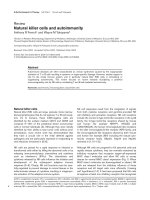
engraftment (Fig. 3). Modulation of tumor antigen
expression, particularly MHC class I and II is a particularly
common component of tumor immune evasion[93]. This
is often accompanied by poor or non-expression of co-
stimulatory molecules, which not only limits clonal
expansion of tumor-specific CD4+ T cells, but also hin-
ders the production of cytokines, and the development of
CTL[44,94,95]. Similarly MSC show no expression of co-
stimulatory molecules (Fig. 2) [28,39]. In addition to
reduced immunogenicity, tumor cells can directly modu-
late DC and T cell function. Studies from patients with
hepatocellular carcinoma showed that neoplasia induced
a defect of DC maturation[96]. This parallels findings by
Beyth et al [42] suggesting that human MSCs interfere
with normal APC maturation, thereby indirectly influenc-
ing T-cell activation. Freshly isolated tumor-infiltrating T
cells are usually inactive against autologous cancer cells
but can be reactivated in-vitro by the addition of IL-2[97].
Studies of MSC by Le Blanc et al showed striking parallels
to this form of suppression[69]. They suggest that MSC act
by preventing expression of CD25 (IL-2 receptor) thereby
limiting T cell activation[69]. Other work has shown that
exogenous IL-2 addition to co-cultures containing MSC
reversed the suppressive effect[19,69]. Similarly, antigen-
specific CD4+ CD25+ regulatory T cells also suppress
tumor-specific CD8 T cell cytotoxicity although this mech-
anism relies on TGF-β secretion by regulatory cells[98,99].
Tumors can suppress CD4+ T cell activity and CTL tumor
lysis directly through secretion of immunosuppressive
factors including TGF-β1 but also PGE-2, and IL-10. Van
der Pouw Kraan et al, showed that tumor-derived prostag-
landins increased the production of inhibitory cytokines
such as IL-10, while suppressing IL-12[100], which is nec-
essary for effective host-cell-mediated anti-tumor immune
response[75,93]. Likewise, TGF-β production has been
Journal of Inflammation 2005, 2:8 />Page 7 of 11
(page number not for citation purposes)
reported from a number of tumors, contributing to
immune evasion. Intriguingly in this context it also inhib-
its CTL differentiation [101]. Although there is little evi-
dence that MSC secrete TGF-β1, the bone marrow is rich
in this cytokine, suggesting that MSC reside in a compart-
ment with immunosuppressive qualities.
Although there are striking parallels between MSC and
some tumor cells, it is not our contention that these cells
are directly related. Indeed there are distinct differences
between the populations (Table 1). The fundamental dif-
ference between the cell types resides in the control of cell
division and apoptosis, which are tightly regulated in
MSC but dysregulated in transformed cells. Furthermore,
it is well documented that some tumors exploit FasL
(CD95L) expression to facilitate immune escape [102-
104]. However, our own studies show that human MSC
do not express FasL (Fig 2) and although there is some evi-
dence from immortalized mini-pig derived MSC to indi-
cate a role for FasL in suppression[105], it seems that
direct induction of apoptotic deletion is not a factor
involved in MSC interaction with T cells in the broader lit-
erature. The parallels between neoplastic cells and MSC lie
in the expressed phenotypes rather than in any direct lin-
eage relation. It appears that MSC retain certain aspects of
the fetal allograft that promote tolerance, some of these
mechanisms may be reactivated in neoplasia, the key dif-
ference being that MSC perform these functions in an
ordered and controlled way whereas tumor cells do so in
a manner that by definition has escaped normal controls
on apoptosis or cell division.
Conclusion
Current research on the interaction between MSC and T
cells support the potential use of allogeneic MSC in regen-
erative medicine. Studies showing enhanced MSC engraft-
ment of bone, muscle, heart etc encourage the translation
of recent research into therapy. The future holds much
promise for the use of allogeneic MSC and whilst obsta-
cles exist, the potential for alloreactivity does not seem to
be a major problem. From the research standpoint, MSC
appear to use a surprising array of mechanisms to avoid
deletion by the host including hypoimmunogenicity,
modulation of DC and T cell function, as well as the crea-
tion of a suppressive microenvironment. The challenge is
now to unravel the timing and control of these mecha-
nisms in an inflammatory situation typical of the recipi-
ent patient.
List of Abbreviations
APC, antigen presenting cells; DC, dendritic cell; ES,
embryonic stem; HGF, hepatocyte growth factor; HSC,
hematopoietic stem cells; IDO, indoleamine 2,3,dioxyge-
nase; KIR, killer inhibitory receptor; MLR, mixed lym-
phocyte-like reaction; MSC, mesenchymal stem cells; OI,
osteogenesis imperfecta; PBMC, peripheral blood mono-
nuclear cells; PGE-2, prostaglandin E2.
Competing interests
JMR and BPM have no competing interests. FPB and JMM
have received salary from an organization and hold stocks
or shares in an organization that may gain or lose finan-
cially from the publication of this manuscript.
Authors' contributions
FPB and BPM conceived the review; JMR performed the
microscopy and flow cytometry. All authors provided
MSC and tumor cells create a suppressive microenvironmentFigure 3
MSC and tumor cells create a suppressive microenviron-
ment. There are fundamental differences between tumor
cells (A) and MSC (B) with respect to control of cell division,
however many mechanisms exploited by the former to evade
immune deletion are also used by MSC to avoid allogeneic
rejection. Details of mechanisms and associated references
are supplied in the body of the text and Table 1.
Journal of Inflammation 2005, 2:8 />Page 8 of 11
(page number not for citation purposes)
interpretation of published stem cell data, and have made
intellectual contributions to the content of the paper. All
authors read and approved the final manuscript.
Additional material
Acknowledgements
This work was supported by the Science Foundation Ireland Centres for
Science Engineering and Technology (CSET) funding of the Regenerative
Medicine Institute (REMEDI). Bernard Mahon is a Wellcome Trust/HRB
"New Blood" Fellow. Ms Karen English is thanked for assistance in prepa-
ration of this manuscript.
References
1. Mosca JD, Hendricks JK, Buyaner D, Davis-Sproul J, Chuang LC,
Majumdar MK, Chopra R, Barry F, Murphy M, Thiede MA, Junker U,
Rigg RJ, Forestell SP, Bohnlein E, Storb R, Sandmaier BM: Mesenchy-
mal stem cells as vehicles for gene delivery. Clin Orthop Relat
Res 2000:S71-90.
2. Barry FP, Murphy JM: Mesenchymal stem cells: clinical applica-
tions and biological characterization. Int J Biochem Cell Biol 2004,
36:568-584.
3. Drukker M, Benvenisty N: The immunogenicity of human
embryonic stem-derived cells. Trends Biotechnol 2004,
22:136-141.
4. Shizuru JA, Negrin RS, Weissman IL: Hematopoietic stem and
progenitor cells: Clinical and Preclinical Regeneration of the
Hematolymphoid System. Annu Rev Med 2005, 56:509-538.
5. Pittenger MF, Mackay AM, Beck SC, Jaiswal RK, Douglas R, Mosca JD,
Moorman MA, Simonetti DW, Craig S, Marshak DR: Multilineage
potential of adult human mesenchymal stem cells. Science
1999, 284:143-147.
6. Friedenstein AJP, Petrokova KV: Osteogenesis in transplants of
bone marrow cells. Journal of Embyological Experimental Morphology
1966, 16:381-390.
7. Jiang Y, Jahagirdar BN, Reinhardt RL, Schwartz RE, Keene CD, Ortiz-
Gonzalez XR, Reyes M, Lenvik T, Lund T, Blackstad M, Du J, Aldrich
S, Lisberg A, Low WC, Largaespada DA, Verfaillie CM: Pluripotency
of mesenchymal stem cells derived from adult marrow.
Nature 2002, 418:41-49.
8. Reyes M, Verfaillie CM: Characterization of multipotent adult
progenitor cells, a subpopulation of mesenchymal stem cells.
Ann N Y Acad Sci 2001, 938:231-233.
9. Jorgensen C, Gordeladze J, Noel D: Tissue engineering through
autologous mesenchymal stem cells. Curr Opin Biotechnol 2004,
15:406-410.
10. Jorgensen C, Djouad F, Apparailly F, Noel D: Engineering mesen-
chymal stem cells for immunotherapy. Gene Ther 2003,
10:928-931.
11. Pawelec G, Rehbein A, Schlotz E, Friccius H, Pohla H: Cytokine
modulation of TH1/TH2 phenotype differentiation in
directly alloresponsive CD4+ human T cells. Transplantation
1996, 62:1095-1101.
12. Wood KJ, Sakaguchi S: Regulatory T cells in transplantation
tolerance. Nat Rev Immunol 2003, 3:199-210.
13. Barry FP, Murphy JM, English K, Mahon BP: Immunogenicity of
adult mesenchymal stem cells: lessons from the fetal
allograft. Stem cells and development 2005, in press:.
14. Niederkorn JY, Peeler JS, Ross J, Callanan D: The immunogenic
privilege of corneal allografts. Reg Immunol 1989, 2:117-124.
15. van den Eynde B, Gaugler B, van der Bruggen P, Coulie P, Brichard V,
Boon T: Human tumor antigens recognised by T cells: per-
spectives for new cancer vaccines. Biochemical Society
Transactions 1995, 23:681-686.
16. Koc ON, Day J, Nieder M, Gerson SL, Lazarus HM, Krivit W: Allo-
geneic mesenchymal stem cell infusion for treatment of
metachromatic leukodystrophy (MLD) and Hurler syn-
drome (MPS-IH). Bone Marrow Transplant 2002, 30:215-222.
17. Koc ON, Gerson SL, Cooper BW, Dyhouse SM, Haynesworth SE,
Caplan AI, Lazarus HM: Rapid hematopoietic recovery after
coinfusion of autologous-blood stem cells and culture-
expanded marrow mesenchymal stem cells in advanced
breast cancer patients receiving high-dose chemotherapy. J
Clin Oncol 2000, 18:307-316.
18. Horwitz EM, Prockop DJ, Gordon PL, Koo WW, Fitzpatrick LA, Neel
MD, McCarville ME, Orchard PJ, Pyeritz RE, Brenner MK: Clinical
responses to bone marrow transplantation in children with
severe osteogenesis imperfecta. Blood 2001, 97:1227-1231.
Table 1: Comparison of MSC and Tumor cells
a
Characteristic MSC Tumor cells References
Cell Division Controlled Uncontrolled [5, 7, 111]
MHC I expression + Variable Fig 2 & [25, 27, 28, 39, 93, 111, 112]
MHC II expression - Variable Fig 2 & [2, 25, 27, 39, 93, 111, 112]
CD80 expression - - [25, 28, 39, 44, 94, 95]
CD86 expression - - Fig 2 & [25, 28, 39, 44, 94, 95]
FasL expression - + Fig. 2 & [102-104]
Prostaglandin secretion + + [21, 28, 82, 100]
IDO expression + Variable [28, 43, 59, 87, 90]
TGF-β secretion Variable + [42, 43, 59, 101, 105]
IL-10 secretion + + [13, 42, 72, 100]
DC modulation + + [24, 26, 42, 96]
Veto effects on T cells + + [23, 112]
a Descriptions of MSC in the literature are diverse and many populations have been described which show different patterns of expression. In
particular work in mice appears to be strain dependent, but further variation arises from differences in isolation, culture, timing and methodology.
Likewise the characteristics of neoplastic cells will vary greatly between different tumors. This table lists those characteristics where at least some
cells from each diverse population show either comparative or contrasting features.
Additional File 1
Ryan et al library.
Click here for file
[ />9255-2-8-S1.enl]
Journal of Inflammation 2005, 2:8 />Page 9 of 11
(page number not for citation purposes)
19. Bartholomew A, Sturgeon C, Siatskas M, Ferrer K, McIntosh K, Patil
S, Hardy W, Devine S, Ucker D, Deans R, Moseley A, Hoffman R:
Mesenchymal stem cells suppress lymphocyte proliferation
in vitro and prolong skin graft survival in vivo. Exp Hematol
2002, 30:42-48.
20. Saito T, Kuang JQ, Bittira B, Al-Khaldi A, Chiu RC: Xenotransplant
cardiac chimera: immune tolerance of adult stem cells. Ann
Thorac Surg 2002, 74:19-24.
21. Aggarwal S, Pittenger MF: Human mesenchymal stem cells
modulate allogeneic immune cell responses. Blood 2005,
105:1815-1822.
22. Deng W, Han Q, Liao L, Li C, Ge W, Zhao Z, You S, Deng H, Zhao
RC: Allogeneic bone marrow-derived flk-1+Sca-1- mesenchy-
mal stem cells leads to stable mixed chimerism and donor-
specific tolerance. Exp Hematol 2004, 32:861-867.
23. Potian JA, Aviv H, Ponzio NM, Harrison JS, Rameshwar P: Veto-like
activity of mesenchymal stem cells: functional discrimina-
tion between cellular responses to alloantigens and recall
antigens. J Immunol 2003, 171:3426-3434.
24. Zhang W, Ge W, Li C, You S, Liao L, Han Q, Deng W, Zhao RC:
Effects of Mesenchymal Stem Cells on Differentiation, Mat-
uration, and Function of Human Monocyte-Derived Den-
dritic Cells. Stem Cells Dev 2004, 13:263-271.
25. Krampera M, Glennie S, Dyson J, Scott D, Laylor R, Simpson E, Dazzi
F: Bone marrow mesenchymal stem cells inhibit the
response of naive and memory antigen-specific T cells to
their cognate peptide. Blood 2003, 101:3722-3729.
26. Jiang XX, Zhang Y, Liu B, Zhang SX, Wu Y, Yu XD, Mao N: Human
mesenchymal stem cells inhibit differentiation and function
of monocyte-derived dendritic cells. Blood 2005, in press:.
27. Le Blanc K, Tammik C, Rosendahl K, Zetterberg E, Ringden O: HLA
expression and immunologic properties of differentiated and
undifferentiated mesenchymal stem cells. Exp Hematol 2003,
31:890-896.
28. Tse WT, Pendleton JD, Beyer WM, Egalka MC, Guinan EC: Suppres-
sion of allogeneic T-cell proliferation by human marrow
stromal cells: implications in transplantation. Transplantation
2003, 75:389-397.
29. Carlin LM, Eleme K, McCann FE, Davis DM: Intercellular transfer
and supramolecular organization of human leukocyte anti-
gen C at inhibitory natural killer cell immune synapses. J Exp
Med 2001, 194:1507-1517.
30. Onfelt B, Nedvetzki S, Yanagi K, Davis DM: Cutting edge: Mem-
brane nanotubes connect immune cells. J Immunol 2004,
173:1511-1513.
31. Vanherberghen B, Andersson K, Carlin LM, Nolte-'t Hoen EN, Wil-
liams GS, Hoglund P, Davis DM: Human and murine inhibitory
natural killer cell receptors transfer from natural killer cells
to target cells. Proc Natl Acad Sci U S A 2004, 101:16873-16878.
32. Ruggeri L, Capanni M, Martelli MF, Velardi A: Cellular therapy:
exploiting NK cell alloreactivity in transplantation. Curr Opin
Hematol 2001, 8:355-359.
33. Hunt JS, Petroff MG, Morales P, Sedlmayr P, Geraghty DE, Ober C:
HLA-G in reproduction: studies on the maternal-fetal
interface. Hum Immunol 2000, 61:1113-1117.
34. Ristich V, Liang S, Zhang W, Wu J, Horuzsko A: Tolerization of
dendritic cells by HLA-G. Eur J Immunol 2005, 35:1133-1142.
35. Moretta L, Moretta A: Killer immunoglobulin-like receptors.
Curr Opin Immunol 2004, 16:626-633.
36. Parham P: Killer cell immunoglobulin-like receptor diversity:
balancing signals in the natural killer cell response. Immunol
Lett 2004, 92:11-13.
37. Gomez-Lozano N, de Pablo R, Puente S, Vilches C: Recognition of
HLA-G by the NK cell receptor KIR2DL4 is not essential for
human reproduction. Eur J Immunol 2003, 33:639-644.
38. Gotherstrom C, Ringden O, Tammik C, Zetterberg E, Westgren M,
Le Blanc K: Immunologic properties of human fetal mesen-
chymal stem cells. Am J Obstet Gynecol 2004, 190:239-245.
39. Majumdar MK, Keane-Moore M, Buyaner D, Hardy WB, Moorman
MA, McIntosh KR, Mosca JD: Characterization and functionality
of cell surface molecules on human mesenchymal stem cells.
J Biomed Sci 2003, 10:228-241.
40. Devine SM, Hoffman R: Role of mesenchymal stem cells in
hematopoietic stem cell transplantation. Curr Opin Hematol
2000, 7:358-363.
41. Djouad F, Plence P, Bony C, Tropel P, Apparailly F, Sany J, Noel D,
Jorgensen C: Immunosuppressive effect of mesenchymal stem
cells favors tumor growth in allogeneic animals. Blood 2003,
102:3837-3844.
42. Beyth S, Borovsky Z, Mevorach D, Liebergall M, Gazit Z, Aslan H,
Galun E, Rachmilewitz J: Human mesenchymal stem cells alter
antigen-presenting cell maturation and induce T-cell
unresponsiveness. Blood 2005, 105:2214-2219.
43. Di Nicola M, Carlo-Stella C, Magni M, Milanesi M, Longoni PD, Mat-
teucci P, Grisanti S, Gianni AM: Human bone marrow stromal
cells suppress T-lymphocyte proliferation induced by cellular
or nonspecific mitogenic stimuli. Blood 2002, 99:3838-3843.
44. Guinan EC, Gribben JG, Boussiotis VA, Freeman GJ, Nadler LM: Piv-
otal role of the B7:CD28 pathway in transplantation toler-
ance and tumor immunity. Blood 1994, 84:3261-3282.
45. Steinman RM, Nussenzweig MC: Avoiding horror autotoxicus:
the importance of dendritic cells in peripheral T cell
tolerance. Proc Natl Acad Sci U S A 2002, 99:351-358.
46. Mills KH: Regulatory T cells: friend or foe in immunity to
infection? Nat Rev Immunol 2004, 4:841-855.
47. Steinman RM: The dendritic cell system and its role in
immunogenicity. Annu Rev Immunol 1991, 9:271-296.
48. Steinman RM, Inaba K, Turley S, Pierre P, Mellman I: Antigen cap-
ture, processing, and presentation by dendritic cells: recent
cell biological studies. Hum Immunol 1999, 60:562-567.
49. Mahon BP, Katrak K, Nomoto A, Macadam AJ, Minor PD, Mills KHG:
Poliovirus-specific CD4+ Th1 clones with both cytotoxic and
helper activity mediate protective humoral immunity
against a lethal poliovirus infection in transgenic mice
expressing the human poliovirus receptor. J Exp Med 1995,
181:1285-1292.
50. Thompson C, Powrie F: Regulatory T cells. Curr Opin Pharmacol
2004, 4:408-414.
51. Wood KJ, Jones ND, Bushell AR, Morris PJ: Alloantigen-induced
specific immunological unresponsiveness. Philos Trans R Soc
Lond B Biol Sci 2001, 356:665-680.
52. Glennie S, Soeiro I, Dyson PJ, Lam EW, Dazzi F: Bone marrow mes-
enchymal stem cells induce division arrest anergy of acti-
vated T cells. Blood 2005, 105:2821-2827.
53. Wells AD, Walsh MC, Bluestone JA, Turka LA: Signaling through
CD28 and CTLA-4 controls two distinct forms of T cell
anergy. J Clin Invest 2001, 108:895-903.
54. Otten GR, Germain RN: Split anergy in a CD8+ T cell: receptor-
dependent cytolysis in the absence of interleukin-2
production. Science 1991, 251:1228-1231.
55. Nash AA, Ashford NP: Split T-cell tolerance in herpes simplex
virus-infected mice and its implication for anti-viral
immunity. Immunology 1982, 45:761-767.
56. Martinez C, Smith JM: Split Tolerance. Nature 1964, 202:508-509.
57. Masson D, Tschopp J: A family of serine esterases in lytic gran-
ules of cytolytic T lymphocytes. Cell 1987, 49:679-685.
58. Ljunggren HG, Karre K: In search of the 'missing self': MHC
molecules and NK cell recognition. Immunol Today 1990,
11:237-244.
59. Rasmusson I, Ringden O, Sundberg B, Le Blanc K: Mesenchymal
stem cells inhibit the formation of cytotoxic T lymphocytes,
but not activated cytotoxic T lymphocytes or natural killer
cells. Transplantation 2003, 76:1208-1213.
60. Dormady SP, Bashayan O, Dougherty R, Zhang XM, Basch RS:
Immortalized multipotential mesenchymal cells and the
hematopoietic microenvironment. J Hematother Stem Cell Res
2001, 10:125-140.
61. Zhang Y, Li CD, Jiang XX, Li HL, Tang PH, Mao N: Comparison of
mesenchymal stem cells from human placenta and bone
marrow. Chin Med J (Engl) 2004, 117:882-887.
62. Haynesworth SE, Baber MA, Caplan AI: Cytokine expression by
human marrow-derived mesenchymal progenitor cells in
vitro: effects of dexamethasone and IL-1 alpha. J Cell Physiol
1996, 166:585-592.
63. Silva WAJ, Covas DT, Panepucci RA, Proto-Siqueira R, Siufi JL, Zan-
ette DL, Santos AR, Zago MA: The profile of gene expression of
human marrow mesenchymal stem cells. Stem Cells 2003,
21:661-669.
64. Kuroiwa T, Kakishita E, Hamano T, Kataoka Y, Seto Y, Iwata N,
Kaneda Y, Matsumoto K, Nakamura T, Ueki T, Fujimoto J, Iwasaki T:
Hepatocyte growth factor ameliorates acute graft-versus-
Journal of Inflammation 2005, 2:8 />Page 10 of 11
(page number not for citation purposes)
host disease and promotes hematopoietic function. J Clin
Invest 2001, 107:1365-1373.
65. Taniguchi F, Harada T, Deura I, Iwabe T, Tsukihara S, Terakawa N:
Hepatocyte growth factor promotes cell proliferation and
inhibits progesterone secretion via PKA and MAPK path-
ways in a human granulosa cell line. Mol Reprod Dev 2004,
68:335-344.
66. Xin X, Yang S, Ingle G, Zlot C, Rangell L, Kowalski J, Schwall R, Fer-
rara N, Gerritsen ME: Hepatocyte growth factor enhances vas-
cular endothelial growth factor-induced angiogenesis in vitro
and in vivo. Am J Pathol 2001, 158:1111-1120.
67. Ono I, Yamashita T, Hida T, Jin HY, Ito Y, Hamada H, Akasaka Y, Ishii
T, Jimbow K: Local administration of hepatocyte growth fac-
tor gene enhances the regeneration of dermis in acute inci-
sional wounds. J Surg Res 2004, 120:47-55.
68. Chunmeng S, Tianmin C, Yongping S, Xinze R, Yue M, Jifu Q, Shufen
L, Hui X, Chengji L: Effects of dermal multipotent cell trans-
plantation on skin wound healing. J Surg Res 2004, 121:13-19.
69. Le Blanc K, Rasmusson I, Gotherstrom C, Seidel C, Sundberg B, Sun-
din M, Rosendahl K, Tammik C, Ringden O: Mesenchymal stem
cells inhibit the expression of CD25 (interleukin-2 receptor)
and CD38 on phytohaemagglutinin-activated lymphocytes.
Scand J Immunol 2004, 60:307-315.
70. Neuss S, Becher E, Woltje M, Tietze L, Jahnen-Dechent W: Func-
tional expression of HGF and HGF receptor/c-met in adult
human mesenchymal stem cells suggests a role in cell mobi-
lization, tissue repair, and wound healing. Stem Cells 2004,
22:405-414.
71. Azuma H, Takahara S, Matsumoto K, Ichimaru N, Wang JD, Moriyama
T, Waaga AM, Kitamura M, Otsuki Y, Okuyama A, Katsuoka Y, Chan-
draker A, Sayegh MH, Nakamura T: Hepatocyte growth factor
prevents the development of chronic allograft nephropathy
in rats. J Am Soc Nephrol 2001, 12:1280-1292.
72. Rasmusson I, Ringden O, Sundberg B, Le Blanc K: Mesenchymal
stem cells inhibit lymphocyte proliferation by mitogens and
alloantigens by different mechanisms. Exp Cell Res 2005, in
press:.
73. Asseman C, Powrie F: Interleukin 10 is a growth factor for a
population of regulatory T cells. Gut 1998, 42:157-158.
74. Ennis DP, Cassidy JP, Mahon BP: Prior Bordetella pertussis infec-
tion modulates allergen priming and the severity of airway
pathology in a murine model of allergic asthma. Clin Exp
Allergy 2004, 34:1488-1497.
75. Mahon BP, Ryan M, Griffin F, McGuirk P, Mills KHG: IL-12 is pro-
duced by macrophages in response to live or killed Borde-
tella pertussis and enhances the efficacy of an acellular
pertussis vaccine by promoting the induction of Th1 cells.
Infect Immun 1996, 64:5295-5301.
76. Flynn MA, Casey DG, Todryk SM, Mahon BP: Efficient delivery of
small interfering RNA for inhibition of IL-12p40 expression
in vivo. Journal of Inflammation 2004, 1:4.
77. Pretolani M, Goldman M: IL-10: a potential therapy for allergic
inflammation. Immunol Today 1997, 18:277-280.
78. Higgins SC, Lavelle EC, McCann C, Keogh B, McNeela E, Byrne P,
O'Gorman B, Jarnicki A, McGuirk P, Mills KH: Toll-like receptor 4-
mediated innate IL-10 activates antigen-specific regulatory
T cells and confers resistance to Bordetella pertussis by
inhibiting inflammatory pathology. J Immunol 2003,
171:3119-3127.
79. McGuirk P, Mills KHG: Direct anti-inflammatory effect of a bac-
terial virulence factor:IL-10-dependent suppression of IL-12
production by filamentous haemagglutinin from Bordetella
pertussis. Eur J Immunol 2000, 30:415-422.
80. Gollnick SO, Cheng HL, Grande CC, Thompson D, Tomasi TB:
Effects of transforming growth factor-beta on bone marrow
macrophage Ia expression induced by cytokines. J Interferon
Cytokine Res 1995, 15:485-491.
81. Ruegemer JJ, Ho SN, Augustine JA, Schlager JW, Bell MP, McKean DJ,
Abraham RT: Regulatory effects of transforming growth fac-
tor-beta on IL-2- and IL-4-dependent T cell-cycle
progression. J Immunol 1990, 144:1767-1776.
82. Arikawa T, Omura K, Morita I: Regulation of bone morphoge-
netic protein-2 expression by endogenous prostaglandin E2
in human mesenchymal stem cells. J Cell Physiol 2004,
200:400-406.
83. Roper RL, Ludlow JW, Phipps RP: Prostaglandin E2 inhibits B
lymphocyte activation by a cAMP-dependent mechanism:
PGE-inducible regulatory proteins. Cell Immunol 1994,
154:296-308.
84. Akasaki Y, Liu G, Chung NH, Ehtesham M, Black KL, Yu JS: Induction
of a CD4+ T regulatory type 1 response by cyclooxygenase-
2-overexpressing glioma. J Immunol 2004, 173:4352-4359.
85. Colgan SP: Lipid mediators in epithelial cell-cell interactions.
Cell Mol Life Sci 2002, 59:754-760.
86. Munn DH, Mellor AL, Rossi M, Young JW: Dendritic cells have the
option to express IDO-mediated suppression or not. Blood
2005, 105:2618.
87. Munn DH, Mellor AL: IDO and tolerance to tumors. Trends Mol
Med 2004, 10:15-18.
88. Mellor AL, Munn DH: Tryptophan catabolism prevents mater-
nal T cells from activating lethal anti-fetal immune
responses. J Reprod Immunol 2001, 52:5-13.
89. Mellor AL, Munn DH: IDO expression by dendritic cells: toler-
ance and tryptophan catabolism. Nat Rev Immunol 2004,
4:762-774.
90. Meisel R, Zibert A, Laryea M, Gobel U, Daubener W, Dilloo D:
Human bone marrow stromal cells inhibit allogeneic T-cell
responses by indoleamine 2,3-dioxygenase-mediated tryp-
tophan degradation. Blood 2004, 103:4619-4621.
91. Frumento G, Rotondo R, Tonetti M, Damonte G, Benatti U, Ferrara
GB: Tryptophan-derived catabolites are responsible for inhi-
bition of T and natural killer cell proliferation induced by
indoleamine 2,3-dioxygenase. J Exp Med 2002, 196:459-468.
92. Strand S, Galle PR: Immune evasion by tumours: involvement
of the CD95 (APO-1/Fas) system and its clinical implications.
Mol Med Today 1998, 4:63-68.
93. Mitra R, Singh S, Khar A: Antitumour immune responses. Expert
Rev Mol Med 2003, 2003:1-22.
94. Fujiwara K, Higashi T, Nouso K, Nakatsukasa H, Kobayashi Y,
Uemura M, Nakamura S, Sato S, Hanafusa T, Yumoto Y, Naito I, Shi-
ratori Y: Decreased expression of B7 costimulatory mole-
cules and major histocompatibility complex class-I in human
hepatocellular carcinoma. J Gastroenterol Hepatol 2004,
19:1121-1127.
95. Jung D, Hilmes C, Knuth A, Jaeger E, Huber C, Seliger B: Gene trans-
fer of the Co-stimulatory molecules B7-1 and B7-2 enhances
the immunogenicity of human renal cell carcinoma to a dif-
ferent extent. Scand J Immunol 1999, 50:242-249.
96. Ninomiya T, Akbar SM, Masumoto T, Horiike N, Onji M: Dendritic
cells with immature phenotype and defective function in the
peripheral blood from patients with hepatocellular
carcinoma. J Hepatol 1999, 31:323-331.
97. Rosenberg SA: The development of new immunotherapies for
the treatment of cancer using interleukin-2. A review. Ann
Surg 1988, 208:121-135.
98. Nakamura K, Kitani A, Fuss I, Pedersen A, Harada N, Nawata H,
Strober W: TGF-beta 1 plays an important role in the mech-
anism of CD4+CD25+ regulatory T cell activity in both
humans and mice. J Immunol 2004, 172:834-842.
99. Ranges GE, Figari IS, Espevik T, Palladino MAJ: Inhibition of cyto-
toxic T cell development by transforming growth factor beta
and reversal by recombinant tumor necrosis factor alpha. J
Exp Med 1987, 166:991-998.
100. van der Pouw Kraan TC, Boeije LC, Snijders A, Smeenk RJ, Wijdenes
J, Aarden LA: Regulation of IL-12 production by human mono-
cytes and the influence of prostaglandin E2. Ann N Y Acad Sci
1996, 795:147-157.
101. Chang HL, Gillett N, Figari I, Lopez AR, Palladino MA, Derynck R:
Increased transforming growth factor beta expression inhib-
its cell proliferation in vitro, yet increases tumorigenicity
and tumor growth of Meth A sarcoma cells. Cancer Res 1993,
53:4391-4398.
102. Walker PR, Saas P, Dietrich PY: Role of Fas ligand (CD95L) in
immune escape: the tumor cell strikes back. J Immunol 1997,
158:4521-4524.
103. O'Connell J, O'Sullivan GC, Collins JK, Shanahan F: The Fas coun-
terattack: Fas-mediated T cell killing by colon cancer cells
expressing Fas ligand. J Exp Med 1996, 184:1075-1082.
104. Niehans GA, Brunner T, Frizelle SP, Liston JC, Salerno CT, Knapp DJ,
Green DR, Kratzke RA: Human lung carcinomas express Fas
ligand. Cancer Res 1997, 57:1007-1012.
Publish with Bio Med Central and every
scientist can read your work free of charge
"BioMed Central will be the most significant development for
disseminating the results of biomedical researc h in our lifetime."
Sir Paul Nurse, Cancer Research UK
Your research papers will be:
available free of charge to the entire biomedical community
peer reviewed and published immediately upon acceptance
cited in PubMed and archived on PubMed Central
yours — you keep the copyright
Submit your manuscript here:
/>BioMedcentral
Journal of Inflammation 2005, 2:8 />Page 11 of 11
(page number not for citation purposes)
105. Liu J, Lu XF, Wan L, Li YP, Li SF, Zeng LY, Zeng YZ, Cheng LH, Lu YR,
Cheng JQ: Suppression of human peripheral blood lym-
phocyte proliferation by immortalized mesenchymal stem
cells derived from bone marrow of Banna Minipig inbred-
line. Transplant Proc 2004, 36:3272-3275.
106. Mackay AM, Beck SC, Murphy JM, Barry FP, Chichester CO, Pittenger
MF: Chondrogenic differentiation of cultured human mesen-
chymal stem cells from marrow. Tissue Eng 1998, 4:415-428.
107. Murphy JM, Fink DJ, Hunziker EB, Barry FP: Stem cell therapy in a
caprine model of osteoarthritis. Arthritis Rheum 2003,
48:3464-3474.
108. Thorpe SJ, Thein SL, Sampietro M, Craig JE, Mahon BP, Huehns ER:
Immunochemical estimation of haemoglobin types in red
blood cells by FACS analysis. British Journal of Haematology 1994,
87:125-131.
109. McGuirk P, Mahon BP, Griffin F, Mills KHG: Compartmentaliza-
tion of T cell responses following respiratory infection with
Bordetella pertussis: hyporesponsiveness of lung T cells is
associated with modulated expression of the costimulatory
molecule CD28. European Journal of Immunology 1998, 28:153-163.
110. Ryan M, McCathy L, Mahon BP, Rappuoli R, Mills KHG: Mechanism
of adjuvanticity of pertussis toxin (PT): PT potentiates Th1
and Th2 responses by stimulating regulatory and accessory
cytokine secretion and enhancing expression of the co-stim-
ulatory molecules B7-1, B7-2 and CD28. International
Immunology 1998, 10:651-662.
111. Yang L, Carbone DP: Tumor-host immune interactions and
dendritic cell dysfunction. Adv Cancer Res 2004, 92:13-27.
112. Mapara MY, Sykes M: Tolerance and cancer: mechanisms of
tumor evasion and strategies for breaking tolerance. J Clin
Oncol 2004, 22:1136-1151.