Báo cáo y học: "Inhibition of neutrophil activity improves cardiac function after cardiopulmonary bypass" pot
Bạn đang xem bản rút gọn của tài liệu. Xem và tải ngay bản đầy đủ của tài liệu tại đây (1.36 MB, 9 trang )
BioMed Central
Page 1 of 9
(page number not for citation purposes)
Journal of Inflammation
Open Access
Research
Inhibition of neutrophil activity improves cardiac function after
cardiopulmonary bypass
Ulf Abdel-Rahman
1
, Stefan Margraf
1
, Tayfun Aybek
1
, Tim Lögters
2
, José Bitu-
Moreno
3
, Ieda Francischetti
3
, Tilmann Kranert
4
, Frank Grünwald
4
,
Joachim Windolf
2
, Anton Moritz
1
and Martin Scholz*
2
Address:
1
Department of Thoracic and Cardiovascular Surgery, Johann Wolfgang Goethe University, Frankfurt am Main, Germany,
2
Department
of Traumatology and Hand Surgery, Heinrich-Heine University, Düsseldorf, Germany,
3
Department of Vascular Surgery, Faculdade Medicina
Marilia (FAMEMA), Marilia, Brasil and
4
Department of Nuclear Medicine, Johann Wolfgang Goethe University, Frankfurt am Main, Germany
Email: Ulf Abdel-Rahman - ; Stefan Margraf - ;
Tayfun Aybek - ; Tim Lögters - ; José Bitu-Moreno - ;
Ieda Francischetti - ; Tilmann Kranert - ;
Frank Grünwald - ; Joachim Windolf - ; Anton Moritz -
frankfurt.de; Martin Scholz* -
* Corresponding author
Abstract
Background: The arterial in line application of the leukocyte inhibition module (LIM) in the
cardiopulmonary bypass (CPB) limits overshooting leukocyte activity during cardiac surgery. We
studied in a porcine model whether LIM may have beneficial effects on cardiac function after CPB.
Methods: German landrace pigs underwent CPB (60 min myocardial ischemia; 30 min reperfusion)
without (group I; n = 6) or with LIM (group II; n = 6). The cardiac indices (CI) and cardiac function
were analyzed pre and post CPB with a Swan-Ganz catheter and the cardiac function analyzer.
Neutrophil labeling with technetium, scintigraphy, and histological analyses were done to track
activated neutrophils within the organs.
Results: LIM prevented CPB-associated increase of neutrophil counts in peripheral blood. In group
I, the CI significantly declined post CPB (post: 3.26 ± 0.31; pre: 4.05 ± 0.45 l/min/m
2
; p < 0.01). In
group II, the CI was only slightly reduced (post: 3.86 ± 0.49; pre 4.21 ± 1.32 l/min/m
2
; p = 0.23).
Post CPB, the intergroup difference showed significantly higher CI values in the LIM group (p <
0.05) which was in conjunction with higher pre-load independent endsystolic pressure volume
relationship (ESPVR) values (group I: 1.57 ± 0.18; group II: 1.93 ± 0.16; p < 0.001). Moreover, the
systemic vascular resistance and pulmonary vascular resistance were lower in the LIM group. LIM
appeared to accelerate the sequestration of hyperactivated neutrophils in the spleen and to reduce
neutrophil infiltration of heart and lung.
Conclusion: Our data provides strong evidence that LIM improves perioperative hemodynamics
and cardiac function after CPB by limiting neutrophil activity and inducing accelerated sequestration
of neutrophils in the spleen.
Published: 10 October 2007
Journal of Inflammation 2007, 4:21 doi:10.1186/1476-9255-4-21
Received: 7 July 2007
Accepted: 10 October 2007
This article is available from: />© 2007 Abdel-Rahman et al; licensee BioMed Central Ltd.
This is an Open Access article distributed under the terms of the Creative Commons Attribution License ( />),
which permits unrestricted use, distribution, and reproduction in any medium, provided the original work is properly cited.
Journal of Inflammation 2007, 4:21 />Page 2 of 9
(page number not for citation purposes)
Background
Cardiac surgery using cardiopulmonary bypass (CPB) is
associated with impaired cardiac function at the end of
surgery [1,2]. However, the underlying pathophysiologi-
cal mechanisms are multifold and unsolved yet. Among
other pathogenic factors the increase in unspecific innate
immune responses seems to play a central role in CPB-
related pathogenicity. It is known that CPB and ischemia/
reperfusion are related to postoperative sequelae due to
aberrant neutrophil activation and inflammatory
responses [3-5]. This unspecific immune activation is
reminiscent of the systemic immune response syndrome
(SIRS) and may be elicited by the contact of patient blood
with artificial surfaces of the extracorporeal circuits [1,2].
Activated neutrophils are known to mediate endothelial
dysfunction via secretion of proteolytic enzymes such as
elastase or oxygen radicals, followed by edema, tissue
destruction [3,4], and impairment of hemodynamics [6].
In addition to these systemic effects, activated neutrophils
may particularly damage the ischemic heart and lung dur-
ing the reperfusion phase after opening of the aortic cross-
clamp [7]. Neutrophils contribute to vascular resistance
and to microvascular blood flow by having to squeeze
through capillaries and forming a temporary obstruction.
During ischemia (and CBP) the pressure that keeps these
cells moving is lost and they appear to become adherent.
When flow is restored they contribute to the "no-reflow"
phenomenon and exacerbate damage [8-15].
Many efforts have been done in the past to limit the CPB-
related inflammatory sequelae. However, strategies such
as leukocyte filtration in the arterial line of the heart-lung
machine were of limited success [16,17]. Recently, we
reported on the effects of a novel leukocyte inhibition
module (LIM) in a porcine model [18]. LIM catalyzes
physiological cellular mechanisms that are important for
the stabilization of the innate immune system. Upon neu-
trophil contact with the biofunctional LIM-matrix consist-
ing of open porous polyurethane foam as a carrier of
stably immobilized anti-Fas (anti-CD95) monoclonal
antibodies, rapid inactivation occurs via Fas-signaling. To
date, the major paradigm of Fas-signaling has been the
induction of apoptosis and the subsequent engulfment of
preapoptotic neutrophils [19,20]. However, we were able
to show earlier, that stimulation of Fas on neutrophils
may also lead to apoptosis-independent inactivation
within minutes after contact with FasL or with respective
agonists [21].
In our recently published experiments [18] we showed
that LIM rapidly inactivated neutrophil function and pre-
vented overshooting immune responses due to CPB. For
example, the proinflammatory cytokine TNF-alpha was
significantly reduced in blood samples over time. Moreo-
ver, the tissue damage markers CK and CK-MB were found
to be reduced when animals were operated with CPB and
LIM [18]. We assumed that hyperactivated neutrophils
perioperatively may participate in the impairment of car-
diac function, a phenomenon that has been related to the
pathogenic features of CPB [1,2]. Therefore, we proposed
that inhibition of neutrophil function by LIM may stabi-
lize cardiac function.
Here, we report on our data showing the effects of LIM on
CPB-related decrease of cardiac function in a porcine
model.
Methods
Porcine model and cardiopulmonary bypass
The investigation conforms to the Guide for the Care and
Use of Laboratory Animals published by the US National
Institutes of Health (NIH Publication NO. 85-23, revised
1996). The study was done after ethical consideration and
approval by the regional government.
Pigs (German landrace; 50.75 +/-1.18 kg) were allocated
to two groups (each n = 6). All pigs were sham-operated
(median sternotomy) with CPB, without (group I; 62 ± 6
min myocardial ischemia and 30 ± 2 min reperfusion) or
with (group II; 63 ± 7 min myocardial ischemia and 30 ±
2 min reperfusion) LIM. Anesthesia was maintained con-
sistently with sufentanyl, pancuronium and propofol.
Ventilation was performed with a FiO
2
of 0.5 and a pCO
2
of 35–40 mmHg. After anticoagulation by systemic
administration of 300 IU/kg heparin (Liquemin™; Roche,
Grenzach-Wyhlen, Germany), CPB was instituted with a
Quadrox™ capillary membrane oxygenator and tubing set
including an arterial filter (Pall, 40 µm, Dreieich, Ger-
many; group I), or in addition the leukocyte inhibition
module (LIM, Leukocare, Munich, Germany; group II).
LIM consists of a thermoplastic housing with a volume of
160 ml. An open porous polyurethane foam carries
immobilized agonistic IgM anti-Fas antibodies (clone
CH11; Coulter-Immunotech, Hamburg, Germany). The
circuit was primed with 1500 ml Ringer's lactate, 500 ml
6% hydroxyethyl starch (HES), 100 ml 20% mannitol,
and 150 U/kg of heparin using a prebypass filter (Pall, 0.2
µm). Additional heparin was administered, when acti-
vated clotting time (ACT) fell below 400 s. A flow of 2.4 l/
min/m
2
body surface was applied. The left ventricle was
vented through the cardioplegic needle in the ascending
aorta. Aortic crossclamp time and reperfusion time were
60 and 30 minutes, respectively in all pigs. Antegrade cold
blood cardioplegia was used (arresting dose: 1000 ml)
and reinfused (400 ml) every 20 min. After 30 minutes of
reperfusion animals were weaned from CPB. Heparin was
fully antagonized with protamine sulphate at the end of
CPB. One hour after end of CPB pigs were euthanized.
Journal of Inflammation 2007, 4:21 />Page 3 of 9
(page number not for citation purposes)
Blood sampling
Blood samples were obtained immediately before onset of
CPB and 10 minutes after weaning from CPB. Blood gas
and leukocyte counts were routinely determined with a
blood gas analyzer, Cell-Dyn 3500R (Abbott, Wiesbaden,
Germany).
Cardiac function analysis
Hemodynamic parameters were measured in steady state
conditions, before CPB and 15 min after weaning from
CPB.
Cardiac index
Left ventricular performance was evaluated with the con-
ductance catheter technique (Leycom CFA-512, Leyden,
Holland) by determination of the end systolic pressure
volume relationship (ESPVR), end diastolic pressure vol-
ume relationship (EDPVR). Pulmonary vascular resist-
ance index (PVRi), systemic vascular resistance index
(SVRi), and cardiac index (CI), were assessed as parame-
ters for myocardial pressure relationships. All indexed
parameters were normalized for body surface area (m
2
).
Cardiac output was determined by duplicate injection at
4°C (10 ml) into the Swan-Ganz catheter in parallel by
the conductance catheter in the left ventricular cavity. The
conductance catheter was calibrated according to the
results measured by the thermo dilution method.
Systemic vascular resistance index (SVRi) was determined
by using the following equation: SVRi = (MAP – CVP)/
CO/body surface area (dyn.sec/cm
5
/m
2
) where CVP is
central venous pressure. Pulmonary vascular resistance
index (PVRi) was calculated accordingly: PVRi = (PAP-
LAP)/CO/body surface area (dyn.sec/cm
5
/m
2
) where PAP
is mean pulmonary artery pressure.
Conductance Catheter Technique
After placement of the conductance catheter to the left
ventricular cavity a 20 kHz, 4 mA current is applied on the
12 catheter electrodes, which divide the ventricle into 6
segments. The electric field generated by the current
applied allows measurement of the electric conductance
within each segment. Differing voltage within a pair of
electrodes is inverse proportional to segmental volume.
Ventricular volume is calculated using the following equa-
tion:
V(t) = ∑
i
= 1–5 V
i
(t) = 1/α)(L
2
/σ) [G
i
(t)-G
i
p]
V (t) left ventricular volume
α correction factor
L distance of electrodes
σ specific conductance of blood
G(t) left ventricular conductance
G(p) parallel conductance
A pressure tip transducer in the conductance catheter
measures left ventricular pressure. Pressure volume loop
relation is plotted in a pressure volume diagram and a
pressure volume loop array of curves is yielded in varying
preload using a clamp for inferior vena cava (IVC) occlu-
sion. The slope of end systolic pressure volume points
result in the end systolic pressure volume relationship
(ESPVR) and describes myocardial contractility. Similarly,
the slope of the end diastolic pressure volume points
yields the end diastolic pressure volume relationship
(EDPVR), and documents myocardial elastance.
ELISA
Serum samples were obtained from porcine blood and
stored at -20°C. Commercial ELISAs were used to deter-
mine serum levels of TNF-α (Becton Dickinson, Heidel-
berg, Germany), CK, and CK-MB (Roche Mannheim,
Germany).
Neutrophil labeling and scintigraphy
Radioactive labelling and scintigraphy was carried out in
the Department of Nuclear Medicine, Johann Wolfgang
Goethe University Frankfurt after approval by the local
commission on radiological protection. The labeling pro-
cedure has been done according to the guidelines of the
German society of Nuclear Medicine (maximum activity
of 740 MBq) and adaptation of the consensus protocol for
the porcine blood [22]. Briefly, fresh full arterial blood
(120 ml) was obtained from the animal for neutrophil
isolation. Neutrophils were isolated from 80 ml blood by
60 min. gravitational sedimentation in citrate buffer (17%
ACD-A) and 17% HES (10%) followed by centrifugation
of the carefully removed supernatant at 150 g for 5 min.
Cell pellet was harvested and resuspended in 1 ml autolo-
gous plasma. Plasma was prepared from 40 ml full blood
by centrifugation in 17% ACD-A at 2000 g for 10 min. Iso-
lated neutrophils were labeled with 1 ml 99mTc-Exam-
etazime (HMPAO) for 10 min. at room temperature. 3 ml
autologous plasma were added and sample was centri-
fuged at 150 g for 5 min. Subsequently, the supernatant
was carefully separated from the cell pellet and stored for
the analysis of cell-free radioactivity. Pellet was washed
with 4 ml plasma and cells were again resuspended in 15
ml plasma. The efficacy of the labelling procedure was cal-
culated as cell-bound radioactivity × 100/total activity
used for labelling. Labelled cells were re-transfused into
the animal at onset of CPB. After euthanizing and washing
out the blood from the vasculature the total body distri-
Journal of Inflammation 2007, 4:21 />Page 4 of 9
(page number not for citation purposes)
bution of the radioactivity was analyzed with scintigraphy
for 30 min.
Histology and staining procedures
Tissue samples were fixed in 4% formaldehyde and
embedded in paraffin according to standard procedures.
Sections (5 µm) were stained with hematoxylin-eosin for
microscopic examination. In addition, chloroacetate este-
rase staining was performed for specific detection of neu-
trophils.
Electron microscopy
Tissue samples were processed for ultrastructural analysis
as described previously [23]. Briefly, tissue was fixed with
2.5% glutaraldehyde, postfixed in 1% osmium tetroxide,
dehydrated in ethanol, and embedded in resin (Dur-
cupan-Epon; Fluka Chemie GmbH, Buchs, Germany).
Thin sections were contrasted with uranyl acetate and lead
citrate, and viewed with a microscope (model JEM 2000
CX; JEOL, Arishima, Japan).
Statistical analysis
Statistical analysis was carried out using the StatView (ver-
sion 5.0) for Windows software (SAS Institute, Inc, Cary,
NC) for repeated assessment of hemodynamic parame-
ters. Wilcoxon test was used to calculate significancies
between groups. Differences were considered significant
at a probablility level less than 0.05. Data are presented as
mean ± standard deviation of mean.
Results
Effects of LIM on leukocyte counts
LIM has been shown earlier to prevent the increase in leu-
kocyte numbers and to reduce the functional neutrophil
activity [18,24]. In order to correlate LIM-related effects
on hemodynamics and cardiac function, leukocyte num-
bers were measured pre- and post CPB. As expected, an
increase of leukocyte numbers has been measured in the
control group but not in the LIM group (Table 1). This
increase was largely due to the increase of neutrophil
numbers but not of lymphocyte numbers. As functional
proinflammatory and tissue damage parameter, TNF-α
and CK/CK-MB, respectively were found to be lower in the
LIM group (Table 1).
Effects of LIM on cardiac function
The cardiac function has been analyzed by the thermodi-
lution and conduction catheter technique.
As shown in Figure 1, the cardiac indices in group I were
significantly reduced after CPB (pre CBP: 4.05 ± 0.67 l/
min/m
2
; post CPB: 3.26 ± 0.56 l/min/m
2
, p < 0.01). In
group II, the cardiac indices were found to be only slightly
decreased post CPB, however the difference between pre
and post CPB was not significant (pre CPB: 4.21 ± 1.14 l/
min/m
2
; post CPB: 3.86 ± 0.71 l/min/m
2
, p = 0.23). The
intergroup difference for CI data post CPB (group I: 3.26
± 0.56 l/min/m
2
; group II: 3.86 ± 0.71 l/min/m
2
) was sta-
tistically significant (p < 0.05).
To explain the LIM-mediated stabilization of CI values,
the slopes of end systolic pressure volume relationship
(ESPVR) and end diastolic pressure volume relationship
(EDPVR) as parameters for myocardial contractility and
elastance, respectively, were measured (Figure 2). Data for
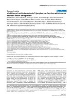
ESPVR (Figure 2A) in group I were significantly lower after
CPB (pre CPB 2.32 ± 0.63 mmHg/ml; post CPB: 1.57 ±
0.42 mmHg/ml, p < 0.001). In the LIM group no signifi-
cant decrease of ESPVR was found (pre CPB: 2.19 ± 0.49
mmHg/ml; post CPB: 1.93 ± 0.4 mmHg/ml, p = 0.06).
Similar data were found for EDPVR values (Figure 2B)
with stabilized EDPVR values in the LIM group. EDPVR
values in group I were found to be significantly decreased
post CPB (pre CPB: 6.19 ± 1.53 mmHg/ml; post CPB: 4.15
± 0.78 mmHg/ml, p < 0.001). For group II the slight
decrease (pre CPB: 6.75 ± 1.5 mmHg/ml; post CPB: 5.92
± 1.04 mmHg/ml) was not significant (p = 0.38). Inter-
group differences for both ESPVR and EDPVR were signif-
icant (p < 0.01).
In order to evaluate a possible beneficial effect of LIM on
systemic and pulmonary hemodynamics, the systemic
vascular resistance index (SVRi) and the pulmonary vascu-
lar resistance index (PVRi) were measured (Figure 3). Fig-
ure 3A depicts the values of the SVRi (n = 6). Post CPB,
Table 1: Perioperative inflammatory and tissue damage markers
Pre-CPB Post-CPB
Control LIM Control LIM
Neutrophils (×10
3
/µl) 5.9 ± 0.8 6.4 ± 0.3 13.4 ± 2.3 7.2 ± 1.8
PBL (×10
3
/µl) 7.5 ± 2.1 7.9 ± 1.1 8.8 ± 0.4 8.2 ± 0.9
TNF-α (pg/ml) 68.4 ± 38.9 89.0 ± 25.3 255.3 ± 64.1 112.4 ± 55.7
CK (U/l) 418.1 ± 39.3 397.6 ± 22.0 727.9 ± 75.7 645.8 ± 89.4
CK-MB (U/l) 339.8 ± 44.7 384.9 ± 77.3 592.6 ± 79.3 517.5 ± 69.6
CK: creatine kinase; CPB: cardiopulmonary bypass; PBL: peripheral blood lymphocytes; TNF: tumor necrosis factor
Journal of Inflammation 2007, 4:21 />Page 5 of 9
(page number not for citation purposes)
SVRi values were slightly lower (pre CPB: 1210 ± 128
dyn.sec/cm
5
/m
2
; post CPB: 795 ± 114 dyn.sec/cm
5
/m
2
)
compared with pre-operative values in both groups. How-
ever, there was no significant intergroup difference. In
contrast, the PVRi values increased up to 3-fold post oper-
atively in group I (pre CPB: 190 ± 72 dyn.sec/cm
5
/m
2
;
post CPB: 375 ± 134 dyn.sec/cm
5
/m
2
) but not in the LIM
group. Post operative PVRi values in the LIM group
remained at baseline level (Figure 3B). The post CPB inter-
group difference was statistically significant (p < 0.01).
Cardiac and pulmonary tissue infiltration
To study the possibility whether LIM may exert its benefi-
cial effects on hemodynamics and cardiac function by
reducing neutrophil tissue infiltration, tissue sections of
heart and lung were stained with neutrophil specific chlo-
racetate-esterase (Figure 4). Semi quantitative evaluation
of tissue sections from CPB-treated pigs revealed neu-
trophil tissue infiltration in heart and lung when com-
pared with sections from untreated control pigs. In tissue
sections from LIM-treated pigs reduced numbers of neu-
trophils in heart and lung were found compared with the
CPB group. High numbers of neutrophils were detected in
the spleen of LIM-treated pigs but not in control pigs.
Electron microscopy qualitatively confirmed that CPB-
mediated neutrophil activation may lead to an accumula-
tion of PMN in the epicardium and to direct interactions
between neutrophils and heart muscle cells within the left
ventricular myocardium (Figure 5). In tissue samples
from LIM-treated animals neutrophils could not be
detected within the myocardium.
Scintigraphy
In order to determine the global distribution of neu-
trophils within the body after passing the LIM, techne-
tium-labeled neutrophils were injected into the blood
circulation before onset of CPB or CPB with LIM (n = 2,
each group). One hour after end of surgery the distribu-
tion of the labeled neutrophils was analyzed by scintigra-
phy (Figure 6). In Figure 6A an example for the total body
distribution of radioactivity is provided. In contrast to the
control animal the depicted scintigraphy of the LIM-
treated animal revealed no or only little radioactive load
Boxplot depiction of pre-load independent (A) end systolic pressure volume relationship (ESPVR) and (B) end diastolic pressure volume relationship (EDPVR) obtained for the con-trol group and for the LIM group, pre- and postoperativelyFigure 2
Boxplot depiction of pre-load independent (A) end systolic
pressure volume relationship (ESPVR) and (B) end diastolic
pressure volume relationship (EDPVR) obtained for the con-
trol group and for the LIM group, pre- and postoperatively.
In the control group but not in the LIM group, the differences
between pre- and post CPB values for ESPVR and EDPVR
were statistically significant (p < 0.001). The post-CPB inter-
group differences were also statistically significant (p < 0.01).
,75
1
1,25
1,5
1,75
2
2,25
2,5
2,75
3
3,25
ESPVR (mmHg/ml)
P<0.01
3,5
4
4,5
5
5,5
6
6,5
7
8,5
9
9,5
EDPVR (mmHg/ml)
P<0.01
A
B
LIM pre CPB
LIM post CPB
Control pre CPB
Control post CPB
Boxplot depiction of Cardiac index values obtained for the control group and for the LIM group, pre- and postopera-tivelyFigure 1
Boxplot depiction of Cardiac index values obtained for the
control group and for the LIM group, pre- and postopera-
tively. In the control group but not in the LIM group, the dif-
ference between pre- and post CPB values was statistically
significant (p < 0.01). The post-CPB intergroup difference
was also statistically significant (p < 0.05).
Control post CPB
Control pre CPB
LIM post CPB
LIM pre CPB
1
2
3
4
5
6
7
CI (l/min/m
2
)
P<0.05
Journal of Inflammation 2007, 4:21 />Page 6 of 9
(page number not for citation purposes)
in heart and lung, whereas the spleen was significantly
loaded. As an internal control, attenuated E.coli were
injected subcutaneously at six different intraoperative
time points (onset of CPB and subsequently each 15 min-
utes) to provoke neutrophil migration to the injection site
(Figure 6B). Black spots indicate that labeled neutrophils
retained their ability to infiltrate the challenged tissues
throughout the entire operation time. Radioactivity deter-
mined in biopsies from heart and muscle (reference tis-
sue) revealed that LIM prevented CPB-mediated
accumulation of labeled neutrophils in the heart (2.69 ×
10
6
± 1.19 and 4.30 ± 1.87 × 10
6
/g, respectively). Data is
shown in percent of the applied radioactivity (Figure 6C)
as the mean ± SD (CPB: n = 7; CPB + LIM: n = 8).
Discussion
Recently, it has been reported that CPB impairs left ven-
tricular contractility and cardiac function [25,26]. Herein,
we showed that LIM when incorporated into the arterial
line of the CPB system effectively stabilized perioperative
cardiac function during CPB in the porcine model.
The pathophysiologic mechanisms underlying CPB-
related impairment of cardiac function are not exactly
known. However, it has been suggested that neutrophil
activation that occurs during cardiac surgery using CPB
may be strongly related with cardiac and pulmonary tissue
damage after opening of the aortic cross clamp [7]. Fol-
lowing reperfusion of the ischemic heart and lung, hyper-
activated neutrophils reach the capillaries of the pre-
damaged tissues where further endothelial leakage and
extracellular matrix destruction may occur due to neu-
trophil adhesion and transendothelial migration [27,28].
The local accumulation of chemokines and proinflamma-
tory cytokines such as TNF-α further attracts and activates
neutrophils that potentially degrade tissue integrity via
oxygen radicals and proteases. Recently, we were able to
show that neutrophil-mediated disruption of microvascu-
lar endothelial cell integrity correlates with prolonged
CPB time [23]. For example, TNF-α seems to catalyze neu-
trophil-mediated tissue damage [29] and has been sus-
pected to directly disturb pulmonary [30] and cardiac
function [31].
From this knowledge it is conceivable, that perioperative
prevention of neutrophil hyperactivity and inflammation
may be an important tool to stabilize pulmonary and car-
diac functionality that would result in better patient out-
come. Therapeutic approaches with immunomodulating
drugs or with leukocyte filtration have not been suffi-
ciently effective to limit perioperative neutrophil activity
in the past [16,17]. In some studies, leukocyte filtration
rather activated proinflammatory responses probably due
to the failure to rapidly inactivate stimulated neutrophils
[32]. It has recently been shown that LIM immediately
inhibits neutrophil function in an experimental porcine
CPB model [18]. We therefore speculated that LIM might
have also beneficial effects on the cardiac outcome follow-
ing CPB.
A feasibility study with cardiac surgery patients already
showed the proof of concept for LIM [24]. In this recent
study LIM significantly prevented the perioperative
increase in leukocyte numbers, neutrophil elastase, and
TNF-α. These elements are known to contribute to the
development of SIRS [33] and epithelial barrier dysfunc-
tion [34]. Moreover, CK and CK-MB values as indicators
for tissue damage and myocardial injury, respectively were
reduced with LIM compared with CPB without LIM [18].
However, the mechanisms by which LIM may protect
heart and lung were unresolved.
From the herein presented data, we conclude that neu-
trophils may affect pulmonary and cardiac function dur-
Boxplot depiction of hemodynamic parameters (A) systemic vascular resistance index (SVRi) and (B) pulmonary vascular resistance index (PVRi) obtained for the control group and for the LIM group, pre- and postoperativelyFigure 3
Boxplot depiction of hemodynamic parameters (A) systemic
vascular resistance index (SVRi) and (B) pulmonary vascular
resistance index (PVRi) obtained for the control group and
for the LIM group, pre- and postoperatively. Post CPB inter-
group differences for PVRi but not for SVRi were statistically
significant (p < 0.01).
Control post CPB
Control pre CPB
LIM post CPB
LIM pre CPB
0
200
400
600
800
1000
1200
1400
1600
1800
SVR
i
(dyn.sec/cm
5
/m
2
)
P>0.05
0
200
400
600
800
1000
1200
1400
1600
1800
SVR
i
(dyn.sec/cm
5
/m
2
)
P>0.05
A
B
Journal of Inflammation 2007, 4:21 />Page 7 of 9
(page number not for citation purposes)
Chloroacetate esterase staining of heart and lung paraffin sectionsFigure 4
Chloroacetate esterase staining of heart and lung paraffin sections. Representative tissue samples for untreated healthy animals,
animals undergoing CPB, and animals undergoing CPB with LIM. Magnification is 200-fold.
Control CPB CPB+LIM
Lung
Heart
Spleen
Electron microscopic microphotographs of accumulated neutrophils within the epicardium (A) and within the left ventricular heart muscle (B) after CPBFigure 5
Electron microscopic microphotographs of accumulated neutrophils within the epicardium (A) and within the left ventricular
heart muscle (B) after CPB.
AB
Journal of Inflammation 2007, 4:21 />Page 8 of 9
(page number not for citation purposes)
ing CPB and thus entail impairment of left ventricular
contractility and increased pulmonary vascular resistance,
both important features of cardiac function. The ESPVR
and EDPVR values as markers for pre-load independent
contractility and elastance of the left ventricle were signif-
icantly stabilized by LIM. Left ventricular outflow tract
accelerated (LVOTacc) velocity, an additional pre-load
independent contractility parameter measured by
echocardiography [35], confirmed the beneficial effects of
LIM (data not shown). The numbers of neutrophils that
infiltrated the cardiac tissue upon CPB were relatively low.
However, the numbers of infiltrated neutrophils were
even lower in the LIM group. In contrast, the lung was
drastically infiltrated by neutrophils after CPB but to a
lesser extent in the LIM group. Although the possibility
that the low number of neutrophils within the heart mus-
cle may directly disturb the contractility of the left ventri-
cle is unlikely, it has been shown that high levels of
cardiac troponine I, MPO, and neutrophil numbers
within the cardiac sinus are related to ischemia/reper-
fusion damage [36]. Moreover, it is rather likely that the
neutrophil infiltration of the pulmonary tissue during
CPB significantly increases the pulmonary vascular resist-
ance (no-reflow phenomenon) [8-15] that in turn may
affect the preload of the left ventricle.
Our preliminary findings obtained by scintigraphy sup-
port our assumption that LIM rapidly prevents hyperacti-
vation of neutrophils and that preapoptotic neutrophils
are effectively recognized by the immune system [20] and
subsequently sequestered by the spleen.
Conclusion
In our porcine model LIM proved to be an effective tool to
limit neutrophil hyperactivation and prevent CPB-associ-
ated impairment of cardiac function. However, the link
between organ neutrophil sequestration and cardiac func-
tion needs to be interpreted in caution, as both the mor-
phological and scintigraphic data were obtained from a
very limited number of animals.
An ongoing clinical study with LIM in patients undergo-
ing cardiopulmonary bypass should confirm clinical effi-
cacy and safety.
Competing interests
SM partly works as a freelancer at Leukocare AG.
MS is CSO at Leukocare AG
The other authors declare that they have no competing
interest.
Authors' contributions
UA-R, JB-M and TA were responsible for the surgical pro-
cedures. IF and TL were responsible for the histological
analyses and electron microscopy. SM, TK, and FG were
responsible for the concept and logistics, as well as for the
neutrophil labeling and measurement of radioactivity.
JW, AM, and MS conceived of the study and were involved
in drafting the manuscript. All authors read and approved
the final manuscript.
Acknowledgements
We appreciate the excellent technical assistance of Mrs. Julia Quathamer
and of Mrs. Kabickova for electron microscopy analyses. For statistical anal-
yses we are grateful to Dr. Sonia Area de Leao Sitals.
Parts of this work were supported by the Deutsche Forschungsgemein-
schaft (DFG).
References
1. Kirklin JK, Westaby S, Blackstone EH, Kirklin JW, Chenoweth DE,
Pacifico AD: Complement and the damaging effects of cardi-
opulmonary bypass. J Thorac Cardiovasc Surg 1983, 86:845-857.
Whole body scintigraphy pictures from an animal without LIM or with LIM following injection of HMPAO-labeled neu-trophils (A)Figure 6
Whole body scintigraphy pictures from an animal without
LIM or with LIM following injection of HMPAO-labeled neu-
trophils (A). High radioactivity was found in the spleen of
LIM-treated animals. An internal control with subcutanously
injected E.coli (control pig with CPB) confirmed the neu-
trophil activity over time (B). Data for the accumulation of
radioactivity in the myocardium and musle tissue of control
and LIM-treated animals is shown (C) as mean ± SD (CPB: n
= 7; CPB + LIM: n = 8).
0,00
20,00
40,00
60,00
80,00
100,00
120,00
140,00
160,00
180,00
200,00
Heart Muscle
Radioactivity (%)
Control
CPB+LIM
CPB
Without LIM
With LIM
75 min
60 min
15 min
onset
45 min
30 min
A
B
C
C
Publish with BioMed Central and every
scientist can read your work free of charge
"BioMed Central will be the most significant development for
disseminating the results of biomedical research in our lifetime."
Sir Paul Nurse, Cancer Research UK
Your research papers will be:
available free of charge to the entire biomedical community
peer reviewed and published immediately upon acceptance
cited in PubMed and archived on PubMed Central
yours — you keep the copyright
Submit your manuscript here:
/>BioMedcentral
Journal of Inflammation 2007, 4:21 />Page 9 of 9
(page number not for citation purposes)
2. Butler J, Rocker GM, Westaby S: Inflammatory response to car-
diopulmonary bypass. Ann Thorac Surg 1993, 55(2):552-559.
3. Carden D, Xiao F, Moak C, Willis BH, Robinson-Jackson S, Alexander
S: Neutrophil elastase promotes lung microvascular injury
and proteolysis of endothelial cadherins. Am J Physiol 1998,
275:H385-H392.
4. Welbourn CR, Goldman G, Paterson IS, Valeri CR, Shepro D, Hech-
tman HB: Neutrophil elastase and oxygen radicals: synergism
in lung injury after hindlimb ischemia. Am J Physiol 1991,
260:H1852-H1856.
5. El Azab SR, Rosseel PM, de Lange JJ, Groeneveld AB, van Strik R, van
Wijk EM, Scheffer GJ: Dexamethasone decreases the pro- to
anti-inflammatory cytokine ratio during cardiac surgery. Br
J Anaesth 2002, 88:496-501.
6. Gohra H, Mikamo A, Okada H, Hamano K, Zempo N, Esato K: Gran-
ulocyte elastase release and pulmonary hemodynamics in
patients with mitral valvular disease. World J Surg 2002,
26:643-647.
7. Rinder C: Cellular inflammatory response and clinical out-
come in cardiac surgery. Curr Opin Anaesthesiol 2006, 19:65-68.
8. Braide M, Amundson B, Chien S, Bragge U: Quantitative studies
on the influence of leukocytes on the vascular resistance in a
skeletal muscle preparation. Microvasc Res 1984, 27(3):331-352.
9. Engler R: Consequences of activation and adenosine-medi-
ated inhibition of granulocytes during myocardial ischemia.
Fed Proc 1987, 46:2407-2412.
10. Engler R: Granulocytes and oxidative injury in myocardial
ischemia and reperfusion. Fed Proc 1987, 46:2395-2396.
11. Engler R, Covell JW: Granulocytes cause reperfusion ventricu-
lar dysfunction after 15 min ischaemia in the dog. Circ Res
1987, 61:20-28.
12. Engler R, Schmid-Schonbein GW, Pavelec R: Role of leukocyte cap-
illary plugging in preventing myocardial reperfusion. Circula-
tion 1981, 64:138.
13. Engler RL, Dahlgren MD, Morris DD, Peterson MA, Schmid-Schon-
bein GW: Role of leukocytes in response to acute myocardial
ischemia and reflow in dogs. Am J Physiol 1986, 251:H314-H322.
14. Engler RL, Schmid-Schonbein GW, Pavelec RS: Leukocyte capillary
plugging in myocardial ischaemia and reperfusion in the dog.
Am J Pathol 1983, 111:98-111.
15. Jordan JE, Zhao Z-Q, Vinten-Johansen J: The role of neutrophils in
myocardial ischemia-reperfusion injury. Cardiovasc Res 1999,
43:860-878.
16. de Vries AJ, Gu YJ, Post WJ, Vos P, Stokroos I, Lip H, van Oeveren
W: Leucocyte depletion during cardiac surgery: a compari-
son of different filtration strategies. Perfusion 2003, 18:31-38.
17. Scholz M, Simon A, Matheis G, Dzemali O, Henrich D, Kleine P, Wim-
mer-Reinecker G, Moritz A: Leukocyte filtration fails to limit
functional neutrophil activity during cardiac surgery. Inflamm
Res 2002, 51(7):363-368.
18. Scholz M, Simon A, Berg M, Schuller AM, Hacibayramoglu M, Margraf
S, Theisen A, Windolf J, Wimmer-Greinecker G, Moritz A: In vivo
inhibition of neutrophil activity by a FAS (CD95) stimulating
module: arterial in-line application in a porcine cardiac sur-
gery model. J Thorac Cardiovasc Surg 2004, 127:1735-1742.
19. Rowe PM: Glimmers of clinical relevance for Fas. Lancet 1996,
347:1398.
20. Cox G, Crossley J, Xing Z: Macrophage engulfment of apoptotic
neutrophils contributes to the resolution of acute pulmo-
nary inflammation in vivo. Am J Respir Cell Mol Biol 1995,
12:232-237.
21. Cinatl J Jr, Blaheta R, Bittoova M, Scholz M, Margraf S, Vogel JU, Cinatl
J, Doerr HW: Decreased neutrophil adhesion to human
cytomegalovirus-infected retinal pigment epithelial cells is
mediated by virus-induced up-regulation of Fas ligand inde-
pendent of neutrophil apoptosis. J Immunol 2000,
165:4405-4413.
22. Roca M, Martin-Comin J, Becker W, Bernardo-Filho M, Gutfilen B,
Moisan A, Peters M, Prats E, Rodrigues M, Sampson C, Signore A, Sin-
zinger H, Thakur M: A consensus protocol for white blood cells
labelling with technetium-99 m hexamethylpropylene amine
oxime. Eur J Nucl Med 1998, 25:797-799.
23. Schuller AM, Windolf J, Blaheta R, Cinatl J, Kreuter J, Wimmer-Grei-
necker G, Moritz A, Scholz M: Degradation of microvascular
brain endothelial cell beta-catenin after co-culture with acti-
vated neutrophils from patients undergoing cardiac surgery
with prolonged cardiopulmonary bypass. Biochem Biophys Res
Commun 2005, 329:616-623.
24. Scholz M, Cinatl J, Barros RT, Lisboa AC, Genevcius CF, Margraf S,
Francischetti I, Oremek G, Windolf J, Simon A, Moritz A, Bitu-
Moreno J: First efficacy and safety results with the antibody
containing leukocyte inhibition module in cardiac surgery
patients with neutrophil hyperactivity. ASAIO J 2005,
51:144-147.
25. Aybek T, Kahn MF, Dogan S, Abdel-Rahman U, Mierdl S, Kessler P,
Wimmer-Greinecker G, Moritz A: Cardiopulmonary bypass
impairs left ventricular function determined by conductance
catheter measurement. Thorac Cardiovasc Surg 2003, 51:301-305.
26. Mavi M, Celkan MA, Ilcol B, Turk T, Yavuz S, Ozdemir A: Hemody-
namic and transesophageal echocardiographic analysis of
global and regional myocardial functions, before and imme-
diately after coronary artery bypass surgery. J Card Surg 2005,
20:147-152.
27. Del Maschio A, Zanetti A, Corada M, Rival Y, Ruco L, Lampugnani
MG, Dejana E: Polymorphonuclear leukocyte adhesion trig-
gers the disorganization of endothelial cell-to-cell adherens
junctions. J Cell Biol 1996, 135:497-510.
28. Scholz M, Wimmer-Greinecker G, Simon A, Dzemali O, Chang H-Y,
Kleine P, Matheis G, Moritz A: Perioperative elastase activity in
cardiac surgery and its role in endothelial leakage. Inflamm
Res 2003, 52:433-438.
29. Scholz M, Nowak P, Schuller A, Margraf S, Blaheta R, Cinatl J, Windolf
J, Moritz A: Cardiac surgery with extracorporeal circulation:
neutrophil ransendothelial migration is mediated by beta1
integrin (CD29) in the presence of TNF-alpha. J Invest Surg
2004, 17:239-247.
30. Chiang CH: Effects of anti-tumor necrosis factor-alpha and
anti-intercellular adhesion molecule-1 antibodies on
ischemia/reperfusion lung injury. Chin J Physiol 2006, 49:266-274.
31. Sun M, Chen M, Dawood F, Zurawska U, Li JY, Parker T, Kassiri Z,
Kirshenbaum LA, Arnold M, Khokha R, Liu PP: Tumor necrosis fac-
tor-alpha mediates cardiac remodeling and ventricular dys-
function after pressure overload state. Circulation 2007,
115:1398-1407.
32. Matheis G, Moritz A, Scholz M: Leukocyte depletion in cardiology and car-
diac surgery Basel: Karger; 2001.
33. Graninger W, Wenisch C: Pentoxifylline in severe inflamma-
tory response syndrome. J Cardiovasc Pharmacol 1995, 25(Suppl
2):S134-S138.
34. Scholz M, Cinatl J, Schädel-Höpfner M, Windolf J: Neutrophils and
the blood-brain barrier dysfunction after trauma. Med Res Rev
2007, 27(3):401-416.
35. Bauer F, Jones M, Shiota T, Firstenberg MS, Qin JX, Tsujino H, Kim YJ,
Sitges M, Cardon LA, Zetts AD, Thomas JD: Left ventricular out-
flow tract mean systolic acceleration as a surrogate for the
slope of the left ventricular end-systolic pressure-volume
relationship. J Am Coll Cardiol 2002, 40:1320-1327.
36. Luo WJ, Qian JF, Jiang HH: Pretreatment with aminophylline
reduces release of Troponin I and neutrophil activation in
the myocardium of patients undergoing cardioplegic arrest.
Eur J Cardiothorac Surg 2007, 31:360-365.