Báo cáo y học: "Serum tumor necrosis factor-alpha concentrations are negatively correlated with serum 25(OH)D concentrations in healthy women" ppt
Bạn đang xem bản rút gọn của tài liệu. Xem và tải ngay bản đầy đủ của tài liệu tại đây (377.01 KB, 9 trang )
BioMed Central
Page 1 of 9
(page number not for citation purposes)
Journal of Inflammation
Open Access
Research
Serum tumor necrosis factor-alpha concentrations are negatively
correlated with serum 25(OH)D concentrations in healthy women
Catherine A Peterson*
†
and Mary E Heffernan
†
Address: Department of Nutritional Sciences, University of Missouri-Columbia, Columbia, MO, 65211, USA
Email: Catherine A Peterson* - ; Mary E Heffernan -
* Corresponding author †Equal contributors
Abstract
Background: Circulating 25 hydroxyvitamin D (25 (OH)D), an accurate measure of vitamin D
status, is markedly greater in individuals with increased exposure to ultraviolet B (UVB) light via
sunlight or the use of artificial UV light. Aside from the known relationship between vitamin D and
bone, vitamin D has also been implicated in immune function and inflammation. Furthermore, a
mass of evidence is accumulating that vitamin D deficiency could lead to immune malfunction. Our
overall objective was to study the relationship between vitamin D status (as determined by serum
25(OH) D concentrations) and inflammatory markers in healthy women.
Methods: This observational study included 69 healthy women, age 25–82 years. Women with
high UVB exposure and women with minimal UVB exposure were specifically recruited to obtain
a wide-range of serum 25(OH)D concentrations. Health, sun exposure and habitual dietary intake
information were obtained from all subjects. Body composition was determined by dual-energy-x-
ray absorptiometry. A fasting blood sample was collected in the morning and analyzed for serum
25(OH)D, parathyroid hormone (iPTH), estradiol (E
2
), cortisol, and inflammatory markers [tumor
necrosis factor -alpha (TNF-α), interleukin-6 and -10 (IL-6, IL-10), and C-reactive protein (CRP)].
Results: Women with regular UVB exposure (Hi-D) had serum 25(OH)D concentrations that
were significantly higher (p < 0.0001) and iPTH concentrations that were significantly lower (p <
0.0001) than women without regular UVB exposure (Lo-D). Although IL-6, IL-10, and CRP did not
have a statistically significant relationship with 25(OH)D concentrations, linear regression models
revealed a significant inverse relationship between serum 25(OH)D and TNF-α concentrations.
This relationship remained significant after controlling for potential covariates such as body fat
mass, menopausal status, age, or hormonal contraceptive use.
Conclusion: Serum 25(OH)D status is inversely related to TNF-α concentrations in healthy
women, which may in part explain this vitamin's role in the prevention and treatment of
inflammatory diseases. Results gleaned from this investigation also support the need to re-examine
the biological basis for determining optimal vitamin D status.
Published: 24 July 2008
Journal of Inflammation 2008, 5:10 doi:10.1186/1476-9255-5-10
Received: 1 November 2007
Accepted: 24 July 2008
This article is available from: />© 2008 Peterson and Heffernan; licensee BioMed Central Ltd.
This is an Open Access article distributed under the terms of the Creative Commons Attribution License ( />),
which permits unrestricted use, distribution, and reproduction in any medium, provided the original work is properly cited.
Journal of Inflammation 2008, 5:10 />Page 2 of 9
(page number not for citation purposes)
Background
Circulating 25 hydroxyvitamin D (25(OH)D), an accurate
measure of vitamin D status, is markedly increased in
individuals who receive regular exposure to ultraviolet B
(UVB) light via sunlight or the use of artificial UV light
(such as tanning beds) [1-6]. Serum 25(OH)D is hydrox-
ylated in the kidney, as well as in numerous other tissues,
to its active form, 1,25-dihydroxyvitamin D
(1,25(OH)
2
D). 1,25(OH)
2
D binds to nuclear vitamin D
receptors in tissues throughout the body. Active vitamin D
is responsible for maintaining calcium homeostasis pri-
marily by increasing the efficiency of intestinal calcium
absorption and by stimulating the differentiation of bone-
resorbing osteoclasts. Furthermore, vitamin D deficiency
increases secretion of parathyroid hormone, which accel-
erates bone breakdown and can lead to decreased bone
formation and density [7,8].
There is a growing body of data supporting the contention
that desirable serum 25(OH)D concentrations in healthy
individuals need to be set higher than the current values
to attain the optimal health benefits of vitamin D [8-11],
especially the benefits beyond calcium homeostasis [12-
14]. For no system does this ring truer than for the influ-
ence of vitamin D status on the immune system.
In the last few years, there has been an effort to under-
stand the possible noncalcemic (i.e. non-calcium regula-
tory) roles of vitamin D, including its role in the immune
system [15,16]. Most of the known biological effects of
1,25(OH)
2
D are mediated through the vitamin D receptor
(VDR); and, within the immune system, the VDR is found
in significant concentrations in the T lymphocyte and
macrophage populations [16]. Moreover, the enzyme
responsible for the final and rate-limiting hydroxylation
step in the synthesis of active vitamin D, 25(OH)D-1-a-
hydroxylase, is expressed by activated macrophages,
allowing these phagocytic cells to synthesize and secrete
1,25(OH)
2
D in a regulated fashion [17]. Additionally, the
major 1,25(OH)
2
D degrading enzyme, 24-hydroxylase, is
also expressed in monocytes/macrophages [18]. All of
these findings, then, suggest a paracrine role for vitamin D
in the immune system [19].
Evidence is accumulating that vitamin D deficiency may
lead to immune dysregulation. The relationship between
low serum 25(OH)D concentrations and autoimmune
disease (especially multiple sclerosis, Type I diabetes and
rheumatoid arthritis) has been appreciated for some time
[5,20,21]. More recently, studies have shown defective
macrophage function, such as impaired chemotaxis,
phagocytosis, and increased production of proinflamma-
tory cytokines, in vitamin D-insufficiency [18]. Vitamin D
has also been shown to downregulate the expression of
monocyte toll-like receptors (TLRs), known inducers of
inflammation that can prompt autoimmune disease exac-
erbation or sepsis [22]. In 2006, a double-blind, rand-
omized, placebo-controlled trial showed that vitamin D
supplementation improved cytokine profiles in patients
with congestive heart failure [12].
Several provocative reports have been published that also
support a role for vitamin D in reducing the risk of certain
infectious diseases [23,24], in part through the induction
of calthelcidin (also known as hCAP18, LL-37 and FALL-
39), an antimicrobial polypeptide [25]. For example, in
two seminal papers, Liu et al demonstrated that poor vita-
min D status may increase susceptibility to Mycobacterium
tuberculosis infection by inefficiently supporting the induc-
tion of cathelcidin mRNA in monocytes [26,27].
On balance, the published literature supports the need for
further inquiry into vitamin D status and its immune sys-
tem implications. Thus, our overall objective was to study
the relationship between vitamin D status (as determined
by serum 25(OH) D concentrations) and inflammatory
markers in healthy women. Women with high UVB expo-
sure and women with minimal UVB exposure were specif-
ically recruited to obtain a wide-range of serum 25(OH)D
concentrations [1,6,28] We hypothesized that serum
25(OH)D concentrations would be inversely correlated
with circulating concentrations of inflammatory markers.
Methods
Subject volunteers
This study used an observational, cross-sectional design to
explore the relationship between serum 25(OH)D con-
centrations and inflammatory marker concentrations in
healthy women. Ethical approval for this study was
received by the University of Missouri Health Sciences
Institutional Review Board (Project number 1069397).
Volunteers were recruited from the University of Missouri-
Columbia campus and surrounding community via email
notices and flyers posted on campus bulletin boards, and
at local tanning salons, fitness and community centers. To
be included in the study, volunteers had to be Caucasian
females who were at least 25 years of age. High vitamin D
women (Hi-D) had to have used a broad-spectrum tan-
ning bed at least once per week for a minimum of four
months. Low vitamin D women (Lo-D) had minimal
daily sunlight exposure, as assessed by a screening ques-
tionnaire, and did not use tanning beds. Volunteers were
excluded from the study if they: took a vitamin D supple-
ment other than a regular multivitamin; had a current or
previous medical condition or took a medication affecting
vitamin D status; had a current or previous medical con-
dition or took a medication affecting immune function;
had implanted metal that would interfere with the dual
energy x-ray absorptiometry (DXA) scan; were undergoing
Journal of Inflammation 2008, 5:10 />Page 3 of 9
(page number not for citation purposes)
ultraviolet radiation as medical therapy; exclusively used
high-pressure (UVA-only) tanning beds; exercised more
than 7 hours per week; were pregnant; or smoked ciga-
rettes.
Following an initial screening for inclusion and exclusion
criteria and after obtaining informed written consent,
qualified volunteers were scheduled for testing. Subjects
were instructed to refrain from exercise and to fast (water
only) for 8 to 10 hours prior to their scheduled morning
visit. On testing day, all subjects of childbearing age took
a urine pregnancy test to confirm non-pregnant status. All
study visits were conducted between late January and
early June of 2007, the predicted seasonal nadir of solar
UVB-produced serum 25(OH)D concentrations in mid-
Missourians [29].
Questionnaires and body composition
Four questionnaires were administered to all subjects: a
one-page health history and medical questionnaire devel-
oped for this study to collect data on previous health con-
ditions or diseases, menopausal status, current or
previous medication use, and exercise habits; a one-page
sun exposure questionnaire developed for this study to
assess tanning bed use, outdoor sun exposure, and sun-
screen use; a Fitzpatrick skin typing questionnaire, a well-
established method of determining skin pigmentation
and response to UVB exposure and thus potential for the
photosynthesis of vitamin D in the skin [30]; and, the 88-
question, self-administered Harvard Semi-quantitative
Food Frequency Questionnaire, a validated tool to assess
habitual dietary intake [31].
Body mass was measured without shoes to the nearest 0.1
kg and height to the nearest 0.5 cm using a medical bal-
ance beam scale. Body fat and lean body mass were meas-
ured by dual energy x-ray absorptiometry (DXA, Hologic
Delphi A bone densitometer, Bedford, MA).
Blood collection
All blood was drawn between the hours of 7:30 am and
11:30 am. Venous blood was collected into vacutainer
tubes and allowed to clot at room temperature for 30 min-
utes. The coagulated blood was centrifuged; the serum
was aliquoted into sterile microcentrifuge tubes, and
stored at -80°C.
Measurement of serum 25(OH)D
25(OH)D serum concentrations were measured using a
125
I radioimmunoassay (RIA) kit (Diasorin, Stillwater,
MN, Intra-assay CV = 10.8%). The 25(OH)D RIA is a two-
step procedure. First, 25(OH)D and other hydroxylated
metabolites are rapidly extracted from serum using ace-
tonitrile. The extracted sample is then assayed using an
antibody with specificity to 25(OH)D.
Measurement of parathyroid hormone, estradiol and
cortisol
Serum intact-PTH (iPTH) was measured using a commer-
cially-available iPTH (1–84) enzyme-linked immuno-
sorbent assay (ELISA) (ALPCO Diagnositics, Salem, NH,
Intra-assay CV = 2.5%). Serum estradiol and cortisol were
also measured using commercially available ELISA kits
(ALPCO Diagnostics, Salem, NH, Intra-assay CV = 7.7%
and 5.8%, respectively).
Measurement of inflammatory markers
Four inflammatory markers were measured: IL-10, C-reac-
tive protein (CRP), IL-6, and TNF-α. IL-10, IL-6, and TNF-
α, were measured using commercially available high sen-
sitivity ELISA kits (R&D Systems Inc., Minneapolis, MN,
Intra-assay CV = 5.3%, 7.4%, and 7.7%, respectively). An
ELISA was also used to measure CRP (R&D Systems Inc.,
Minneapolis, MN, Intra-assay CV = 5.5%).
Statistical analysis
Unpaired two-tailed t-tests were used to determine differ-
ences in subject characteristics and measured outcomes;
for data not normally distributed or of unequal variance,
a rank-sum test was performed. Linear regression and uni-
variate multiple regression models were used to deter-
mine the relationships between serum 25(OH)D and
serum inflammatory markers. All statistics were per-
formed using SAS statistical software version 9.1 (SAS Inc,
Cary, NC). Statistical significance was accepted when P <
0.05.
Results
Vitamin D status
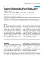
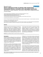
Serum 25(OH)D concentrations of all subjects are pre-
sented in Figure 1. Sixty-nine women between the ages of
25 and 82 years participated in the study. Forty-nine of the
women were classified as Lo-D and 20 women were clas-
sified as Hi-D based on UVB exposure. The mean serum
25(OH)D status (nmol/L) of the Hi-D women (129.6 ±
11.0 nmol/L) was significantly higher than that of the Lo-
D women (74.4 ± 4.0 nmol/L) (P < 0.0001).
Subject characteristics and serum hormone concentrations
Subject characteristics and serum hormone concentra-
tions by vitamin D status are presented in Table 1. There
were no significant differences in age, height, weight, BMI,
percent body fat, hormonal contraceptive use or serum
estradiol or cortisol concentrations between vitamin D
status groups. The mean iPTH concentration of the Hi-D
women was significantly lower than that of the Lo-D
women (P < 0.0001). Furthermore, there was a significant
inverse relationship between 25(OH)D and iPTH concen-
trations (R
2
= 0.2498; P = 0.0001). The skin type of the Hi-
D was significantly higher than that of the Lo-D group (P
= 0.0031). The Fitzpatrick skin typing method determines
Journal of Inflammation 2008, 5:10 />Page 4 of 9
(page number not for citation purposes)
Table 1: Subject characteristics and serum hormone concentrations.
Characteristic/Hormone Lo-D (n = 49) Hi-D (n = 20) P Value
Age (years) 39.8 ± 1.8 41.7 ± 3.5 0.5733
Height (m) 1.70 ± 0.01 1.65 ± 0.01 0.6894
Weight (kg) 65.9 ± 1.6 67.9 ± 2.7 0.3800
Body Mass Index (kg/m
2
) 23.8 ± 0.5 25.0 ± 1.1 0.2488
Body Fat (%) 30.1 ± 1.0 30.6 ± 1.7 0.7665
Skin Type 2.4 ± 0.1 3.1 ± 0.2* 0.0031
Contraceptive Use (%) 31% 20% 0.3780
Estradiol (pg/mL) 158.0 ± 15.6 151.3 ± 15.3 0.7974
Cortisol (μg/dL) 8.4 ± 0.5 9.4 ± 1.2 0.3129
iPTH (pg/mL) 48.1 ± 3.1 26.2 ± 2.6* <0.0001
Subject characteristics and serum hormone concentrations of healthy women, age 25–82 years, categorized as low vitamin D status (Lo-D) or high
vitamin D status (Hi-D) based on UVB exposure. Data are expressed as means ± SEM. *Significantly different from Lo-D, P < 0.05.
skin type based on pigmentation and ability to burn and/
or tan with sun exposure (Type I-IV, lighter-darker) [30].
Thus, it is not surprising that the Hi-D women had a
higher-level skin type because their skin is capable of tan-
ning; while women with lower-level skin types would not
be expected to use a tanning bed since their skin is less
able to tan. There were no differences between vitamin D
status groups for dietary intakes of energy, macronutrients
including omega-3 fatty acids, alcohol or caffeine (data
not shown).
Inflammatory marker outcomes
Mean serum TNF-α was significantly lower in the Hi-D
than the Lo-D women (1.22 ± 0.11 vs. 0.79 ± 0.11, P =
0.0200. IL-10, CRP and IL-6 did not significantly differ
between groups.
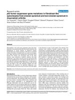
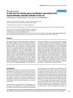
Figure 2 shows the relationships between 25 (OH)D and
IL-10, CRP, IL-6, and TNF-α. Serum 25(OH)D concentra-
tions were negatively correlated with TNF-α (R
2
= 0.0605,
P = 0.0463). Thus, serum 25(OH)D status explained
6.05% of the variation in TNF-α concentrations. IL-10,
CRP and IL-6 concentrations were not significantly associ-
ated with the concentration of 25(OH)D in serum.
When controlling for percent body fat, menopausal sta-
tus, age, serum estradiol, serum cortisol, and hormonal
contraceptive use, a significant relationship (P < 0.05)
remained between 25(OH)D and TNF-α. Controlling for
percent body fat, menopausal status, age, serum estradiol,
serum cortisol, and hormonal contraceptive use did not
change the relationship between 25(OH)D concentra-
tions and IL-6, IL-10, and CRP. Analysis of potential cov-
ariates revealed a significant positive association between
age and IL-6 (R
2
= 0.09413, P = 0.0116); and menopausal
status and IL-6 (R
2
= 0.0764, P = 0.0246).
Discussion
The objective of the present study was to determine the
relationship between 25(OH)D concentrations and
inflammatory marker concentrations in healthy women.
Although IL-6, IL-10, and CRP did not have a statistically
significant relationship with 25(OH)D concentrations,
linear regression models revealed a significant inverse
relationship between serum 25(OH)D and serum TNF-α
concentrations. This relationship remained significant
after controlling for potential covariates such as body fat
mass, menopausal status, age, or hormonal contraceptive
use. Hinton et al. found that hormonal contraceptive use
was associated with greater TNF-α concentrations in
Serum 25(OH)D concentrations of Lo-D and Hi-D status womenFigure 1
Serum 25(OH)D concentrations of Lo-D and Hi-D
status women. Mean (± SEM) serum 25(OH)D concentra-
tions of healthy women, age 25–82 years, categorized as low
vitamin D status (Lo-D; n = 49) or high vitamin D status (Hi-
D; n = 20) based on UVB exposure. Single points for each
category are means (± SEMS). *Significantly different from
Lo-D, P < 0.0001.
0.0
50.0
100.0
150.0
200.0
250.0
Serum 25(OH)D (nmol/L)
Lo-D Hi-D
*
Journal of Inflammation 2008, 5:10 />Page 5 of 9
(page number not for citation purposes)
young female athletes [32]. Our data from healthy female
non-athletes representing a much wider age range did not
reveal such a relationship with TNF-α (P = 0.2336); how-
ever, like Hinton et al., there was a significant relationship
between hormonal contraceptive and serum cortisol level
(P = 0.0030). Interestingly, in our study serum 25(OH)D
remained a significant predictor of TNF-α even after con-
trolling for contraceptive use and cortisol concentrations.
The lack of significance between serum estradiol and any
of the inflammatory markers (data not shown) supports
previous research indicating that, in premenopausal
women, menstrual phase may affect circulating cytokine
concentrations, but the impact is generally not detectable
[33].
TNF-α is produced by numerous cell types, including
macrophages, monocytes, T-cells, smooth muscle cells,
adipocytes, and fibroblasts [34] many of which also have
VDR [14,15,35]. Thus, it is difficult to discern the specific
mechanisms by which elevations in systemic 25(OH)D
attenuate circulating TNF-α concentrations. Nonetheless,
our results agree with experimental data showing that
vitamin D is capable of suppressing TNF-α production
[36-39]. Zhu et al. recently showed that in the colonic tis-
sue of mice with inflammatory bowel disease,
1,25(OH)
2
D was capable of down-regulating several
genes associated with TNFα, including proteins involved
in the transcription of TNFα, one of its primary receptors,
and TNF-α itself [39].
Human studies of diseased populations have also shown
beneficial effects of vitamin D status on TNF-α concentra-
tions. Serum concentrations of TNF-α increased in unsup-
plemented congestive heart failure patients over a period
of 9 months, whereas serum TNF-α concentrations in
patients receiving daily supplementation of vitamin D
(2000 IU) remained constant [12]. Calcitriol
(1,25(OH)
2
D
3
) supplementation for 6 months in post-
menopausal women with osteoporosis resulted in a sig-
nificant reduction in serum TNF-α concentrations and an
The relationship between serum 25(OH)D concentrations and inflammatory marker concentrationsFigure 2
The relationship between serum 25(OH)D concentrations and inflammatory marker concentrations. The rela-
tionship between serum 25(OH)D concentrations and serum IL-10, C-reactive protein (CRP), IL-6 and TNF-a concentrations
in healthy women, ages 25–82 years (n = 69). Linear regression equations for each inflammatory marker are shown. * Slope of
regression line significantly less than zero, P < 0.05.
0.00
2.00
4.00
6.00
8.00
10.00
12.00
14.00
16.00
0.0 50.0 100.0 150.0 200.0 250.0
25(OH)D (nmol/L)
Serum IL-10 pg/m
L
y = 2.45715 – 0.00080588x
R
2
= 0.0003
P = 0.8906
0.0
0.5
1.0
1.5
2.0
2.5
0.0 50.0 100.0 150.0 200.0 250.0
25(OH)D (nmol/L)
Serum CRP (mg/L)
y = 0.62864 + 0.00072368x
R
2
= 0.0028
P = 0.6770
0.00
0.50
1.00
1.50
2.00
2.50
3.00
3.50
4.00
0.0 50.0 100.0 150.0 20 0.0 250.0
25(OH)D (nmol/L)
Serum IL-6 (pg/mL)
y = 1.48246 – 0.00339x
R
2
= 0.0440
P = 0.0909
0.00
0.50
1.00
1.50
2.00
2.50
3.00
3.50
4.00
0.0 50.0 100.0 150.0 200.0 250.0
25(OH)D (nmol/L)
Serum TNF-alpha (pg/mL)
y = 1.45095 – 0.00393x
R
2
= 0.0605
P = 0.0463*
TNF-α
αα
α
IL-6
CRP
IL-10
Journal of Inflammation 2008, 5:10 />Page 6 of 9
(page number not for citation purposes)
increase in bone mineral density [40]. Additionally, six
months of calcitriol supplementation in hemodialysis
patients also caused significant decreases in serum TNF-α
[41]. Our study is the first to show a significant inverse
relationship between serum 25(OH)D and TNF-α con-
centration in a healthy population.
TNF-α concentrations are increased in several disease
states such multiple sclerosis (MS), inflammatory bowel
disease (IBD), rheumatoid arthritis (RA), heart disease,
and osteoporosis; and are often correlated with clinical
impairment [42,43]. Therefore, attenuating the concen-
trations of circulating TNF-α has the potential to posi-
tively impact the risk for or treatment of such conditions.
Our data suggest that serum 25(OH)D status explains
~6% of the variation in TNF-α concentrations in healthy
women, thus a mild relationship.
Even a slight drop in circulating TNF-α due to improved
vitamin D status may have clinical significance. MS
patients with < 2 active brain lesions visible on magnetic
resonance imagery were shown to have serum TNF-α con-
centrations that were slightly but significantly (1.6 pg/
mL) less than those with ≥ 2 active brain lesions [44].
Patients with active ulcerative colitis were found to have
41% greater mean TNF-α concentrations than those with
inactive disease (9.46 and 5.54 pg/mL, respectively);
while, those with active Crohn's disease had TNF-α con-
centrations that were only 18% greater than patients with
inactive Crohn's (14.0 and 11.5 pg/mL, respectively) [45].
Increases in circulating TNF-α concentrations have been
associated with heart disease progression. Koller-Strametz
reported that TNF-α concentrations were 3.2 ± 0.2 pg/mL
in patients with New York Heart Association (NYHA)
function class II, 4.0 ± 0.3 pg/mL in NYHA function class
III patients, and 5.3 ± 0.9 pg/mL in NYHA function class
IV patients [46].
Anti-TNF-α medications are efficacious in the manage-
ment of IBD [47]. Martinez-Borra et al. found that patients
with lower TNF-α concentrations (14 ± 25 pg/mL) prior to
treatment with the anti-TNF drug, infliximab, responded
to the treatment, whereas non-responders had signifi-
cantly higher baseline serum concentrations (201 ± 362
pg/mL) [48]. Therefore, it is possible vitamin D supple-
mentation may be a viable adjunct to anti-TNF therapy.
Human studies involving diseased populations have
shown positive relationships between 25(OH)D concen-
trations and IL-10 [12]. Despite this evidence, in the
present study, serum IL-10 was not significantly correlated
with serum 25(OH)D, suggesting that in healthy adults,
vitamin D status does not affect IL-10 secretion into sys-
temic circulation.
Similarly, serum 25(OH)D and serum CRP were not cor-
related in the present study. As a non-specific inflamma-
tory marker of general wellness, CRP increases with mild
chronic infection, aging, and tissue damage [49]. Research
in diseased populations, such as diabetes [50], arthritis
[51,52], prolonged chronic illness [53], and clinical vita-
min D deficiency (25(OH)D <27.5 nmol/L) [54] have
demonstrated negative associations between vitamin D
status and CRP concentrations. Nevertheless, intervention
studies of healthy post-menopausal women [55] and
patients with congestive heart failure [12] failed to see
changes in CRP concentrations after vitamin D supple-
mentation.
Although the result of the linear regression analysis was
not statistically significant, there appears to be a slight ten-
dency towards an inverse relationship between 25(OH)D
concentrations and serum IL-6 (P = 0.0909). Several in
vitro studies have shown that 1,25(OH)
2
D and several of
its analogs are capable of inhibiting the production of IL-
6 in various cell types [38,56-59]; while most published in
vivo studies have failed to show an effect of vitamin D sta-
tus on circulating IL-6 concentrations in humans
[12,52,60,61]. One report, however, involving hemodial-
ysis patients with elevated parathyroid hormone (PTH)
demonstrated that both oral and intravenous
1,25(OH)
2
D supplementation were capable of signifi-
cantly decreasing serum IL-6 concentrations following 6
months of treatment [62]. It has been well documented
that PTH induces the production of IL-6 by osteoblasts
[63,64], thus, it is likely that the effects of vitamin D sup-
plementation on serum IL-6 in this population were
mediated primarily through the inverse relationship
between 25(OH)D and PTH. In our study, there was no
relationship between intact PTH and IL-6 concentrations
(P = 0.8039). The significant relationship found between
age and IL-6 (P = 0.0116) in this study was anticipated
due to several reports showing that circulating IL-6 con-
centrations increase with advancing age [65-68]. Further,
IL-6 has been implicated in age-associated diseases (such
as lymphoproliferative disorders, multiple myeloma,
osteoporosis, and Alzheimer's disease) and frailty; and, it
is postulated that certain clinically important late-life
changes are due to an inappropriate presence of IL-6.
Therefore, our results indicating a trend for a negative rela-
tionship between vitamin D status and IL-6 concentra-
tions warrants further investigation. The lowering of
circulating IL-6 through the improvement of vitamin D
nutriture may have the potential to decrease disability and
mortality in older populations in addition to helping
maintain muscle strength and bone health.
The range of serum 25 (OH)D concentrations observed in
our healthy female subjects are in accordance with the
overwhelming number of reports documenting the preva-
Journal of Inflammation 2008, 5:10 />Page 7 of 9
(page number not for citation purposes)
lence of vitamin D deficiency and insufficiency in the gen-
eral population [69-75]. In recent years, mounting data
has highlighted the need to re-examine vitamin D status
and recommendations [76]. Bischoff-Ferrari et al. summa-
rized results from randomized controlled trials, prospec-
tive and cross-sectional epidemiologic studies, strong
mechanistic evidence, and dose-response relationships to
determine an optimal serum 25(OH)D concentration
[77]. They showed that for all endpoints (bone mineral
density, lower-extremity function, dental health, and risk
of falls, fractures, and colorectal cancer), optimal
25(OH)D status began at 75 nmol/L. Our study demon-
strates that like these other health outcomes, circulating
TNF-α concentrations continue to be associated with
serum 25(OH)D concentrations above this point, in a
manner consistent with decreased disease risk/progres-
sion (i.e. lower TNF-α concentrations).
The primary limitation of this study was sample size.
Women who were regularly exposed to UVB light and
qualified to participate based on our inclusion and exclu-
sion criteria were far more difficult to recruit than women
with minimal UVB exposure. Additionally, women who
tan regularly are inherently different from non-tanners.
Frequent tanning bed use is associated with high risk
behaviours, including frequent dieting, laxative use or
vomiting to control weight, cigarette smoking, binge
drinking, and recreational drug use. [78]. In light of this,
the present study was designed to control or account for
these behaviors through subject inclusion/exclusion crite-
ria and inclusion of pertinent questionnaire data in the
multiple regression analysis.
Conclusion
Serum TNF-α concentrations are negatively correlated
with vitamin D status in healthy women. This study is the
first known report to show this inverse relationship in a
non-diseased population. Results gleaned from this inves-
tigation also support the need to re-examine the biologi-
cal basis for determining optimal vitamin D status. More
studies are needed to fully characterize the relationship
between vitamin D and TNF-α relationship; but if proven
effective, vitamin D therapy may show promise as adjunct
to anti-TNF therapy in inflammatory disease states.
Abbreviations
1,25(OH)
2
D: 1,25-dihydroxyvitamin D; 25(OH)D: 25-
hydroxyvitamin D; CRP: C-reactive protein; DXA: dual-
energy x-ray absorptiometry; Hi-D: high vitamin D status;
Lo-D: low vitamin D status; IL-6: interleukin-6; IL-10:
interleukin 10; TNF-α: tumor necrosis factor-alpha.
Competing interests
The authors declare that they have no competing interests.
Authors' contributions
CAP developed the project idea and study design;
obtained IRB approval; and wrote the manuscript. MEH,
the graduate student under CAP's mentorship, coordi-
nated the project including subject recruitment, testing,
sample collection and analyses. Both authors contributed
to the final edits of the manuscript.
Acknowledgements
This work was supported by the University Of Missouri Department Of
Nutritional Sciences and the University of Missouri Research Council (grant
#C2250048). The authors would like to thank the Schade family for their
generous support of MEH through the establishment of the Maxine Sea-
baugh Schade Graduate Fellowship. The authors also wish to thank Laura
Hillman for her assistance with the 25(OH)D assay and Dr. Mark Ellersieck
for his assistance with the statistical analysis
References
1. Grant WB, Garland CF, Holick MF: Comparisons of estimated
economic burdens due to insufficient solar ultraviolet irradi-
ance and vitamin D and excess solar UV irradiance for the
United States. Photochem Photobiol 2005, 81:1276-1286.
2. Grant WB, Garland CF: The association of solar ultraviolet B
(UVB) with reducing risk of cancer: multifactorial ecologic
analysis of geographic variation in age-adjusted cancer mor-
tality rates. Anticancer Res 2006, 26:2687-2699.
3. Gronowitz E, Larko O, Gilljam M, Hollsing A, Lindblad A, Mellstrom
D, Strandvik B: Ultraviolet B radiation improves serum levels
of vitamin D in patients with cystic fibrosis. Acta Paediatr 2005,
94:547-552.
4. Koutkia P, Lu Z, Chen TC, Holick MF: Treatment of vitamin D
deficiency due to Crohn's disease with tanning bed ultravio-
let B radiation. Gastroenterology 2001, 121:1485-1488.
5. Ponsonby AL, Lucas RM, vanderMei IA: UVR, vitamin D and three
autoimmune diseases multiple sclerosis, type 1 diabetes,
rheumatoid arthritis. Photochem Photobiol 2005, 81:1267-1275.
6. Tangpricha V, Turner A, Spina C, Decastro S, Chen TC, Holick MF:
Tanning is associated with optimal vitamin D status (serum
25-hydroxyvitamin D concentration) and higher bone min-
eral density. Am J Clin Nutr 2004, 80:1645-1649.
7. Holick MF: The role of vitamin D for bone health and fracture
prevention. Curr Osteoporos Rep 2006, 4:96-102.
8. Heaney RP: The Vitamin D requirement in health and disease.
J Steroid Biochem Mol Biol 2005, 97:13-19.
9. Holick MF: High prevalence of vitamin D inadequacy and
implications for health. Mayo Clin Proc 2006, 81:353-373.
10. Holick MF: Resurrection of vitamin D deficiency and rickets. J
Clin Invest 2006, 116:2062-2072.
11. Chapuy MC, Preziosi P, Maamer M, Arnaud S, Galan P, Hercberg S,
Meunier PJ: Prevalence of vitamin D insufficiency in an adult
normal population. Osteoporos Int 1997, 7:
439-443.
12. Schleithoff SS, Zittermann A, Tenderich G, Berthold HK, Stehle P,
Koerfer R: Vitamin D supplementation improves cytokine
profiles in patients with congestive heart failure: a double-
blind, randomized, placebo-controlled trial. Am J Clin Nutr
2006, 83:754-759.
13. Holick MF: Vitamin D: its role in cancer prevention and treat-
ment. Prog Biophys Mol Biol 2006, 92:49-59.
14. Cantorna MT: Vitamin D and autoimmunity: is vitamin D sta-
tus an environmental factor affecting autoimmune disease
prevalence? Proc Soc Exp Biol Med 2000, 223:230-233.
15. Cantorna MT, Zhu Y, Froicu M, Wittke A: Vitamin D status, 1,25-
dihydroxyvitamin D3, and the immune system. Am J Clin Nutr
2004, 80:1717S-1720S.
16. Deluca HF, Cantorna MT: Vitamin D: its role and uses in immu-
nology. FASEB J 2001, 15:2579-2585.
17. Mathieu C, Adorini L: The coming of age of 1,25-dihydroxyvita-
min D(3) analogs as immunomodulatory agents. Trends Mol
Med 2002, 8:174-179.
18. Overbergh L, Decallonne B, Valckx D, Verstuyf A, Depovere J, Lau-
reys J, Rutgeerts O, Saint-Arnaud R, Bouillon R, Mathieu C: Identifi-
Journal of Inflammation 2008, 5:10 />Page 8 of 9
(page number not for citation purposes)
cation and immune regulation of 25-hydroxyvitamin D-1-
alpha-hydroxylase in murine macrophages. Clin Exp Immunol
2000, 120:139-146.
19. vanEtten E, Mathieu C: Immunoregulation by 1,25-dihydroxyvi-
tamin D3: basic concepts. J Steroid Biochem Mol Biol 2005,
97:93-101.
20. Cutolo M, Otsa K, Uprus M, Paolino S, Seriolo B: Vitamin D in
rheumatoid arthritis. Autoimmun Rev 2007, 7:59-64.
21. Young AR, Walker SL: UV radiation, vitamin D and human
health: an unfolding controversy introduction. Photochem Pho-
tobiol 2005, 81:1243-1245.
22. Sadeghi K, Wessner B, Laggner U, Ploder M, Tamandl D, Friedl J,
Zugel U, Steinmeyer A, Pollak A, Roth E, Boltz-Nitulescu G, Spittler
A: Vitamin D3 down-regulates monocyte TLR expression
and triggers hyporesponsiveness to pathogen-associated
molecular patterns. Eur J Immunol 2006, 36:361-370.
23. Aloia JF, Li-Ng M: Re: epidemic influenza and vitamin D. Epide-
miol Infect 2007, 135:1095-1096.
24. Zasloff M: Fighting infections with vitamin D. Nat Med 2006,
12:388-390.
25. Grant WB: Hypothesis ultraviolet-B irradiance and vitamin
D reduce the risk of viral infections and thus their sequelae,
including autoimmune diseases and some cancers. Photochem
Photobiol 2008, 84:356-365.
26. Liu PT, Stenger S, Li H, Wenzel L, Tan BH, Krutzik SR, Ochoa MT,
Schauber J, Wu K, Meinken C, Kamen DL, Wagner M, Bals R, Stein-
meyer A, Zugel U, Gallo RL, Eisenberg D, Hewison M, Hollis BW,
Adams JS, Bloom BR, Modlin RL: Toll-like receptor triggering of
a vitamin D-mediated human antimicrobial response. Science
2006, 311:1770-1773.
27. Liu PT, Stenger S, Tang DH, Modlin RL: Cutting edge: vitamin D-
mediated human antimicrobial activity against Mycobacte-
rium tuberculosis is dependent on the induction of cathelici-
din. J Immunol 2007, 179:2060-2063.
28. Rajakumar K, Greenspan SL, Thomas SB, Holick MF: SOLAR ultra-
violet radiation and vitamin D: a historical perspective. Am J
Public Health 2007,
97:1746-1754.
29. Webb AR, Engelsen O: Calculated ultraviolet exposure levels
for a healthy vitamin D status. Photochem Photobiol 2006,
82:1697-1703.
30. Fitzpatrick TB: The validity and practicality of sun-reactive skin
types I through VI. Arch Dermatol 1988, 124:869-871.
31. Salvini S, Hunter DJ, Sampson L, Stampfer MJ, Colditz GA, Rosner B,
Willett WC: Food-based validation of a dietary questionnaire:
the effects of week-to-week variation in food consumption.
Int J Epidemiol 1989, 18:858-867.
32. Hinton PS, Rector.R.S., Peppers JE, Imhoff RD, Hillman LS: Serum
markers of inflammation and endothelial function are ele-
vated by hormonal contraceptive use but not exercise-asso-
ciated menstrual disorders in physically active young
women. J Sports Sci Med 2006, 5:235-242.
33. Banks RE: Measurement of cytokines in clinical samples using
immunoassays: problems and pitfalls. Crit Rev Clin Lab Sci 2000,
37:131-182.
34. Popa C, Netea MG, van Riel PL, van der Meer JW, Stalenhoef AF: The
role of TNF-alpha in chronic inflammatory conditions, inter-
mediary metabolism, and cardiovascular risk. J Lipid Res 2007,
48:751-762.
35. Cantorna MT, Mahon BD: D-hormone and the immune system.
J Rheumatol Suppl 2005, 76:11-20.
36. Cantorna MT, Woodward WD, Hayes CE, Deluca HF: 1,25-dihy-
droxyvitamin D3 is a positive regulator for the two anti-
encephalitogenic cytokines TGF-beta 1 and IL-4. J Immunol
1998, 160:5314-5319.
37. Cohen ML, Douvdevani A, Chaimovitz C, Shany S: Regulation of
TNF-alpha by 1alpha,25-dihydroxyvitamin D3 in human
macrophages from CAPD patients. Kidney Int 2001, 59:69-75.
38. Evans KN, Nguyen L, Chan J, Innes BA, Bulmer JN, Kilby MD, Hewi-
son M: Effects of 25-hydroxyvitamin D3 and 1,25-dihydroxyvi-
tamin D3 on cytokine production by human decidual cells.
Biol Reprod 2006, 75:816-822.
39. Zhu Y, Mahon BD, Froicu M, Cantorna MT: Calcium and 1
alpha,25-dihydroxyvitamin D3 target the TNF-alpha path-
way to suppress experimental inflammatory bowel disease.
Eur J Immunol 2005, 35:217-224.
40. Inanir A, Ozoran K, Tutkak H, Mermerci B: The effects of calcitriol
therapy on serum interleukin-1, interleukin-6 and tumour
necrosis factor-alpha concentrations in post-menopausal
patients with osteoporosis. J Int Med Res 2004, 32:570-582.
41. Borazan A, Ustun H, Cefle A, Sekitmez N, Yilmaz A: Comparative
efficacy of oral and intravenous calcitriol treatment in
haemodialysis patients: effects on serum biochemistry and
cytokine levels. J Int Med Res 2003, 31:489-496.
42. Kieseier BC, Giovannoni G, Hartung HP: Immunological surro-
gate markers of disease activity in multiple sclerosis. Electro-
encephalogr Clin Neurophysiol Suppl 1999, 50:570-583.
43. Prince HE: Biomarkers for diagnosing and monitoring autoim-
mune diseases. Biomarkers 2005, 10 Suppl 1:S44-S49.
44. Giovannoni G, Miller DH, Losseff NA, Sailer M, Lewellyn-Smith N,
Thompson AJ, Thompson EJ: Serum inflammatory markers and
clinical/MRI markers of disease progression in multiple scle-
rosis. J Neurol 2001, 248:487-495.
45. Komatsu M, Kobayashi D, Saito K, Furuya D, Yagihashi A, Araake H,
Tsuji N, Sakamaki S, Niitsu Y, Watanabe N: Tumor necrosis fac-
tor-alpha in serum of patients with inflammatory bowel dis-
ease as measured by a highly sensitive immuno-PCR. Clin
Chem 2001, 47:1297-1301.
46. Koller-Strametz J, Pacher R, Frey B, Kos T, Woloszczuk W, Stanek B:
Circulating tumor necrosis factor-alpha levels in chronic
heart failure: relation to its soluble receptor II, interleukin-6,
and neurohumoral variables. J Heart Lung Transplant 1998,
17:356-362.
47. Kam LY, Targan SR: Cytokine-based therapies in inflammatory
bowel disease. Curr Opin Gastroenterol 1999, 15:302-307.
48. Martinez-Borra J, Lopez-Larrea C, Gonzalez S, Fuentes D, Dieguez A,
Deschamps EM, Perez-Pariente JM, Lopez-Vazquez A, de FR, Rodrigo
L: High serum tumor necrosis factor-alpha levels are associ-
ated with lack of response to infliximab in fistulizing Crohn's
disease. Am J Gastroenterol 2002, 97:2350-2356.
49. Kao PC, Shiesh SC, Wu TJ: Serum C-reactive protein as a
marker for wellness assessment. Ann Clin Lab Sci 2006,
36:163-169.
50. Targher G, Bertolini L, Padovani R, Zenari L, Scala L, Cigolini M,
Arcaro G: Serum 25-hydroxyvitamin D3 concentrations and
carotid artery intima-media thickness among type 2 diabetic
patients. Clin Endocrinol (Oxf) 2006, 65:593-597.
51. Patel S, Farragher T, Berry J, Bunn D, Silman A, Symmons D: Associ-
ation between serum vitamin D metabolite levels and dis-
ease activity in patients with early inflammatory
polyarthritis. Arthritis Rheum 2007, 56:2143-2149.
52. Oelzner P, Franke S, Muller A, Hein G, Stein G: Relationship
between soluble markers of immune activation and bone
turnover in post-menopausal women with rheumatoid
arthritis. Rheumatology (Oxford) 1999, 38:841-847.
53. Vanden Berghe G, VanRoosbroeck D, Vanhove P, Wouters PJ,
DePourcq L, Bouillon R: Bone turnover in prolonged critical ill-
ness: effect of vitamin D. J Clin Endocrinol Metab 2003,
88:4623-4632.
54. Timms PM, Mannan N, Hitman GA, Noonan K, Mills PG, Synder-
combe-Court, Aganna E, Price CP, Boucher BJ: Circulating MMP9,
vitamin D and variation in the TIMP-1 response with VDR
genotype: mechanisms for inflammatory damage in chronic
disorders? QJM 2002, 95:787-796.
55. Pittas AG, Harris SS, Stark PC, Dawson-Hughes B: The effects of
calcium and vitamin D supplementation on blood glucose
and markers of inflammation in nondiabetic adults. Diabetes
Care 2007, 30:980-986.
56. Muller K, Heilmann C, Poulsen LK, Barington T, Bendtzen K: The
role of monocytes and T cells in 1,25-dihydroxyvitamin D3
mediated inhibition of B cell function in vitro. Immunopharma-
cology 1991, 21:121-128.
57. Equils O, Naiki Y, Shapiro AM, Michelsen K, Lu D, Adams J, Jordan S:
1,25-Dihydroxyvitamin D inhibits lipopolysaccharide-
induced immune activation in human endothelial cells. Clin
Exp Immunol 2006, 143:58-64.
58. Komine M, Watabe Y, Shimaoka S, Sato F, Kake K, Nishina H, Ohtsuki
M, Nakagawa H, Tamaki K: The action of a novel vitamin D3
analogue, OCT, on immunomodulatory function of kerati-
nocytes and lymphocytes. Arch Dermatol Res 1999, 291:500-506.
59. Riachy R, Vandewalle B, Belaich S, Kerr-Conte J, Gmyr V, Zerimech
F, d'Herbomez M, Lefebvre J, Pattou F: Beneficial effect of 1,25
Publish with BioMed Central and every
scientist can read your work free of charge
"BioMed Central will be the most significant development for
disseminating the results of biomedical research in our lifetime."
Sir Paul Nurse, Cancer Research UK
Your research papers will be:
available free of charge to the entire biomedical community
peer reviewed and published immediately upon acceptance
cited in PubMed and archived on PubMed Central
yours — you keep the copyright
Submit your manuscript here:
/>BioMedcentral
Journal of Inflammation 2008, 5:10 />Page 9 of 9
(page number not for citation purposes)
dihydroxyvitamin D3 on cytokine-treated human pancreatic
islets. J Endocrinol 2001, 169:161-168.
60. Gannage-Yared MH, Azoury M, Mansour I, Baddoura R, Halaby G,
Naaman R: Effects of a short-term calcium and vitamin D
treatment on serum cytokines, bone markers, insulin and
lipid concentrations in healthy post-menopausal women. J
Endocrinol Invest 2003, 26:748-753.
61. Puts MT, Visser M, Twisk JW, Deeg DJ, Lips P: Endocrine and
inflammatory markers as predictors of frailty. Clin Endocrinol
(Oxf) 2005, 63:403-411.
62. Turk S, Akbulut M, Yildiz A, Gurbilek M, Gonen S, Tombul Z, Yeksan
M: Comparative effect of oral pulse and intravenous calcitriol
treatment in hemodialysis patients: the effect on serum IL-1
and IL-6 levels and bone mineral density. Nephron 2002,
90:188-194.
63. Greenfield EM, Shaw SM, Gornik SA, Banks MA: Adenyl cyclase
and interleukin 6 are downstream effectors of parathyroid
hormone resulting in stimulation of bone resorption. J Clin
Invest 1995, 96:1238-1244.
64. Li NH, Ouchi Y, Okamoto Y, Masuyama A, Kaneki M, Futami A, Hosoi
T, Nakamura T, Orimo H: Effect of parathyroid hormone on
release of interleukin 1 and interleukin 6 from cultured
mouse osteoblastic cells. Biochem Biophys Res Commun 1991,
179:236-242.
65. Ershler WB, Keller ET: Age-associated increased interleukin-6
gene expression, late-life diseases, and frailty. Annu Rev Med
2000, 51:245-270.
66. Cappola AR, Xue QL, Ferrucci L, Guralnik JM, Volpato S, Fried LP:
Insulin-like growth factor I and interleukin-6 contribute syn-
ergistically to disability and mortality in older women. J Clin
Endocrinol Metab 2003, 88:2019-2025.
67. Cohen HJ, Pieper CF, Harris T, Rao KM, Currie MS: The associa-
tion of plasma IL-6 levels with functional disability in com-
munity-dwelling elderly. J Gerontol A Biol Sci Med Sci 1997,
52:M201-M208.
68. Ferrucci L, Harris TB, Guralnik JM, Tracy RP, Corti MC, Cohen HJ,
Penninx B, Pahor M, Wallace R, Havlik RJ: Serum IL-6 level and
the development of disability in older persons. J Am Geriatr Soc
1999, 47:639-646.
69. Hanley DA, Davison KS:
Vitamin D insufficiency in North
America. J Nutr 2005, 135:332-337.
70. Hypponen E, Power C: Hypovitaminosis D in British adults at
age 45 y: nationwide cohort study of dietary and lifestyle pre-
dictors. Am J Clin Nutr 2007, 85:860-868.
71. Rockell JE, Skeaff CM, Williams SM, Green TJ: Serum 25-hydroxy-
vitamin D concentrations of New Zealanders aged 15 years
and older. Osteoporos Int 2006, 17:1382-1389.
72. Stein EM, Laing EM, Hall DB, Hausman DB, Kimlin MG, Johnson MA,
Modlesky CM, Wilson AR, Lewis RD: Serum 25-hydroxyvitamin
D concentrations in girls aged 4-8 y living in the southeastern
United States. Am J Clin Nutr 2006, 83:75-81.
73. Vieth R, Cole DE, Hawker GA, Trang HM, Rubin LA: Wintertime
vitamin D insufficiency is common in young Canadian
women, and their vitamin D intake does not prevent it. Eur J
Clin Nutr 2001, 55:1091-1097.
74. Visser M, Deeg DJ, Puts MT, Seidell JC, Lips P: Low serum concen-
trations of 25-hydroxyvitamin D in older persons and the risk
of nursing home admission. Am J Clin Nutr 2006, 84:616-622.
75. Zadshir A, Tareen N, Pan D, Norris K, Martins D: The prevalence
of hypovitaminosis D among US adults: data from the
NHANES III. Ethn Dis 2005, 15(4 Suppl 5):S5–97-101.
76. Vieth R, Bischoff-Ferrari H, Boucher BJ, Dawson-Hughes B, Garland
CF, Heaney RP, Holick MF, Hollis BW, Lamberg-Allardt C, McGrath
JJ, Norman AW, Scragg R, Whiting SJ, Willett WC, Zittermann A:
The urgent need to recommend an intake of vitamin D that
is effective. Am J Clin Nutr 2007, 85:649-650.
77. Bischoff-Ferrari HA, Giovannucci E, Willett WC, Dietrich T, Dawson-
Hughes B: Estimation of optimal serum concentrations of 25-
hydroxyvitamin D for multiple health outcomes. Am J Clin
Nutr 2006, 84:18-28.
78. O'Riordan DL, Field AE, Geller AC, Brooks DR, Aweh G, Colditz GA,
Frazier AL: Frequent tanning bed use, weight concerns, and
other health risk behaviors in adolescent females (United
States). Cancer Causes Control 2006,
17:679-686.