Báo cáo y học: "Prevalence, clinical relevance and characterization of circulating cytotoxic CD4+CD28– T cells in ankylosing spondylitis" ppt
Bạn đang xem bản rút gọn của tài liệu. Xem và tải ngay bản đầy đủ của tài liệu tại đây (444.97 KB, 9 trang )
R292
Introduction
The immunogenetic link between HLA-B27 and ankylosing
spondylitis (AS) is the strongest reported association of an
HLA class I molecule with a disease to date. HLA-B27 pre-
sents specific peptides to CD8
+
T lymphocytes in the pres-
ence of β
2
-microglobulin [1,2], and much work has
concentrated on characterizing B27 restricted cytotoxic
T lymphocytes in spondyloarthropathy patients [3,4]. CD8
+
cytotoxic T lymphocytes identified by the CD28
–
pheno-
type account for up to 78% of peripheral CD8
+
T cells [5].
Several more recent studies have raised the possibility
that HLA-B27 may be more than a restriction element of
CD8
+
T cells in AS, and that HLA-B27 may be recognized
even by CD4
+
T cells [6,7]. In HLA-B27 transgenic rats
CD4
+
T cells were used to transfer AS disease [8,9], and
in autoimmune MRL/lpr mice CD4
+
T cell depletion pre-
vented the development of arthritis [10]. MHC class II mol-
ecules, which usually present antigenic structures to
CD4
+
T cells, are not necessarily required for develop-
ment of AS, and MHC class II negative, B27
+
transgenic
mice may still develop spontaneous AS-like disease [11].
In humans, CD4
+
T cells are more frequently present than
CD8
+
T cells in both the peripheral blood and in biopsies
of sacroiliac joints [12,13], and T cell clones with speci-
ficity for arthritogenic bacteria exhibited the CD4
+
pheno-
type when derived from the synovial fluids of patients with
Yersinia, Salmonella, and Chlamydia-induced reactive
arthritis [14–16]. However, bacteria and/or autoantigen
specific CD8
+
and HLA-B27 restricted T cells are also
AS = ankylosing spondylitis; BASFI = Bath Ankylosing Spondylitis Functional Index; BASMI = Bath Ankylosing Spondylitis Metrology Index; CMV =
cytomegalovirus; EBV = Epstein–Barr virus; ESR = erythrocyte sedimentation rate; FACS = fluorescence activated cell sorting; FITC = fluorescein
isothiocyanate; HAQ-S = Health Assessment Questionnaire for the Spondyloarthropathies; HLA = human leucocyte antigen; IFN = interferon; IL =
interleukin; NK = natural killer; MHC = major histocompatibility complex; PBMC = peripheral blood mononuclear cell.
Arthritis Research & Therapy Vol 5 No 5 Duftner et al.
Research article
Prevalence, clinical relevance and characterization of circulating
cytotoxic CD4
+
CD28
–
T cells in ankylosing spondylitis
Christina Duftner
1
, Christian Goldberger
1
, Albrecht Falkenbach
2
, Reinhard Würzner
3
,
Barbara Falkensammer
3
, Karl P Pfeiffer
4
, Elisabeth Maerker-Hermann
5
and Michael Schirmer
1
1
Department of Internal Medicine, University of Innsbruck, Innsbruck, Austria
2
Gasteiner Heilstollen Hospital, Bad Gastein-Böckstein, Austria
3
Institute of Hygiene and Social Medicine, University of Innsbruck, Innsbruck, Austria
4
Institute of Biostatistics, University of Innsbruck, Innsbruck, Austria
5
HSK-Aukammallee, Wiesbaden, Germany
Correspondence: M Schirmer (e-mail: )
Received: 19 Mar 2003 Revisions requested: 22 Apr 2003 Revisions received: 5 Jun 2003 Accepted: 24 Jun 2003 Published: 16 Jul 2003
Arthritis Res Ther 2003, 5:R292-R300 (DOI 10.1186/ar793)
© 2003 Duftner et al., licensee BioMed Central Ltd (Print ISSN 1478-6354; Online ISSN 1478-6362). This is an Open Access article: verbatim
copying and redistribution of this article are permitted in all media for any purpose, provided this notice is preserved along with the article's original
URL.
Abstract
Circulating CD3
+
CD4
+
CD28
–
cells exhibit reduced
apoptosis and were found to be more enriched in patients
with ankylosing spondylitis than in age-matched healthy
control individuals (7.40 ± 6.6% versus 1.03 ± 1.0%;
P < 0.001). Levels of CD4
+
CD28
–
T cells correlate with
disease status as measured using a modified metrology
score, but they are independent of age and duration of
ankylosing spondylitis. CD4
+
CD28
–
T cells produce IFN-γ
and perforin, and thus they must be considered
proinflammatory and cytotoxic. These T cells share
phenotypic and functional properties of natural killer cells,
strongly expressing CD57 but lacking the lymphocyte marker
CD7. MHC class I recognizing and activating natural killer cell
receptors on the surface of CD4
+
CD28
–
T cells may be
involved in a HLA-B27 mediated co-stimulation of these
proinflammatory and cytotoxic cells.
Keywords: ankylosing spondylitis, CD28 molecule, CD4
+
T cells, cytotoxicity, HLA-B27
Open Access
Available online />R293
thought to contribute to the pathogenesis and regulation
of the spondyloarthropathies [3].
In other autoimmune diseases, including rheumatoid arthri-
tis, Wegener’s granulomatosis, and multiple sclerosis, an
unusual subset of proinflammatory, cytotoxic CD4
+
T cells
was described that is rare in healthy individuals [17–20].
These T cells are clonally expanded and lack the important
CD28 co-stimulatory molecule on their surface. This char-
acteristic phenotype provides a means by which they may
be distinguished from normal CD4
+
T cells. Instead of the
CD28-mediated co-stimulation, several alternate co-stimu-
latory pathways have been examined in these
CD4
+
CD28
–
T cells [21,22]. Because CD4
+
CD28
–
T cells share phenotypic as well as functional features with
natural killer (NK) cells and express NK receptors on their
surface [23–25], these specific T cells should receive co-
stimulatory signals by recognition of ubiquitous MHC
class I molecules such as HLA-B27 [26].
The aim of the present study was to examine the preva-
lence and clinical relevance of CD4
+
CD28
–
T cells in a
cohort of AS patients in comparison with healthy control
individuals, and to characterize NK cell features and func-
tional properties of these unusual T cells with respect to
HLA-B27 mediated mechanisms.
Materials and method
Patient characteristics
Patients with definite AS, as defined by the modified New
York criteria [27], were recruited from the Gasteiner Heil-
stollen Hospital (Bad Gastein-Böckstein, Austria), as was
recently described [5]. In brief, 95 AS patients (age
49.1 ± 11.4 years) and 65 healthy volunteers (age
51.4 ± 15.2 years) were enrolled in the study. Time from
onset of symptoms was 16.6 ± 12.2 years and time from
diagnosis of AS was 9.9 ± 9.3 years. In AS patients the
erythrocyte sedimentation rate (ESR) was elevated to
31.4 ± 20.3 mm/hour. The Health Assessment Question-
naire for the Spondyloarthropathies (HAQ-S; n = 55) [28],
Bath Ankylosing Spondylitis Metrology Index (BASMI,
n = 55) [29] and Bath Ankylosing Spondylitis Functional
Index (BASFI, n = 75) [30] scores were 0.89 ± 0.51,
4.67 ± 2.05 and 5.04 ± 2.20, respectively (normal 0–3,
0–10 and 0–10). In the control individuals, inflammatory
and neoplastic diseases were excluded by physical exami-
nation and detailed history.
Cell preparation and cell culture
After informed consent had been obtained, peripheral
venous blood was drawn and peripheral blood mono-
nuclear cells (PBMCs) were isolated by Ficoll density
gradient centrifugation. Short-term cell lines were estab-
lished from fresh PBMCs stimulated with immobilized anti-
CD3 (OKT3; Dako, Copenhagen, Denmark) for 18 hours.
Cells were then maintained in logarithmic growth with
densities between 0.5 and 2 × 10
6
cells/ml in RPMI 1640
containing 10% foetal calf serum, 2 mmol/l
L-glutamine
and 20 U/ml recombinant human IL-2 (Sigma, St. Louis,
MO, USA). Experiments were performed 7 days after initi-
ation of the culture.
Immunostaining and flow cytometry
Surface staining of PBMCs was performed using FITC-
conjugated anti-CD4, anti-CD57, anti-CD7, anti-CD94,
anti-NKB1, anti-CD158a/h (anti-KIR2DL1/anti-KIR2DS1)
and anti-CD158b/j (anti-KIR2DL2/anti-KIR2DL3/anti-
KIRD2S2), phycoerythrin-conjugated anti-CD28 and peri-
dinin chlorophyll protein-conjugated anti-CD3 or anti-CD4
monoclonal antibodies (all from Becton Dickinson, San
Diego, CA, USA). For detection of subdiploid apoptotic
cells, cells were permeabilized with 0.05% Tween 20 and
stained with 7-aminoactinomycin D (Sigma). For intracellu-
lar staining, cells were stimulated with 25 ng/ml phorbol
12-myristate 13-acetate and 1 µg/ml ionomycin in the pres-
ence of 1 µg/ml brefeldin A for 4 hours (Sigma). After cell
surface staining and permeabilization, cells were stained
with FITC-conjugated antiperforin, anti-IFN-γ and control
immunoglobulin, respectively (Becton Dickinson). After fixa-
tion with 4% paraformaldehyde, cells were analyzed on a
FACS-Calibur flow cytometer (Becton Dickinson). Gating
was performed on CD4 and CD28, as appropriate, to
analyze further the phenotypical and functional features of
the CD4
+
CD28
+
and CD4
+
CD28
–
T cells. Thus, the intra-
cellular production of cytokines and the surface expression
of NK receptors could be directly compared between the
CD4
+
CD28
+
and CD4
+
CD28
–
T cell cohorts. Data were
analyzed using WinMDI software (Joseph Trotter, Scripps
Research Institute, La Jolla, CA, USA).
Co-incubation of CD4
+
T cells with HLA-B27 transfected
cell lines
Short-term cell lines from HLA-B27 positive AS patients
(1×10
5
cells) were incubated with the HLA-B*2705
transfected cell line C1R-B27 and the nontransfected cell
line C1R (5 × 10
4
cells), and cells were either stimulated
by cross-linking the T cell receptor with anti-CD3 (OKT3)
or not. The original C1R cell line is of lymphoblastoid
origin and was selected for loss of HLA class I antigen
expression. C1R derived cells express no HLA-A or HLA-
B products, but they do express small amounts of HLA-
Cw4. For blocking experiments the unconjugated
monoclonal antibodies specifically directed against CD94
and CD158b/j (KIR2DL2/KIR2DL3/KIR2DS2; Becton
Dickinson) were each used at concentrations of 20 µg/ml.
Single antibodies were not tested. After 24 hours parallel
cultures were harvested and the expression of CD25 was
determined by three-colour fluorescence activated cell
sorting (FACS) analysis.
In order to identify a possible HLA-B27 mediated enrich-
ment of CD28
–
T cells in the CD4
+
CD25
+
T cell compart-
Arthritis Research & Therapy Vol 5 No 5 Duftner et al.
R294
ment, fresh PBMCs (1 × 10
5
) from three HLA-B27 positive
AS patients were incubated in RPMI 1640 and 10% fetal
calf serum together with the Tap 1 and 2 and MHC class II
deficient HLA-B*2705 transfected cell line T2-B27 or the
nontransfected control cell line T2 (5 × 10
4
) in the pres-
ence or absence of stimulation by cross-linking the T cell
receptor with immobilized anti-CD3 (OKT3). The T2-B27
cell line expresses a variety of different forms of HLA-B27,
including free B27 H chain monomers, homodimers and
low levels of B27 heterodimers, but no HLA class II mole-
cules [31]. After 36 hours parallel cultures were harvested
and the expression of IL-2 receptor α chain (CD25) was
determined, together with CD4 and CD28 expression, by
three-colour FACS.
Serological assays
Investigators assessing Epstein–Barr virus (EBV) and
cytomegalovirus (CMV) seropositivity were blinded to the
sera of 30 AS patients of different ages. Anti-EBV and
anti-CMV IgG antibodies were identified using enzyme-
linked immunosorbent assay kits from Aventis Behring
(Vienna, Austria), in accordance with the manufacturer’s
instructions.
Statistical analysis
The two-sided paired t-test, the Wilcoxon ranking test, the
Kruskal–Wallis test and regression analysis by receiver
operating curves were performed using the SPSS program,
version 11.0 (Chicago, IL, USA). Bonferroni adjustment was
performed in case of multiple testing of clinical measure-
ments. P≤ 0.05 was considered statistically significant.
Values are expressed as mean ±standard deviation.
Results
Prevalence of CD4
+
CD28
–
T cells in ankylosing
spondylitis patients and healthy control individuals
Percentages of CD3
+
CD4
+
CD28
–
T cells were deter-
mined in the peripheral blood from 95 consecutive AS
patients and 65 age-matched healthy control individuals
by flow cytometry analysis. In AS patients the percentage
of CD3
+
CD4
+
T cells lacking the co-stimulatory molecule
CD28 was significantly increased as compared with the
control individuals (7.40 ± 6.6% versus 1.03 ± 1.0%;
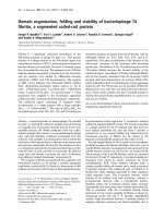
P < 0.001; Fig. 1a). Logarithmic transformation of the per-
centages of CD4
+
CD28
–
T cells was used to correct for
data skewing and to detect different populations of CD4
+
T cells. A cumulative frequency distribution showed an
underlying bimodal distribution of the frequencies of
CD4
+
CD28
–
T cells (Fig. 1b). The cutoff value determined
at the intersection of the two bimodal distribution curves
was 1.7%. Using this cutoff value, 70.3% of the AS
patients had increased levels versus 6.5% in the control
group.
As regression models for AS disease and age, receiver
operating curves were applied to display sensitivity and
specificity of CD4
+
CD28
–
levels. The area under the
curve was calculated to be 0.912 for AS disease and
0.540 for the age of those patients who had 1.7% or more
CD4
+
CD28
–
T cells in peripheral blood (Fig. 1c, d). These
findings reflect high sensitivity and specificity of
CD4
+
CD28
–
T cell levels for AS disease when compared
with healthy control individuals, but demonstrate the lack
of correlation between the levels of CD4
+
CD28
–
T cells
and age.
Clinical relevance of CD4
+
CD28
–
T cells in ankylosing
spondylitis
To detect a possible association between CD4
+
CD28
–
T cells and disease status, patients were grouped accord-
ing to their movement restrictions and functional measure-
ments. The nonparametric Kruskal–Wallis test was used
to compare patient groups with minor, mean and major
restrictions. Out of the clinical and serological measure-
ments, a correlation between CD4
+
CD28
–
T cells and
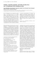
reduction in height since onset of disease (P = 0.037) and
increased ESR (P = 0.047) was detected (Fig. 2a, b;
Table 1). With respect to movement restrictions, a trend
was found only for the correlation between the percentage
of CD4
+
CD28
–
T cells and patient groups with minor,
mean and major restrictions according to the BASMI
score (P = 0.063; Fig. 2c).
Based on the results with CD8
+
CD28
–
T cells in AS
patients, we proposed a modified metrology score sum-
marizing measurements for cervical rotation in sitting posi-
tion, chin to jugulum distance, thoracic Schober test,
chest expansion and fingers to floor distance, but not
tragus to wall distance, intermalleolar distance, modified
Schober test and lumbar side flexion, as included in the
BASMI score [5]. When patients were grouped according
to their restriction as measured using this modified metrol-
ogy index, the percentage of CD4
+
CD28
–
T cells corre-
lated with the disease status (P = 0.02; Fig. 2d). No
correlation was detected between the percentage of
CD4
+
CD28
–
T cells and time since onset of symptoms,
duration of disease, HAQ-S score, BASFI score, levels of
C-reactive protein and blood cell counts.
Apoptosis and functional characterization of
CD4
+
CD28
–
T cells
Spontaneous apoptosis in CD4
+
CD28
+
and CD4
+
CD28
–
T cells from short-term cell lines of healthy control individu-
als and AS patients was identified from the fraction of sub-
diploid cells in CD4
+
cells after staining of DNA using
7-aminoactinomycin D. Three days after the last addition of
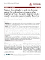
recombinant human IL-2, 16.2 ± 5.9% of the CD4
+
CD28
+
but only 0.7 ± 1.1% of the CD4
+
CD28
–
T cells were apop-
totic (n = 4; P < 0.001; Fig. 3a). Because CD4
+
CD28
+
and
CD4
+
CD28
–
T cells were maintained under identical con-
ditions, differences in the apoptotic rate cannot be attrib-
uted to tissue culture conditions. In the healthy control
individuals, 15.8 ± 0.8% of the CD4
+
CD28
+
T cells were
subdiploid cells (n = 3). There was no CD4
+
CD28
–
T cell
population in the healthy control individuals tested.
Intracellular staining for perforin and IFN-γ was performed
in activated cells to determine the number of cytokine pro-
ducing cells. Production of perforin was more frequent in
CD4
+
CD28
–
T cells than in their CD28
+
counterparts
(35.9 ± 18.7% versus 1.0 ± 0.6% perforin-producing cells;
n = 12; P < 0.001). Nonspecific staining with IgG control
antibodies was negligible (0.5 ± 0.4% IgG positive cells in
both CD4
+
CD28
+
and CD4
+
CD28
–
T cells; Fig. 3c). Intra-
cellular staining of IFN-γ showed that production of IFN-γ
was also more frequent in CD4
+
CD28
–
T cells
(37.9 ± 20.4% IFN-γ positive cells versus 0.3 ± 0.2% cells
stained with IgG control antibodies) than in their CD28
+
counterparts (9.3 ± 3.1% IFN-γ positive cells versus
0.3 ± 0.2% cells stained with IgG control antibodies;
n = 7; P = 0.006; Fig. 3b).
Expression of natural killer cell surface markers on
CD4
+
CD28
–
T cells
For phenotypic characterization of CD4
+
CD28
–
T cells,
surface expressions of CD57 and CD7 were compared
between the CD28
+
and the CD28
–
CD4
+
T cell compart-
ments. The CD57 molecule is a 110 kDa glycoprotein that
is presented by NK cells. The CD7 molecule, which is
involved in T cell activation, is present in most normal
human T cells under physiological conditions, but not on
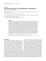
NK cells [32]. CD57 surface expression was higher on
CD4
+
CD28
–
T cells than on CD4
+
CD28
+
T cells
(63.0 ± 29.5% versus 1.0 ± 0.6%; n = 7; P = 0.001),
whereas the expression of CD7 was 72.4 ± 11.2% on
CD28
+
but only 3.4 ± 2.1% on CD28
–
CD4
+
T cells
(n = 7; P < 0.001; Fig. 4a).
Available online />R295
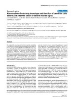
Figure 1
Levels of CD3
+
CD4
+
CD28
–
T cells in patients with ankylosing
spondylitis and healthy control individuals. (a) Accumulation of
CD3
+
CD4
+
CD28
–
cells in peripheral blood mononuclear cells of
95 patients with ankylosing spondylitis (AS; ᭹) and 65 age-matched
healthy control individuals (᭺). The Mann–Whitney test was used to
determine statistical difference. (b) Logarithmic transformation of
percentages of CD3
+
CD4
+
CD28
–
T cells was performed to detect
different populations of CD4
+
T cells and to correct for data skewing.
We found a bimodal distribution of frequencies of CD3
+
CD4
+
CD28
–
T cells (line for healthy control individuals, boxes for patients with AS).
The cutoff value, which was determined at the intersection of the two
bimodal distribution curves, was 1.7%. Using this value as cutoff,
70.3% of the patients had increased levels as compared with only
6.5% of the control group. (c, d) As a regression model, receiver
operating curves were used to display sensitivity and specificity of
CD28
–
T cell levels for AS disease and age. The area under the curve
(AUC) was determined for both independent parameters.
(c)
(b)
CD4
+
CD28
–
[%; log]
1.40.80.1–0.5
–1.1
Relative frequency (%)
10
20
30
0
controls
CD4
+
CD28
–
[%]
AS patients
(a)
(d)
1 – specificity
Ankylosing spondylitis
sensitivity
0
1.00
0.75
0.50
0.25
1.000.750.500.25
0
AUC = 0.912
Ag
e
AUC = 0.540
1.000.750.500.25
0
Figure 2
Associations between CD3
+
CD4
+
CD28
–
T cell levels and
(a) decrement in height, (b) erythrocyte sedimentation rate (ESR),
(c) Bath Ankylosing Spondylitis Metrology Index (BASMI) and (d) a
newly calculated metrology index. The Kruskal–Wallis test was used to
compare levels of CD4
+
CD28
–
T cells from patient groups with minor,
mean or major restrictions. Whiskers boxblots show 50% of cases
within the boxes and 80% between the end-points of the whiskers
(lines). P ≤0.05 was considered statistically significant.
CD4
+
CD28
–
[% ]
BASMI score
30
20
10
0
controls
Metrology scorecontrols
P
= 0.02
(c)
(d)
CD4
+
CD28
–
[% ]
<20 <40 >40controls
ESR [mm/h]
P
= 0.047
Decrement of height [cm]
>7.5<7.5<2.5
30
20
10
0
controls
P
= 0.037
(b)(a)
Surface expressions of NK receptors, including killer cell
immunoglobulin like receptors (NKB1, CD158a/h
[KIR2DL1/KIR2DS1], CD158b/j [KIR2DL2/KIR2DL3/
KIR2DS2]) and the C-type lectin receptor CD94, were
examined in 11 subsequent patients with increased levels
of CD4
+
CD28
–
T cells. Of the NK receptors, NKB1 is
considered to be inhibitory, whereas CD94, CD158a/h
and CD158b/j are considered inhibitory or activating NK
receptors [33]. All 11 patients expressed at least one of
these NK receptors on the surface of their CD4
+
CD28
–
T
cells. As shown in Fig. 4b, all NK receptors were exclu-
sively found on the CD4
+
T cells that lacked the CD28
surface molecule. Low levels of inhibitory NKB1 were
detected on CD4
+
CD28
–
T cells (2.8 ± 4.4% versus
0.2 ± 0.1%; P = 0.003), whereas CD158a/h (KIR2DL1/
KIR2DS1) was detected neither on CD4
+
CD28
–
nor on
CD4
+
CD28
+
T cells (0.7 ± 1.9% versus 0.1 ± 0.1%). The
expression of the other NK receptors CD94
(10.2 ± 10.0%) and CD158b/j (KIR2DL2/KIR2DL3/
KIR2DS2; 12.4 ± 18.6%) was increased on CD4
+
CD28
–
cells as compared with their CD28
+
counterparts
(0.6 ± 0.9% versus 0.4 ± 0.3%; n = 9; P = 0.006 and
0.003, respectively; Fig. 4b).
HLA-B27-mediated effects on CD4
+
CD28
–
T cells
To examine possible NK receptor-mediated effects of
HLA-B27 on activation of CD4
+
CD28
–
T cells, short term
cell lines were co-cultured with the HLA-B*2705 trans-
fected cell line C1R-B27 or the nontransfected cell line
C1R in the presence or absence of CD3 mediated stimu-
lation. This co-incubation with HLA-B27 transfected C1R
cells resulted in an increased expression of CD25 on
CD4
+
CD28
–
T cells in the presence of cross-linking of
T cell receptors as compared with co-incubation with
nontransfected C1R cells (P = 0.012) and cross-linking of
T cell receptors alone (P = 0.012; Fig.5a). No changes in
CD25 expression were seen on the CD4
+
CD28
+
T cells.
To examine whether this effect was mediated by the NK
receptors on CD4
+
CD28
–
T cells, antibodies specifically
directed against the NK receptors (anti-CD94 and anti-
CD158b/j [KIR2DL2/KIR2DL3/KIR2DS2]) were added
to parallel cultures. Indeed, the HLA-B27 mediated effect
was reversed by blockade of NK receptors (P = 0.047),
thus supporting a possible co-stimulatory role of NK
receptors in CD4
+
CD28
–
T cells.
To investigate whether these HLA-B27 mediated mecha-
nisms are also detectable in fresh PBMCs and would
result in an enrichment of CD28
–
T cells in the CD25
+
T helper cell compartment, fresh PBMCs were co-cultured
with the HLA-B27 transfected Tap deficient cell line T2
(T2-B27) in the presence or absence of immobilized anti-
CD3. Co-stimulation of T cells with anti-CD3 and T2-B27
over 36 hours resulted in an increased percentage of
CD28
–
cells in the compartment of CD4
+
CD25
+
cells
when compared with parallel assays with anti-CD3 and T2
(P = 0.008), T2 or T2-B27 cells alone (Fig. 5b).
Association with serological Epstein–Barr
virus and cytomegalovirus seropositivity
Of the AS patients tested, 96.7% were positive for EBV
IgG and 60% were positive for CMV IgG [5]. There was no
correlation between the levels of CD4
+
CD28
–
T cells and
the EBV IgG titres. However, levels of CD4
+
CD28
–
T cells
correlated positively with the CMV IgG titres (r = 0.542;
P = 0.002). On the other hand, levels of CD4
+
CD28
–
T cells did not differ between patients who were positive or
negative for CMV IgG (7.9 ± 8.5% and 6.4 ±5.5%, respec-
tively). The CMV IgG negative AS patients had levels of
CD4
+
CD28
–
T cells ranging up to 18.1%. The only patient
who was seronegative for EBV was seropositive for CMV,
and had 21.4% CD4
+
CD28
–
T cells. Taking 1.7% as a
cutoff level between normal and pathological percentages
of CD4
+
CD28
–
T cells, we did not find a difference in EBV
specific or CMV specific IgG titres between patients with
low and high levels of CD4
+
CD28
–
T cells.
Arthritis Research & Therapy Vol 5 No 5 Duftner et al.
R296
Table 1
Association between CD3
+
CD4
+
CD28
–
T cells and clinical
measurements
% CD4
+
CD28
–
grouped according to
grade of movement restriction
Minor Mean Major
Cervical rotation (sitting) 3.6 ±3.4 7.6 ±6.5 8.9 ±8.6
Cervical rotation (lying) 4.1 ±3.5 7.9 ±6.6 8.7 ±7.6
Tragus to wall 7.5 ±5.6 7.7 ±7.7 8.5 ±6.7
Chin to jugulum 6.0 ±6.2 6.6 ±6.7 9.1 ±6.6
Head to wall 7.9 ±6.9 9.2 ±6.7 6.6 ±6.0
Chest expansion 4.5 ±3.8 6.4 ±5.7 8.5 ±7.4
Thoracic Schober test 7.1 ±6.6 7.8 ±6.6 7.0 ±6.7
Modified Schober test 4.8 ± 4.4 7.4 ± 6.2 9.0 ± 7.3
Lumbar side flexion 4.2 ±3.8 8.1 ±6.5 8.8 ±7.3
Fingers to floor 5.8 ±6.9 7.5 ±6.4 8.9 ±7.4
Intermalleolar distance 5.9 ±5.4 8.9 ±6.9 8.3 ±7.1
Using the Kruskal–Wallis test with subsequent Bonferroni adjustment,
there was no association between the levels of CD3
+
CD4
+
CD28
–
T cells and grade of clinical restriction in ankylosing spondylitis
patients. Patients were grouped into those with minor restrictions
(cervical rotation in sitting and lying position > 70°, tragus to wall
distance < 15 cm, chin to jugulum distance <3 cm, head to wall
distance < 5 cm, chest expansion > 6 cm, thoracic Schober test
> 32 cm, modified Schober test >6 cm, lumbar side flexion >10 cm,
fingers to floor distance < 20 cm, intermalleolar distance >100 cm),
those with mean restrictions, and those with major restrictions (cervical
rotation in sitting and lying position < 15°, tragus to wall distance
> 22 cm, chin to jugulum distance >6 cm, head to wall distance
> 20 cm, chest expansion <2 cm, thoracic Schober test <30.5 cm,
modified Schober test < 3 cm, lumbar side flexion < 5 cm, fingers to
floor distance > 50 cm, intermalleolar distance <70 cm).
Available online />R297
Discussion
The present study shows that circulating CD3
+
CD4
+
CD28
–
cells were expanded in the peripheral blood of AS patients
but not in age-matched healthy control individuals. The per-
centages of CD4
+
CD28
–
T cells were clearly lower than
those of CD8
+
CD28
–
T cells in AS patients (7.40 ± 6.6%
and 41.1 ± 17.7%, respectively) [5]. Increased levels of
CD4
+
CD28
–
T cells have been described in patients with
rheumatoid arthritis, Wegener’s granulomatosis and multi-
ple sclerosis [17–20]. Although they also occur in unse-
lected elderly individuals [34], the expansion of these
cytotoxic and proinflammatory CD4
+
T cells in AS disease
was unexpected. AS is clearly associated with the MHC
class I molecule HLA-B27, and not with MHC class II mol-
ecules. Until now, elevated percentages of CD4
+
CD28
–
T cells have only been described in autoimmune diseases
with established associations with specified MHC class II
molecules. Our findings support a possible role for CD4
+
T cells even in AS – a MHC class I associated disease –
as suggested by animal studies [8–10] and immunohisto-
logical studies of sacroiliac biopsies [13]. Further studies
are needed to establish the role of IFN-γ in AS, but the
rapid release of this T-helper-1 type cytokine by
CD4
+
CD28
–
T cells may be important in sustaining syno-
vitis, which is comparable to its role in rheumatoid synovi-
tis [35]. In addition, CD4
+
CD28
–
T cells produce perforin,
a membranolytic protein that is expressed in the cytoplas-
mic granules of cytotoxic T cells and NK cells, providing
them with the ability to lyse target cells. Thus,
CD4
+
CD28
–
T cells are distinct from classic T helper cells
in several aspects.
From the clinical perspective, the presence of
CD4
+
CD28
–
T cells in AS patients did not correlate with
disease parameters such as time from onset of symptoms
or disease duration, with serological parameters such as
levels of C-reactive protein and blood cell counts, and with
established clinical measurements such as scores for
functional impairment (BASFI), disease status (BASMI)
and general health (HAQ-S) [28–30]. However, after
grouping patients into those with minor, mean and major
decrement in height (from onset of the disease), and those
with minor, mean and major elevations in ESR, a correla-
tion was found between these parameters and the periph-
Figure 3
(a) Spontaneous apoptosis, and activation-induced intracellular production of (b) IFN-γ and (c) perforin in CD28
+
and CD28
–
CD4
+
T cells from
patients with ankylosing spondylitis (AS). For determination of subdiploidy, peripheral blood mononuclear cells of healthy control individuals and AS
patients were surface stained with monoclonal antibodies directed against CD4 and CD28, and then intracellularly stained with 7-aminoactinomycin D.
Peripheral blood mononuclear cells were stimulated with phorbol 12-myristate 13-acetate and ionomycin in the presence of brefeldin A. Cells were
stained with fluorescence-labelled monoclonal antibodies (mAb) directed against CD4, CD28 and either IFN-γ or perforin, and counted by flow
cytometry. The number of positive cells were compared between CD28
+
and CD28
–
CD4
+
T cells using the two-sided paired t-test. P ≤0.05 was
considered statistically significant. PE, phycoerythrin; PerCP, peridinin chlorophyll protein.
Arthritis Research & Therapy Vol 5 No 5 Duftner et al.
R298
eral levels of CD4
+
CD28
–
T cells (Fig. 2a, b). These find-
ings support the clinical relevance of CD4
+
CD28
–
T cell
levels to disease status and chronic inflammation, as
found in patients with rheumatoid arthritis and Wegener’s
granulomatosis [19,36]. Interestingly, there was no corre-
lation between CD3
+
CD4
+
CD28
–
cells and established
BASMI score, but again there was a correlation with our
recently described modified metrology index [5]. Thus, the
percentage of CD4
+
CD28
–
T cells in AS patients appears
to reflect the anatomical restrictions and status of disease,
but not functional and quality of life restrictions. Other
factors, including age, were not correlated with levels of
CD4
+
CD28
–
T cells in our cohorts of AS patients and
healthy control indivuduals, although others observed an
accumulation of CD4
+
CD28
–
T cells in unselected elderly
patients [34]. This observation can be explained by the
fact that more than 80% of all probands were younger
than 60 years and all control individuals were preselected
for a history not suspicious for an acute or chronic inflam-
matory disease. We suspect that increased levels of
CD3
+
CD4
+
CD28
–
T cells in the elderly may be a conse-
quence of reduced apoptosis and persistence of these
cells after an inflammatory disease over the years. Taken
Figure 4
Phenotypic characterization of CD4
+
CD28
–
T cells. (a) Surface
staining of CD4
+
T cells was performed using monoclonal antibodies
directed against the natural killer cell marker CD57 and the lymphocyte
marker CD7 (n =7). (b) Further staining was performed using the
specific antibodies directed against the natural killer cell
immunoglobulin-like receptors CD158a/h (KIR2DL1/KIR2DS1),
CD158b/j (KIR2DL2/KIR2DL3/KIR2DS2) and NKB1 and the C-type
lectin receptor CD94. Whiskers box blots show the results of
11 independent experiments, with 50% of cases within the boxes and
80% between the end-points of the whiskers (lines). The Wilcoxon test
was used to determine statistical differences.
control NKB1 CD158a/h CD158b/jCD94
CD28
+++ ++
–
–––
–
KI R
KAR
++
++
–+++
30
20
10
0
P
= 0.003
P
= 0.006
P
= 0.006
positive cells [%]
(b)
(a)
CD57 CD7
80
60
40
20
0
CD28 +
–
+
–
P
< 0.001
P
= 0.001
100
positive cells [%]
Figure 5
HLA-B27 mediated expression of the α chain of the IL-2 receptor
(CD25) as an activation marker on CD4
+
CD28
–
T cells. (a) Short term
cell lines from HLA-B27 positive patients with ankylosing spondylitis
were cross-linked to anti-CD3 directed immobilized antibodies
(αCD3), incubated with HLA-B27 transfected C1R cells (C1R-B27) or
untransfected cells (C1R) and controls, and exposed to antibodies
directed against the NK receptors CD94 and CD158b/j
(KIR2DL2/KIR2DL3/KIR2DS2), as indicated. (b) Fresh peripheral
blood mononuclear cells were incubated together with anti-CD3 and
HLA-B27 transfected T2 cells (T2-B27) or untransfected cells (T2) as
indicated, harvested after 36 hours, and surface stained for CD4,
CD28 and CD25. Whiskers box blots show the results of the
independent experiments, with 50% of cases within the boxes and
80% between the end-points of the whiskers (lines). The two-sided
paired t-test was used to determine statistical differences.
αCD3
+
T2B27
αCD3
+
T2
αCD3
T2B27T2
30
20
10
0
control
P
= 0.008
%
C
D
2
8
–
o
f
a
l
l
C
D
4
+
C
D
2
5
+
c
e
l
l
s
C1R-B27
αCD94
αCD158b/j
C1R
αCD94
αCD158b/j
C1R-B27C1Rcontrol
20
10
P
= 0.012
P
= 0.047
P
= 0.018
P
= 0.012
0
C
D
2
5
+
o
u
t
o
f
C
D
4
+
C
D
2
8
–
c
e
l
l
s
[
%
]
(a)
(b)
Available online />R299
together, we believe that a specific metrology index is
superior for describing disease status as an integral func-
tion of AS duration and activity.
A possible involvement of EBV in the pathogenesis of AS
was recently suggested because different HLA-B27 sub-
types are able to present the same EBV peptide [37]. In
our study the levels of CD4
+
CD28
–
T cells did not corre-
late with EBV IgG titres, suggesting a minor role of EBV in
relation to the CD4
+
CD28
–
T cells. Irrespective of EBV,
CD4
+
CD28
–
T cells were more expanded in CMV
seropositive AS patients, as was described for healthy
individuals and seropositive patients with RA [38].
Interestingly, HLA-B27 interacts with CD4
+
T cells via
several different modes of action. First, Boyle and cowork-
ers [6] and other investigators [7] described human HLA-
B27 specific CD4
+
T cells that proliferated in response to
B27-transfected cells without cross-linking of the T cell
receptor. Most of the HLA-B27 mediated proliferative
effect could be inhibited by CD4 specific monoclonal anti-
bodies, suggesting a CD4 mediated recognition of the
HLA-B27 molecule. In addition, CD4
+
T cells can recog-
nize ubiquitous MHC class I molecules by NK receptors on
their cell surface [26]. Recent studies suggest a role for
abnormal HLA-B27 molecules, especially HLA-B27 heavy
chain homodimers, in the pathogenesis of spondyl-
arthropathies [7,31]. These aberrant forms of HLA-B27
can be recognized by immunomodulatory killer cell
immunoglobulin receptors, such as KIR3DL1 and
KIR3DL2, and the immunoglobulin-like transcript ILT-4.
Indeed, the CD4
+
CD28
–
T cells from AS patients that we
tested expressed high levels of CD57 and NK receptors,
but they lacked expression of CD7, thus sharing typical NK
cell features [39]. The expression of NK receptors on
CD4
+
CD28
–
T cells in AS patients resembles that in
rheumatoid arthritis patients, even though these two dis-
eases are associated with different classes of MHC mole-
cules. It is clear that not only CD4
+
CD28
–
T cells from
rheumatoid arthritis and melanoma patients but also those
from AS patients show NK cell features and represent a
hybrid lineage of NK T cells [40]. Activating NK receptors
on the cell surface may recognize HLA-B27 [41]. Thus, an
activating effect of HLA-B27 on CD4
+
CD28
–
T cells may
be expected, even in AS disease. As in rheumatoid arthritis,
NK receptor mediated recognition of MHC class I mole-
cules without the obligatory presence of specific peptides
may be important, especially during a prolonged course of
disease, independent of concurrent antigen presentation
[42]. At least in part, CD4
+
CD28
–
T cells appear to be
under peptide-independent control of MHC class I mole-
cules by signalling through activating NK receptors.
Conclusion
Cytotoxic, proinflammatory CD4
+
T cells are enriched in
the peripheral blood of AS patients. These unusual
CD4
+
CD28
–
T cells share phenotypic and functional
properties of NK and T cells. NK receptor mediated recog-
nition of HLA-B27 may be responsible for peptide inde-
pendent activation of these CD4
+
T cells in AS disease.
Thus, the model of CD4
+
CD28
–
NK T cells initiating and
sustaining immune responses, and providing a link
between the adaptive and the innate immune systems,
may hold true for AS disease.
Competing interests
Not declared.
Acknowledgements
The study was supported by the ‘Verein zur Förderung der Hämatolo-
gie, Onkologie und Immunologie’, Innsbruck, and by the Gasteiner Heil-
stollen GesmbH, Badgastein, Austria.
References
1. Khare SD, Luthra HS, David CS: Spontaneous inflammatory
arthritis in HLA-B27 transgenic mice lacking beta 2-
microglobulin: a model of human spondyloarthropathies. J
Exp Med 1995, 182:1153-1158.
2. Scofield RH, Kurien B, Gross T, Warren WL, Harley JB: HLA-B27
binding of peptide from its own sequence and similar pep-
tides from bacteria: implications for spondyloarthropathies.
Lancet 1995, 345:1542-1544.
3. Hermann E, Yu DT, Meyer zum Buschenfelde KH, Fleischer B:
HLA-B27-restricted CD8 T cells derived from synovial fluids of
patients with reactive arthritis and ankylosing spondylitis.
Lancet 1993, 342:646-650.
4. Ugrinovic S, Mertz A, Wu P, Braun J, Sieper J: A single nonamer
from the Yersinia 60-kDA heat shock protein is the target of
HLA-B27 restricted CTL response in Yersinia-induced reactive
arthritis. J Immunol 1997, 159:5715-5723.
5. Schirmer M, Goldberger C, Wurzner R, Duftner C, Pfeiffer KP,
Clausen J, Neumayr G, Falkenbach A: Circulating cytotoxic
CD8+ CD28- T cells in ankylosing spondylitis. Arthritis Res
2002, 4:71-76.
6. Boyle LH, Goodall JC, Opat SS, Gaston JS: The recognition of
HLA-B27 by human CD4+ T lymphocytes. J Immunol 2001,
167:2619-2624.
7. Turner MJ, Colbert RA: HLA-B27 and pathogenesis of spondy-
loarthropathies. Curr Opin Rheumatol 2002, 14:367-372.
8. Breban M, Hammer RE, Richardson JA, Taurog JD: Transfer of
the inflammatory disease of HLA-B27 transgenic rats by bone
marrow engraftment. J Exp Med 1993, 178:1607-1616.
9. Breban M, Fernandez-Sueiro JL, Richardson JA, Hadavand RR,
Maika SD, Hammer RE, Taurog JD. T cells, but not thymic expo-
sure to HLA-B27, are required for the inflammatory disease of
HLA-B27 transgenic rats. J Immunol 1996, 157:794-803.
10. O’Sullivan FX, Vogelweid CM, Besch-Williford CL, Walker SE:
Differential effects of CD4+ T cell depletion on inflammatory
central nervous system disease, arthritis and sialadenitis in
MRL/lpr mice. J Autoimmun 1995, 8:163-175.
11. Khare SD, Bull MJ, Hanson J, Luthra HS, David CS: Spontaneous
inflammatory disease in HLA-B27 transgenic mice is indepen-
dent of MHC class II molecules: a direct role for B27 H chains
and not B27-derived peptides. J Immunol 1998, 160:101-106.
12. Brand JM, Neustock P, Kruse A, Alvarez-Ossorio L, Schnabel A,
Kirchner H: Stimulation of whole blood cultures in patients
with ankylosing spondylitis by a mitogen derived from
Mycoplasma arthritidis (MAS) and other mitogens. Rheumatol
Int 1997, 16:207-211.
13. Braun J, Bollow M, Neure L, Seipelt E, Seyrekbasan F, Herbst H,
Eggens U, Distler A, Sieper J: Use of immunohistologic and in
situ hybridization techniques in the examination of sacroiliac
joint biopsy specimens from patients with ankylosing
spondylitis. Arthritis Rheum 1995, 38:499-505.
14. Hermann E, Fleischer B, Mayet WJ, Poralla T, Meyer zum
Buschenfelde KH: Response of synovial fluid T cell clones to
Yersinia enterocolitica antigens in patients with reactive
Yersinia arthritis. Clin Exp Immunol 1989, 75:365-370.
Arthritis Research & Therapy Vol 5 No 5 Duftner et al.
R300
15. Viner NJ, Bailey LC, Life PF, Bacon PA, Gaston JS: Isolation of
Yersinia-specific T cell clones from the synovial membrane
and synovial fluid of a patient with reactive arthritis. Arthritis
Rheum 1991, 34:1151-1157.
16. Hermann E, Mayet WJ, Thomssen H, Sieper J, Poralla T, Meyer
zum Buschenfelde KH, Fleischer B: HLA-DP restricted Chlamy-
dia trachomatis specific synovial fluid T cell clones in Chlamy-
dia-induced Reiter’s disease. J Rheumatol 1992, 19:1243-1246.
17. Schmidt D, Goronzy JJ, Weyand CM: CD4+CD7-CD28- T cells
are expanded in rheumatoid arthritis and are characterized by
autoreactivity. J Clin Invest 1996, 97:2027-2037.
18. Markovic-Plese S, Cortese I, Wandinger KP, McFarland HF,
Martin R: CD4+CD28- costimulation-independent T cells in
multiple sclerosis. J Clin Invest 2001, 108:1185-1194.
19. Lamprecht P, Moosig F, Csernok E, Seitzer U, Schnabel A,
Mueller A, Gross WL: CD28 negative T cells are enriched in
granulomatous lesions of the respiratory tract in Wegener’s
granulomatosis. Thorax 2001, 56:751-757.
20. Moosig F, Csernok E, Wang G, Gross WL: Costimulatory mole-
cules in Wegener’s granulomatosis (WG): lack of expression
of CD28 and preferential up-regulation of its ligands B7-1
(CD80) and B7-2 (CD86) on T cells. Clin Exp Immunol 1998,
114:113-118.
21. Park W, Weyand CM, Schmidt D, Goronzy JJ: Co-stimulatory
pathways controlling activation and peripheral tolerance of
human CD4+ CD28- T cells. Eur J Immunol 1997, 27:1082-
1090.
22. Vallejo AN, Mugge LO, Klimiuk PA, Weyand CM, Goronzy JJ:
Central role of thrombospondin-1 in the activation and clonal
expansion of inflammatory T cells. J Immunol 2000, 164:2947-
2954.
23. Namekawa T, Snyder MR, Yen JH, Goehring BE, Leibson PJ,
Weyand CM, Goronzy JJ: Killer cell activating receptors func-
tion as costimulatory molecules on CD4+CD28null T cells
clonally expanded in Rheumatoid Arthritis. J Immunol 2000,
165:1138-1145.
24. Yen JH, Moore BE, Nakajima T, Scholl D, Schaid DJ, Weyand CM,
Goronzy JJ: Major histocompatibility complex class I recogniz-
ing receptors are disease risk genes in RA. J Exp Med 2001,
193:1159-1167.
25. Snyder MR, Muegge LO, Offord C, O’Fallon WM, Bajzer Z,
Weyand CM, Goronzy JJ: Formation of the killer Ig-like recep-
tor repertoire on CD4+CD28 null T cells. J Immunol 2002,
168:3839-3846.
26. Mandelboim O, Davis DM, Reyburn HT, Vales-Gomez M, Sheu
EG, Pazmany L, Strominger JL: Enhancement of class II-
restricted T cell responses by co-stimulatory NK receptors for
class I MHC proteins. Science 1996, 274:2097-2100.
27. van der Linden S, Valkenburg HA, Cats A: Evaluation of diag-
nostic criteria for ankylosing spondylitis. A proposal for modi-
fication of the New York criteria. Arthritis Rheum 1984,
27:361-368.
28. Daltroy LH, Larson MG, Roberts NW, Liang, MH: A modification
of the Health Assessment Questionnaire for the spondy-
loarthropathies. J Rheumatol 1990, 17:946-950.
29. Jenkinson TR, Mallorie PA, Whitelock HC, Kennedy LG, Garrett
SL, Calin A: Defining spinal mobility in ankylosing spondylitis
(AS). The Bath AS Metrology Index. J Rheumatol 1994,
21:1694-1698.
30. Calin A, Garrett S, Whitelock H, Kennedy LG, O’Hea J, Mallorie P,
Jenkinson T: A new approach to defining functional ability in
ankylosing spondylitis: the development of the Bath Ankylos-
ing Spondylitis Functional Index. J Rheumatol 1994, 21:2281-
2285.
31. Kollnberger S, Bird L, Sun MY, Retiere C, Braud VM, McMichael
A, Bowness P: Cell-surface expression and immune receptor
recognition of HLA-B27 homodimers. Arthritis Rheum 2002,
46:2972-2982.
32. Reinhold U, Abken H: CD4+CD7- T cells: a separate subpopu-
lation of memory T cells? J Clin Immunol 1997, 17:265-271.
33. Brumbaugh KM, Perez-Villar JJ, Dick CJ, Schoon RA, Lopez-Botet
M, Leibson PJ: Clonotypic differences in signaling from CD94
(kp43) on NK cells lead to divergent cellular responses. J
Immunol 1996, 157:2804-2812.
34. Weyand CM, Brandes JC, Schmidt D, Fulbright JW, Goronzy JJ:
Functional properties of CD4+CD28- T cells in the aging
immune system. Mech Ageing Dev 1998, 102:131-147.
35. O’Shea JJ, Ma A, Lipsky P: Cytokines and autoimmunity. Nat
Rev Immunol 2002, 2:37-45.
36. Martens PB, Goronzy JJ, Schaid D, Weyand CM: Expansion of
unusual CD4+ T cells in severe rheumatoid arthritis. Arthritis
Rheum 1997, 40:1106-1114.
37. Brooks JM, Murray RJ, Thomas WA, Kurilla MG, Rickinson AB:
Different HLA-B27 subtypes present the same immunodomi-
nant Epstein–Barr virus peptide. J Exp Med 1993, 178:879-
887.
38. Hooper M, Kallas EG, Coffin D, Campbell D, Evans TG, Looney
RJ: Cytomegalovirus seropositivity is associated with the
expansion of CD4+CD28- and CD8+CD28- T cells in rheuma-
toid arthritis. J Rheumatol 1999, 26:1452-1457.
39. Namekawa T, Wagner UG, Goronzy JJ, Weyand CM: Functional
subsets of CD4 T cells in rheumatoid synovitis. Arthritis
Rheum 1998, 41:2108-2116.
40. Bix M, Locksley RM: Natural T cells. Cells that co-express
NKRP-1 and TCR. J Immunol 1995, 155:1020-1022.
41. Dulphy N, Rabian C, Douay C, Flinois O, Laoussadi S, Kuipers J,
Tamouza R, Charron D, Toubert A: Functional modulation of
expanded CD8+ synovial fluid T cells by NK cell receptor
expression in HLA-B27-associated reactive arthritis. Int
Immunol 2002, 14:471-479.
42. Warrington KJ, Takemura S, Goronzy JJ, Weyand CM: CD4+,
CD28- T cells in rheumatoid arthritis patients combine fea-
tures of the innate and adaptive immune systems. Arthritis
Rheum 2001, 44:13–20.
Correspondence
M Schirmer, Department of Internal Medicine, Innsbruck University
Hospital, Anichstraße 35, A-6020 Innsbruck, Austria. Tel: +43 512
504 0; fax: +43 512 580435; e-mail: