Báo cáo y học: "Does carbon monoxide treatment alter cytokine levels after endotoxin infusion in pigs? A randomized controlled study" pptx
Bạn đang xem bản rút gọn của tài liệu. Xem và tải ngay bản đầy đủ của tài liệu tại đây (306.97 KB, 8 trang )
BioMed Central
Page 1 of 8
(page number not for citation purposes)
Journal of Inflammation
Open Access
Research
Does carbon monoxide treatment alter cytokine levels after
endotoxin infusion in pigs? A randomized controlled study
Anna-Maja Åberg*, Pernilla Abrahamsson, Göran Johansson, Michael Haney,
Ola Winsö and Jan Erik Larsson
Address: Division of Anaesthesiology and Intensive Care Medicine, Department of Surgical and Perioperative Sciences, Umeå University Hospital,
Umeå, Sweden
Email: Anna-Maja Åberg* - ; Pernilla Abrahamsson - ;
Göran Johansson - ; Michael Haney - ;
Ola Winsö - ; Jan Erik Larsson -
* Corresponding author
Abstract
Background: Carbon monoxide (CO) has recently been suggested to have anti-inflammatory
properties, but data seem to be contradictory and species-specific. Thus, in studies on macrophages
and mice, pretreatment with CO attenuated the inflammatory response after endotoxin exposure.
On the other hand, human studies showed no effect of CO on the inflammatory response. Anti-
inflammatory efficacy of CO has been shown at concentrations above 10% carboxyhaemoglobin.
This study was undertaken to elucidate the possible anti-inflammatory effects of CO at lower CO
concentrations.
Methods: Effects of CO administration on cytokine (TNF-alpha, IL-6, IL-1beta and IL-10) release
were investigated in a porcine model in which a systemic inflammatory response syndrome was
induced by endotoxin infusion. Endotoxin was infused in 20 anaesthetized and normoventilated
pigs. Ten animals were targeted with inhaled CO to maintain 5% COHb, and 10 animals were
controls.
Results: In the control group, mean pulmonary artery pressure increased from a baseline value of
17 mmHg (mean, n = 10) to 42 mmHg (mean, n = 10) following 1 hour of endotoxin infusion. Similar
mean pulmonary artery pressure values were found in animals exposed to carbon monoxide.
Plasma levels of all of the measured cytokines increased in response to the endotoxin infusion. The
largest increase was observed in TNF-alpha, which peaked after 1.5 hours at 9398 pg/ml in the
control group and at 13395 pg/ml in the carbon monoxide-exposed group. A similar peak was
found for IL-10 while the IL-6 concentration was maximal after 2.5 hours. IL-1beta concentrations
increased continuously during the experiment. There were no significant differences between
carbon monoxide-exposed animals and controls in any of the measured cytokines.
Conclusion: Our conclusion is that 5% COHb does not modify the cytokine response following
endotoxin infusion in pigs.
Published: 7 August 2008
Journal of Inflammation 2008, 5:13 doi:10.1186/1476-9255-5-13
Received: 28 February 2008
Accepted: 7 August 2008
This article is available from: />© 2008 Åberg et al; licensee BioMed Central Ltd.
This is an Open Access article distributed under the terms of the Creative Commons Attribution License ( />),
which permits unrestricted use, distribution, and reproduction in any medium, provided the original work is properly cited.
Journal of Inflammation 2008, 5:13 />Page 2 of 8
(page number not for citation purposes)
Background
Carbon monoxide (CO) is recognized as a toxic gas in
humans, originating from tobacco smoke, car exhaust and
fire. CO bound to haemoglobin (Hb) can lead to injury
related to impaired oxygen delivery, since the affinity of
Hb for CO is much greater than for oxygen. CO also inter-
feres with cellular respiration through the electron trans-
port chain by inhibition of cytochrome c oxidase.
However, some studies suggest that CO also has positive
biological effects such as a vasodilative action [1,2]. Many
in vitro studies, as well as studies in rodents postulate
anti-inflammatory effects of CO [3-7]. A conflicting lack
of effect of CO was found in humans after endotoxin
exposure, where no protective or anti-inflammatory
effects were demonstrated [8].
Our hypothesis was that a low dose of CO has protective
anti-inflammatory effects during sepsis. We aimed to test
this using a model of endotoxin-induced systemic inflam-
mation in pigs. Further, we aimed to test this at CO levels
below concentrations that may be toxic.
Methods
The study was approved by the Animal Experimental Eth-
ics Committee and performed in accordance with the NIH
Institutional animal care and use committee guidebook. A
total of 20 female pigs weighing 23–40 kg were used. They
were delivered from the breeder to the University stable
and kept overnight.
Anaesthesia
For premedicination, a mixture of ketamine 10 mg/kg
(Ketalar
®
, Pfizer, Morris Plains, New Jersey, USA), azaper-
one 4 mg/kg (Stresnil
®
, Janssen-Cilag, Neuss, Germany)
and atropine sulphate 0.05 mg/kg (Atropin, NM Pharma,
Stockholm, Sweden) was given intramuscularly. Anaes-
thesia was induced by an intravenous bolus dose of 10
mg/kg sodium pentobarbital (Pentobarbitalnatrium,
Apoteksbolaget, Stockholm, Sweden). Infusion of fenta-
nyl (Fentanyl, Braun, Melsungen, Germany) 20 μg/kg/h,
midazolam (Dormicum, Roche, Basel, Switzerland) 0.3
mg/kg/h and sodium pentobarbital 5 mg/kg/h was used
for maintenance of anaesthesia. The animals were trache-
otomized (7.0 OP endotracheal tube, Rusch, Kernen, Ger-
many) and mechanically ventilated with air containing
30% oxygen (Evita 4, Dräger, Germany). The ventilator
was set to give a positive end-expiratory pressure of 3 cm
H
2
O. Ventilation was adjusted to obtain normoventila-
tion, as determined by the goal of P
a
CO
2
levels between
4.5 and 5.5 kPa, as measured with intermittent arterial
blood gas analyses (ABL5 autoanalyzer, Radiometer,
Copenhagen, Denmark). During the protocol, the frac-
tion of inspired oxygen (FiO
2
) was adjusted to avoid
hypoxia (FiO
2
varied between 30–100%), as measured by
the arterial oxygen saturation (S
a
O
2
) of haemoglobin and
the Hb concentration (OSM3 hemoximeter, Radiometer,
Denmark). A S
a
O
2
of more than 90% and a Hb concentra-
tion of more than 90 g/l were considered sufficient for this
purpose. One litre of Ringer's acetate was given to the ani-
mals during the first hour of the preparation and stabilisa-
tion period, and was followed by an infusion that started
at 15 ml/kg/h and was increased during the day to main-
tain normovolemia, as determined by the goal to achieve
a CVP between 5 and 10 mmHg.
Instrumentation
All vascular catheterisations were conducted by vessel cut-
downs in the neck. An arterial catheter was placed in a
small neck artery. A central venous catheter was inserted
in the external jugular vein. A 7F, 4-lumen, balloon-
tipped pulmonary artery catheter (Optimetrix, Abbot Inc.
Illinois, USA) was placed to an occlusion position in the
pulmonary vascular tree, where the balloon was deflated
and the catheter secured. Measurements included heart
rate (HR), mean arterial pressure (MAP), central venous
pressure (CVP) and mean pulmonary arterial pressure
(MPAP). Cardiac output was measured by thermodilution
with 5 ml iced saline as indicator (WTI, Wetenskappwlijk,
Technische Instituut, Rotterdam, The Netherlands). All
pressures were measured using fluid filled catheters and
pressure transducers (Ohmeda Inc., USA) at the mid-axil-
lary level. HR and all pressure measurements were contin-
uously recorded using a computer based multi-channel
signal acquisition and analysis system (Acqknowledge,
Biopac systems Inc., CA, USA).
Experimental Protocol
The animals were randomized following pre-medication
to receive CO or not until equal numbers of CO-infused
and control pigs were obtained. The treatment was open
to all personnel performing the experiment. One hour
after the preparation, CO (5% in nitrogen) was adminis-
trated to the low-pressure circuit of the ventilator. First, a
bolus of CO was given with the goal to obtain 5% COHb
in the blood, as determined by hemoxiometry (OSM3
hemoximeter, Radiometer, Denmark). This was followed
by delivery of CO at a flow rate of 4–50 ml/min through-
out the protocol to match a predicted clearance of 25 ml/
min [9] and to maintain a stable CO level, as measured by
COHb concentrations. Ten animals were used as controls
and were not given CO. Two hours after the preparation,
endotoxin (lipopolysaccharides from Escherichia coli,
0111:B4, Sigma, USA) was infused intravenously, begin-
ning at 0.05 μg/kg/h and reaching 0.25 μg/kg/h after 30
minutes, which was maintained during the remaining
protocol. This infusion rate aimed at a total dose of 1.175
μg/kg to each animal. The endotoxin dose was not
adjusted when the animals demonstrated respiratory or
circulatory dysfunction. Blood samples were taken every
Journal of Inflammation 2008, 5:13 />Page 3 of 8
(page number not for citation purposes)
30 minutes. The total protocol time was 6 hours, includ-
ing 5 hours of endotoxin infusion.
Analysis
A total of 13 blood samples were collected from each ani-
mal. All arterial and mixed venous blood samples were
analysed immediately for P
a
O
2
, P
a
CO
2
(ABL5 auto ana-
lyzer, Radiometer, Denmark), Hb and Hb-saturation
(OSM3). Double samples of all 13 arterial blood samples
were collected in gas tight tubes and kept at 4°C until they
were analysed for CO. CO analysis was performed using
gas chromatography (GC) with a nickel catalyst and flame
ionization detection (HP 5790A, Agilent Technologies
Sweden AB, Stockholm, Sweden), as described elsewhere
[10]. The concentration from the gas chromatograph was
also calculated to COHb fraction using the transforma-
tion [9]:
Where C is the CO concentration expressed in M, COHb
is the carboxyhaemoglobin fraction, Hb is the haemo-
globin concentration (g/l), 64400 is the molecular mass
of haemoglobin in mammals and the constant 4 repre-
sents the four binding sites of haemoglobin to carbon
monoxide.
Ten of the arterial blood samples were collected in EDTA
tubes (BD Vacutainer
®
, NJ, USA) and centrifuged at 4°C,
3000 G, for 20 minutes. The plasma was collected and
stored at -80°C. These plasma samples were analysed for
cytokines (TNF-a, IL-6, IL-10 and IL-1beta) using ELISA
with porcine antibody kits (R&D Systems Inc., USA) in
accordance with the instructions delivered by the manu-
facturer. The absorbance was read on a spectrophotometer
(Labsystems Multiskan MS, Triad Scientific Inc., USA).
Statistical analysis
A two-sample power analysis was performed using data
from an in vivo study in mice where the difference in TNF-
alpha concentration between CO exposed animals and
controls was 30% in the group exposed to 10 ppm CO [5].
The standard deviation was calculated using SEM values
presented in the article and n = 7. Based on these results,
an experimental design with 10 animals in each group
would give a power of 99%, with an alfa p-level of 0.05
and a beta p-level of 0.007. For each measurement point
in each group, the one-sample Kolmogorov-Smirnov test
for normality was performed (SPSS 12.0, SPSS Inc. Chi-
cago, USA) for the parameters; MPAP, CO concentrations
and plasma cytokine concentrations. No significant differ-
ences from normality were found at a p-level of 0.05, indi-
cating that these data were normally distributed. The
effect of CO on MPAP, plasma cytokine concentrations
and CO concentrations were analysed by SPSS 12.0 (SPSS
Inc., Chicago, USA) using mixed between-within subjects
analysis of variance for repeated measures (ANOVA). A p-
value of less than 0.05 was considered to be a statistically
significant difference.
Results
Seventeen of 20 animals completed the endotoxin proto-
col and all measurement points. One animal in the CO
group died during the 4
th
hour of endotoxin infusion
resulting in missing values at 270 and 300 minutes. Two
animals from the control group died before the protocol
was completed, one after 2 hours of endotoxin infusion
and one after 3.5 hours of endotoxin infusion.
General circulatory and blood gas data
General circulatory and blood gas data from selected
measurement points are presented in Table 1. MPAP
increased to a first peak of almost 50 mmHg after 60 min-
utes of endotoxin infusion and reached a second peak at
approximately 180 minutes indicating a severe systemic
inflammatory response. There were no differences in this
pattern related to CO (Figure 1). Cardiac output decreased
during the protocol (Table 1). Levels of P
a
CO
2
increased
COHb
C
Hb
=
•
•
64400
4
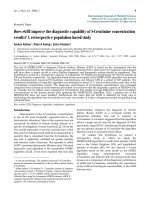
Mean pulmonary artery pressure in pigs after endotoxin induced systemic inflammationFigure 1
Mean pulmonary artery pressure in pigs after endo-
toxin induced systemic inflammation. Values are repre-
sented as means ± SEM for CO treated animals (open circles,
n = 10 except at 270 and 300 min where n = 9) and controls
(closed circles, n = 10 except at 150, 180 and 210 min where
n = 9 and at 240, 270 and 300 min where n = 8). Endotoxin
was administered (0.05 μg/kg/h) just after time 0, reaching
maximum infusion rate (0.25 μg/kg/h) at 30 min. CO was
administrated just after time -60 min. No significant differ-
ence between the groups (ANOVA F(1, 9) = 0.158).
Journal of Inflammation 2008, 5:13 />Page 4 of 8
(page number not for citation purposes)
during the experimental procedure, but remained within
the normocapnic range.
Carbon Monoxide
Results from blood analyses of CO concentrations are pre-
sented in Figure 2, where 250 μM corresponds to approx-
imately 5% COHb according to the transformation. The
control group showed very low CO concentrations
(approximately 50 μM) with small inter individual varia-
bility. CO administration to 10 animals resulted in steady
CO levels throughout the protocol, where 250 μM in
blood was the target concentration.
Cytokines
Plasma cytokine measurements are shown in Figure 3.
TNF-alpha concentrations increased after 60 minutes of
endotoxin infusion and decreased after approximately
150 minutes. There was no difference between the groups
regarding TNF-alpha concentrations. There was a large
variation between individuals, especially at peak levels.
Two animals in the CO-treated group had much higher
TNF-alpha peak concentrations than the others. Concen-
trations of IL-6 increased in response to endotoxin infu-
sion, with a peak at 150 minutes followed by a decrease,
but not to baseline levels. The two animals with extreme
TNF-alpha levels also had relatively high IL-6 concentra-
tions. The individuals with the highest IL-6 concentra-
tions were in the control group and died before the
protocol was completed. There was no statistically signifi-
cant difference in IL-6 concentrations between the groups.
The IL-10 concentration peaked at 90 minutes after which
it quickly decreased to near baseline levels and no differ-
ence was observed between groups. IL-1beta increased
continuously during the protocol with the highest levels
after 5 hours of endotoxin infusion. One of the animals
with the highest IL-6 concentrations also had the highest
IL-1beta concentrations. This animal died before the pro-
tocol was completed. IL-1beta concentrations were not
statistically significant different in CO-treated animals
compared with controls.
Discussion
We were unable to show that administration of CO had
any effect on cytokine release during endotoxin-induced
inflammatory response. Pro-inflammatory cytokines
(TNF-alpha, IL-6 and IL-1beta) were neither attenuated in
CO-treated animals, nor did the anti-inflammatory
cytokine (IL-10) increase. These results were unexpected
and contrasted to findings in an endotoxin mouse model,
where lower TNF-alpha and IL-1beta and higher IL-10 lev-
els in CO-treated animals compared with controls were
found [5]. In the present study, 3 animals died before
completing the whole duration of the protocol, 2 control
animals and 1 animal in the CO exposed group. These
animals are not included in the statistical calculations due
to the limitations of ANOVA, resulting in the fact that the
animals that may have had the most powerful inflamma-
tory response may have been excluded from comparison.
Analysis of the data shows that the 3 animals that died
before completing the protocol did not have the highest
TNF-alpha or IL-10 concentrations. However, the highest
IL-1beta concentration was found in a control animal that
Table 1: Circulatory and respiratory data from pigs during endotoxin infusion.
-60 (min) 0 (min) 60 (min) 120 (min) 210 (min) 300 (min)
group mean ± sem mean ± sem mean ± sem mean ± sem mean ± sem mean ± sem
HR Control 102 ± 7 90 ± 4 93 ± 6 96 ± 8 91 ± 8 a 97 ± 8 b
(bpm) CO 103 ± 4 94 ± 7 88 ± 4 79 ± 4 88 ± 5 92 ± 8 b
MAP Control 101 ± 5 95 ± 4 88 ± 5 91 ± 8 93 ± 11 a 95 ± 6 b
(mmHg) CO 100 ± 4 89 ± 3 84 ± 5 94 ± 3 89 ± 7 86 ± 7 a
CVP Control 3 ± 0.6 4 ± 0.6 7 ± 0.6 8 ± 0.8 8 ± 0.8 a 6 ± 0.6 b
(mmHg) CO 4 ± 0.7 4 ± 0.8 6 ± 0.8 7 ± 0.7 6 ± 0.7 7 ± 0.6 a
Cardiac Control 5.0 ± 0.4 a 4.8 ± 0.4 3.7 ± 0.4 4.1 ± 0.4 a 2.9 ± 0.3 a 3.5 ± 0.5 b
output (l/min) CO 5.8 ± 0.3 5.2 ± 0.3 3.8 ± 0.2 a 3.7 ± 0.3 2.9 ± 0.2 a 3.0 ± 0.2 a
P
a
CO
2
Control 4.5 ± 0.3 b 5.0 ± 0.2 a 5.3 ± 0.2 a 5.8 ± 0.1 b 5.9 ± 0.3 c 5.9 ± 0.3 b
(kPa) CO 4.5 ± 0.2 4.9 ± 0.1 5.4 ± 0.2 5.7 ± 0.2 6.0 ± 0.3 6.2 ± 0.9 a
P
a
O
2
Control 19.3 ± 0.7 b 18.3 ± 0.4 a 29.2 ± 3.0 a 28.3 ± 6.3 b 25.0 ± 5.7 c 19.4 ± 3.6 b
(kPa) CO 20.4 ± 0.5 20.0 ± 0.7 38.2 ± 5.7 38.7 ± 5.8 22.8 ± 5.1 28.5 ± 5.8 a
Hb Control 92 ± 1.9 a 89 ± 1.5 95 ± 2.7 100 ± 2.2 108 ± 1.2 a 103 ± 2.2 b
(g/l) CO 93 ± 2.8 88 ± 2.1 91 ± 1.6 101 ± 1.8 108 ± 2.9 105 ± 3.5 a
FiO
2
Control 30 ± 0 30 ± 0 55 ± 5.8 62 ± 8.0 68 ± 7.0 70 ± 6.6
(%) CO 30 ± 0 30 ± 0 56 ± 5.9 64 ± 8.4 78 ± 8.3 81 ± 7.5
Administration of CO began 1 hour before the endotoxin infusion was started, whereas control animals received endotoxin infusion but no CO
inhalation. Values are presented as means ± SEM, n = 10 in each group (Control and CO), except otherwise stated (a, b, c; n = 9, 8, 7 respectively,
as indexed). Endotoxin was administered (0.05 μg/kg/h) just after time 0, reaching maximum infusion rate (0.25 μg/kg/h) at 30 min. CO was
administrated just after time -60 min.
Journal of Inflammation 2008, 5:13 />Page 5 of 8
(page number not for citation purposes)
died following 4 hours of endotoxin exposure. The 2 ani-
mals from the control group that died had the highest IL-
6 concentrations. If these 3 animals would have survived
and been included the statistical analysis, this could imply
a difference in the interpretation of the IL-6 and IL-1beta
concentrations. However, these missing data do not have
any effect on the conclusion regarding TNF-alpha and IL-
10 response which remains contradictory to the mouse
study [5]. Published data on inflammatory effects of CO
in pigs is limited to only one other study, where higher
levels of TNF-alpha were found in CO-treated animals
compared with controls [11]. It was concluded [11] that
although the TNF-alpha levels were higher in the CO
treated group, CO ameliorated several of the acute patho-
logical changes. They also found a suppression of IL-1beta
in the CO-treated group, resulting in a significantly higher
level of IL-1beta in the control group. This is in contrast to
our findings, which show no differences in IL-1beta con-
centrations as a result of CO administration. One explana-
tion for this conflicting result could be that the other study
[11] only included 4 animals in each group. In a study in
man, where CO was administered before a bolus of endo-
toxin was injected, there were no differences in plasma
cytokines (TNF-alpha, IL-6, IL-8, IL-10), cytokine mRNA
(IL-1 alpha, IL-1 beta), heart rate, MAP or SpO
2
when the
CO-treated group was compared with controls [8]. These
clinical findings also support the interpretation that CO
does not help to improve the inflammatory response after
endotoxin infusion. Our interpretation of previous stud-
ies together with our findings is that CO may have an anti-
inflammatory effect in mice but not in humans or pigs.
The cytokine levels following endotoxin infusion in our
study were high, and individual TNF-alpha levels were
found up to 46000 pg/ml. In comparison, other endo-
toxin studies in pigs reported maximum levels of TNF-
alpha of 3500 pg/ml [11], 4000 pg/ml [12], 9000 pg/ml
[13] or 20000 pg/ml [14], respectively. The cytokine
response for TNF-alpha, IL-6 and IL-10 following endo-
toxin infusion shows the same pattern over time in our
study as has been observed by others [14], but the IL-
1beta response was different. Our findings show an
increase in IL-1beta concentration during endotoxin infu-
sion, whereas the other study [14] showed no change in
IL-1beta response.
In order to further evaluate possible anti-inflammatory
effects of CO, we have used a porcine model of human
sepsis. Pig sensitivity to endotoxin and tissue antigenicity
has been found to be similar to humans [15]. Further-
more, pigs also have similar cardiac anatomy and physiol-
ogy as humans [16]. The endotoxin infusion model
appeared to provide a highly stable and predictable circu-
latory and pathophysiological state for our study, as dem-
onstrated by a consistent biphasic MPAP pattern. The
endotoxin infusion rate was 0.25 μg/kg/h, corresponding
to a total dose of 1.175 μg/kg. The same dose has been
used in one other study investigating central haemody-
namics [17]. This is a low dose compared with other pig
studies [11,13]. Since there are different serotypes of
endotoxin, there may be a wide range of potency. Com-
pared with other studies, which have employed the same
lipopolysaccharide serotype as in the present study
(0111:B4), we still have a low dose of endotoxin. Endo-
toxin dosing regimens for the same serotype have been the
following; a bolus of 100 μg/kg [12], a bolus of 75 μg/kg
[18], and an infusion of a total dose of 250 μg/kg [19].
Different batches of endotoxin probably have different
potency. Also, different breeds of pigs probably have dif-
ferent sensitivity to endotoxin. The MPAP levels in our
study were high in comparison with other authors [11,20]
or similar [21]. This acute increase in MPAP associated
with endotoxin administration (Figure 1) was close or
similar to levels found in cardiovascular decompensation.
Given this perspective of wide variation in endotoxin dos-
ing for pig sepsis models, our interpretation is that the low
endotoxin dose in our study resulted in large cytokine
release as well as high MPAP levels, indicating a massive
systemic inflammatory activation.
Carbon monoxide concentrations in the two groups after endotoxin induced systemic inflammation in pigsFigure 2
Carbon monoxide concentrations in the two groups
after endotoxin induced systemic inflammation in
pigs. Values are represented as means ± SEM, for CO
treated animals (open circles, n = 10 except at 270 and 300
min where n = 9) and controls (closed circles, n = 10 except
at 150, 180 and 210 min where n = 9 and at 240, 270 and 300
min where n = 8). Endotoxin was administered (0.05 μg/kg/h)
just after time 0, reaching maximum infusion rate (0.25 μg/kg/
h) at 30 min. CO was administrated just after time -60 min.
Journal of Inflammation 2008, 5:13 />Page 6 of 8
(page number not for citation purposes)
The administration rate of CO in this study was chosen
with the aim to quickly achieve constant blood CO levels
and to avoid toxic effects. In contrast to a fixed CO dose,
the rate of delivery was modulated in order to maintain
relatively constant blood CO concentrations. An increase
in the CO administration rate was necessary during the
experiment, which we interpret as a result of reduced pul-
monary gas exchange due to the severe inflammatory
response. Constant CO levels were achieved, which is a
strength in this study compared to other studies, in which
the CO concentration decreased during the experiment
[8,11] or never was measured [5]. The chosen target con-
centration of CO (5% COHb) in the present study was
determined to be a clinically relevant dose, since higher
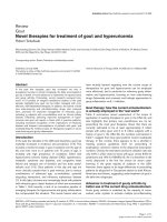
Plasma cytokine concentrations in pigs after endotoxin-induced systemic inflammation with or without CO treatmentFigure 3
Plasma cytokine concentrations in pigs after endotoxin-induced systemic inflammation with or without CO
treatment. Values are presented as individual measurements for CO treated animals (open circles) and controls (closed cir-
cles). A dotted (CO group) and solid (controls) line represents means for the two groups (n = 10 except for the CO-group at
270 and 300 min where n = 9 and for controls at 150, 180 and 210 min where n = 9 and at 240, 270 and 300 min where n = 8).
Endotoxin was administered (0.05 μg/kg/h) just after time 0, reaching maximum infusion rate (0.25 μg/kg/h) at 30 min. No sig-
nificant differences were detected between the groups for any of the cytokines (TNF: ANOVA F(1, 8) = 1.074, IL-6: ANOVA
F(1, 8) = 0.892, IL-10: ANOVA F(1, 8) = 1.347, IL-1beta: ANOVA F(1, 8) = 1.716).
0
50
100
150
200
250
300
350
0 60 120 180 240 300
Time (min)
IL-10 (pg/ml
)
0
100
200
300
400
500
600
700
0 60 120 180 240 300
Time (min)
IL-1beta (pg/ml
)
0
10000
20000
30000
40000
50000
0 60 120 180 240 300
Time (min)
TNF-alpha (pg/ml
0
1000
2000
3000
4000
5000
6000
0 60 120 180 240 300
Time (min)
IL-6 (pg/ml
)
Mean CO inhalationMean Controls
Journal of Inflammation 2008, 5:13 />Page 7 of 8
(page number not for citation purposes)
doses may induce toxic symptoms. A CO concentration of
20% in the blood may lead to unconsciousness [22,23].
Negative effects on performance during exercise after car-
bon monoxide inhalation in healthy men can be seen at
CO levels from 4.8% COHb [24]. Studies on patients with
angina pectoris show that carbon monoxide at levels from
2.7% to 4.5% COHb shortens the time to pain during
exercise and also induces a longer duration of pain [25-
27]. Performance during exercise in patients with chronic
anaemia is reduced at 2.0% COHb [28]. The relation
between CO dose and inflammatory response may be
important. Effects in pigs have been described at 10–12%
COHb [11], but no effects in humans have been reported
at 7% COHb [8]. If the previously suggested anti-inflam-
matory effect of CO is found at these higher CO concen-
trations, this may imply that the therapeutic potential of
CO is limited due to the risk of toxic side effects.
An important consideration regarding the animal model
is that the affinity of Hb for CO is dependent upon the
studied animal species. For example, mouse Hb has lower
affinity for CO compared with human Hb [8]. Pig Hb has
lower affinity for CO than some other mammals, e.g. rat
and hamster [29]. A lower affinity of Hb for CO could
result in a higher unbound or free fraction of CO, eliciting
a greater biological response at similar COHb fractions.
Elimination time for CO may also vary in different spe-
cies, as well as by differences in oxygenation. It has been
shown that the affinity of Hb for CO increases at low oxy-
gen tension [30]. All of this has to be considered when
evaluating the proper dose of CO. This also points out
why it is of great importance to measure CO concentra-
tions in the studied subjects, in contrast to measurements
of ambient or inhaled CO levels.
Conclusion
In summary, no clear effects of CO on the systematic
inflammatory process were shown in this study conducted
in endotoxin administered pigs, as evaluated by measured
concentrations of plasma cytokines (TNF-alpha, IL-6, IL-
1beta and IL-10). The model was characterised by massive
inflammation and a stable and controlled CO level. We
conclude that 5% COHb in the blood does not appear to
demonstrate any potential therapeutic effects on the mod-
ulation of systemic inflammation in this porcine model.
Competing interests
The authors declare that they have no competing interests.
Authors' contributions
AMÅ participated in the design of the study, the practical
work, the result discussion the statistical calculations and
writing the manuscript. PA participated in the practical
work, the result discussion and the revision of the manu-
script. GJ participated in the practical work, the statistical
calculations, the result discussion and the revision of the
manuscript. MH participated in the practical work, the
result discussion and helped to draft the manuscript. OW
participated in the design of the study, the result discus-
sion, revision of the manuscript and financial support. JEL
participated in the design of the study, the practical work,
the result discussion, the statistical calculations and in
writing the manuscript. All authors (AMÅ, PA, GJ, MH,
OW and JEL) have read and approved the final manu-
script.
Acknowledgements
Financial support from the Medical Faculty, Umeå University is gratefully
acknowledged.
References
1. Motterlini R, Gonzales A, Foresti R, Clark JE, Green CJ, Winslow RM:
Heme oxygenase-1-derived carbon monoxide contributes to
the suppression of acute hypertensive responses in vivo. Circ
Res 1998, 83(5):568-577.
2. Suematsu M, Goda N, Sano T, Kashiwagi S, Egawa T, Shinoda Y,
Ishimura Y: Carbon monoxide: an endogenous modulator of
sinusoidal tone in the perfused rat liver. J Clin Invest 1995,
96(5):2431-2437.
3. Nakao A, Kimizuka K, Stolz DB, Seda Neto J, Kaizu T, Choi AM, Uch-
iyama T, Zuckerbraun BS, Bauer AJ, Nalesnik MA, Otterbein LE, Gel-
ler DA, Murase N: Protective effect of carbon monoxide
inhalation for cold-preserved small intestinal grafts. Surgery
2003, 134(2):285-292.
4. Otterbein LE, Otterbein SL, Ifedigbo E, Liu F, Morse DE, Fearns C,
Ulevitch RJ, Knickelbein R, Flavell RA, Choi AM: MKK3 mitogen-
activated protein kinase pathway mediates carbon monox-
ide-induced protection against oxidant-induced lung injury.
Am J Pathol 2003, 163(6):2555-2563.
5. Otterbein LE, Bach FH, Alam J, Soares M, Tao Lu H, Wysk M, Davis
RJ, Flavell RA, Choi AM: Carbon monoxide has anti-inflamma-
tory effects involving the mitogen-activated protein kinase
pathway. Nat Med 2000, 6(4):422-428.
6. Zuckerbraun BS, McCloskey CA, Gallo D, Liu F, Ifedigbo E, Otterbein
LE, Billiar TR: Carbon monoxide prevents multiple organ
injury in a model of hemorrhagic shock and resuscitation.
Shock 2005, 23(6):527-532.
7. Hoetzel A, Dolinay T, Schmidt R, Choi AM, Ryter SW: Carbon
monoxide in sepsis. Antioxid Redox Signal 2007, 9(11):2013-2026.
8. Mayr FB, Spiel A, Leitner J, Marsik C, Germann P, Ullrich R, Wagner
O, Jilma B: Effects of carbon monoxide inhalation during
experimental endotoxemia in humans. Am J Respir Crit Care
Med 2005, 171(4):354-360.
9. Aberg AM, Hultin M, Abrahamsson P, Larsson JE: Circulatory
effects and kinetics following acute administration of carbon
monoxide in a porcine model. Life Sci 2004, 75(9):1029-1039.
10. Sundin AM, Larsson JE: Rapid and sensitive method for the anal-
ysis of carbon monoxide in blood using gas chromatography
with flame ionisation detection.
Journal of chromatography 2002,
766(1):115-121.
11. Mazzola S, Forni M, Albertini M, Bacci ML, Zannoni A, Gentilini F,
Lavitrano M, Bach FH, Otterbein LE, Clement MG: Carbon monox-
ide pretreatment prevents respiratory derangement and
ameliorates hyperacute endotoxic shock in pigs. Faseb J 2005,
19(14):2045-2047.
12. Tuchscherer M, Kanitz E, Puppe B, Tuchscherer A, Stabenow B:
Effects of postnatal social isolation on hormonal and
immune responses of pigs to an acute endotoxin challenge.
Physiol Behav 2004, 82(2-3):503-511.
13. Brix-Christensen V, Gjedsted J, Andersen SK, Vestergaard C, Nielsen
J, Rix T, Nyboe R, Andersen NT, Larsson A, Schmitz O, Tonnesen E:
Inflammatory response during hyperglycemia and hyperin-
sulinemia in a porcine endotoxemic model: the contribution
of essential organs. Acta Anaesthesiol Scand 2005, 49(7):991-998.
14. Myers MJ, Farrell DE, Palmer DC, Post LO: Inflammatory media-
tor production in swine following endotoxin challenge with
Publish with Bio Med Central and every
scientist can read your work free of charge
"BioMed Central will be the most significant development for
disseminating the results of biomedical research in our lifetime."
Sir Paul Nurse, Cancer Research UK
Your research papers will be:
available free of charge to the entire biomedical community
peer reviewed and published immediately upon acceptance
cited in PubMed and archived on PubMed Central
yours — you keep the copyright
Submit your manuscript here:
/>BioMedcentral
Journal of Inflammation 2008, 5:13 />Page 8 of 8
(page number not for citation purposes)
or without co-administration of dexamethasone. Int Immunop-
harmacol 2003, 3(4):571-579.
15. Goldfarb RD, Dellinger RP, Parrillo JE: Porcine models of severe
sepsis: emphasis on porcine peritonitis. Shock 2005, 24 Suppl
1:75-81.
16. Swindle MM, Smith AC, Hepburn BJ: Swine as models in experi-
mental surgery. J Invest Surg 1988, 1(1):65-79.
17. Konrad D, Haney M, Johansson G, Wanecek M, Weitzberg E, Oldner
A: Cardiac effects of endothelin receptor antagonism in
endotoxemic pigs. American journal of physiology 2007,
293(2):H988-96.
18. Frank JW, Carroll JA, Allee GL, Zannelli ME: The effects of ther-
mal environment and spray-dried plasma on the acute-phase
response of pigs challenged with lipopolysaccharide. J Anim Sci
2003, 81(5):1166-1176.
19. Bergmann M, Gornikiewicz A, Tamandl D, Exner R, Roth E, Fugger R,
Gotzinger P, Sautner T: Continuous therapeutic epinephrine
but not norepinephrine prolongs splanchnic IL-6 production
in porcine endotoxic shock. Shock 2003, 20(6):575-581.
20. Javeshghani D, Magder S: Regional changes in constitutive nitric
oxide synthase and the hemodynamic consequences of its
inhibition in lipopolysaccharide-treated pigs. Shock 2001,
16(3):232-238.
21. Nalos M, Vassilev D, Pittner A, Asfar P, Bruckner UB, Schneider EM,
Georgieff M, Radermacher P, Froeba G: Tin-mesoporphyrin for
inhibition of heme oxygenase during long-term hyperdy-
namic porcine endotoxemia. Shock 2003, 19(6):526-532.
22. Kondo A, Saito Y, Seki A, Sugiura C, Maegaki Y, Nakayama Y, Yagi K,
Ohno K: Delayed neuropsychiatric syndrome in a child fol-
lowing carbon monoxide poisoning. Brain Dev 2007,
29(3):174-177.
23. Mannaioni PF, Vannacci A, Masini E: Carbon monoxide: the bad
and the good side of the coin, from neuronal death to anti-
inflammatory activity. Inflamm Res 2006, 55(7):261-273.
24. Ekblom B, Huot R: Response to submaximal and maximal exer-
cise at different levels of carboxyhemoglobin. Acta Physiol
Scand 1972, 86(4):474-482.
25. Anderson EW, Andelman RJ, Strauch JM, Fortuin NJ, Knelson JH:
Effect of low-level carbon monoxide exposure on onset and
duration of angina pectoris. A study in ten patients with
ischemic heart disease. Ann Intern Med 1973, 79(1):46-50.
26. Aronow WS, Isbell MW: Carbon monoxide effect on exercise-
induced angina pectoris. Ann Intern Med 1973, 79(3):392-395.
27. Aronow WS, Stemmer EA, Isbell MW: Effect of carbon monoxide
exposure on intermittent claudication. Circulation 1974,
49(3):415-417.
28. Aronow WS: Aggravation of angina pectoris by two percent
carboxyhemoglobin. Am Heart J 1981, 101(2):154-157.
29. Klimisch HJ, Chevalier HJ, Harke HP, Dontenwill W: Uptake of car-
bon monoxide in blood of miniture pigs and other mammals.
Toxicology 1975, 3(3):301-310.
30. Westphal M, Weber TP, Meyer J, von Kegler S, Van Aken H, Booke
M: Affinity of carbon monoxide to hemoglobin increases at
low oxygen fractions. Biochem Biophys Res Commun 2002,
295(4):975-977.