Báo cáo y học: "Cigarette smoke regulates the expression of TLR4 and IL-8 production by human macrophages" pdf
Bạn đang xem bản rút gọn của tài liệu. Xem và tải ngay bản đầy đủ của tài liệu tại đây (605.99 KB, 9 trang )
BioMed Central
Page 1 of 9
(page number not for citation purposes)
Journal of Inflammation
Open Access
Research
Cigarette smoke regulates the expression of TLR4 and IL-8
production by human macrophages
Hadi Sarir
1,2
, Esmaeil Mortaz*
1,3,4
, Khalil Karimi
5
, Aletta D Kraneveld
1
,
Irfan Rahman
6
, Eric Caldenhoven
7
, Frans P Nijkamp
1
and Gert Folkerts
1
Address:
1
Division of Pharmacology and Pathophysiology, Departement of Pharmaceutical Sciences, Faculty of Sciences, Utrecht University, the
Netherlands,
2
Department of Animal Science, Birjand University, Iran,
3
Department of Clinical Biochemistry, Faculty of Medical Sciences, Tarbiat
Modarres University, Tehran, Iran,
4
Department of Basic Science, Section of Biochemistry, Faculty of Veterinary Medicine, Urmia University, Iran,
5
Department of Pathology and Molecular Medicine, Centre for Gene Therapeutics, McMaster University, Ontario, Canada,
6
Department of
Environmental Medicine, Division of Lung Biology and Disease, University of Rochester Medical Center, USA and
7
Danone Research Centre for
Specialised Nutrition, Wageningen, the Netherlands
Email: Hadi Sarir - ; Esmaeil Mortaz* - ; Khalil Karimi - ;
Aletta D Kraneveld - ; Irfan Rahman - ; Eric Caldenhoven - ;
Frans P Nijkamp - ; Gert Folkerts -
* Corresponding author
Abstract
Background: Toll-like receptors (TLRs) are present on monocytes and alveolar macrophages that
form the first line of defense against inhaled particles. The importance of those cells in the
pathophysiology of chronic obstructive pulmonary disease (COPD) has well been documented.
Cigarette smoke contains high concentration of oxidants which can stimulate immune cells to
produce reactive oxygen species, cytokines and chemokines.
Methods: In this study, we evaluated the effects of cigarette smoke medium (CSM) on TLR4
expression and interleukin (IL)-8 production by human macrophages investigating the involvement
of ROS.
Results and Discussion: TLR4 surface expression was downregulated on short term exposure
(1 h) of CSM. The downregulation could be explained by internalization of the TLR4 and the
upregulation by an increase in TLR4 mRNA. IL-8 mRNA and protein were also increased by CSM.
CSM stimulation increased intracellular ROS-production and decreased glutathione (GSH) levels.
The modulation of TLR4 mRNA and surface receptors expression, IRAK activation, IκB-α
degradation, IL-8 mRNA and protein, GSH depletion and ROS production were all prevented by
antioxidants such as N-acetyl-L-cysteine (NAC).
Conclusion: TLR4 may be involved in the pathogenesis of lung emphysema and oxidative stress
and seems to be a crucial contributor in lung inflammation.
Introduction
Macrophages play a central role in both specific and non-
specific immunity against bacterial, viral, and fungal
infections. The unique localization of alveolar macro-
phages in the alveoli (between air and lung tissue) [1],
represent them as the first line of defense against inhaled
microorganisms or particles [2]. The role of these cells in
the pathophysiology of chronic obstructive pulmonary
Published: 1 May 2009
Journal of Inflammation 2009, 6:12 doi:10.1186/1476-9255-6-12
Received: 5 November 2008
Accepted: 1 May 2009
This article is available from: />© 2009 Sarir et al; licensee BioMed Central Ltd.
This is an Open Access article distributed under the terms of the Creative Commons Attribution License ( />),
which permits unrestricted use, distribution, and reproduction in any medium, provided the original work is properly cited.
Journal of Inflammation 2009, 6:12 />Page 2 of 9
(page number not for citation purposes)
disease (COPD) has been well documented [3,4]. Ciga-
rette smoke (CS) stimulates various immune cells to
increase the production of cytokines and generate of reac-
tive oxygen species [1]. CS causes lung damage by oxida-
tive stress either by itself or due to oxidants released by
inflammatory cells that are recruited as a result of smoke-
induced injury. CS is a major source of oxidants/free rad-
icals and a complex of over 4700 chemical compounds
[5]. This huge amount of oxidants from CS and those
formed endogenously cause an imbalance between oxi-
dants and antioxidants which are considered to be impor-
tant in the pathogenesis of COPD [6,7]. Multiple
intracellular signaling events occur by CS, which ulti-
mately leads to the synthesis and release of pro-inflamma-
tory mediators, such as interlukine-8 (IL-8), IL-1β, and
tumor necrosis factor-α (TNF-α) [8,9].
The function of the innate immune system is the discrim-
ination of invading pathogens and self-cells by utilizing
signals from the Toll-like receptors (TLRs). TLRs recognize
specific patterns of microbial components [10] and sig-
nals to initiate a range of host defense mechanisms [11].
TLR4 is a crucial component of the signaling receptor
complex which is involved in recognition of a major inte-
gral glycolipid component of the outer membrane of
gram-negative bacteria (lipopolysaccharide or LPS) [12].
Downstream signaling of TLR4 pathway includes myeloid
differentiation factor 88 (MyD88), IL-1 receptor associ-
ated kinases (IRAKs), and TNF receptor-activated factor 6
(TRAF6). TRAF6 activates various kinases, which leads to
I-κB degradation and NF-κB activation. Activated NF-κB
translocates into the nucleus and increases the production
of pro-inflammatory mediators like IL-8 [13-15]. The
redox status of cells contributes to the modulation of NF-
κB. Moreover, ROS regulate immune-inflammatory cellu-
lar signaling via TLR4 by activation of NF-κB [16,17].
Intracellular reduced glutathione (GSH), an efficient thiol
antioxidant system in the lung, provides protection
against oxidants. GSH may be crucial for oxidant-induced
NF-κB response [18]. At present, the only antioxidant
widely available for patients with COPD is N-acetyl-L-
cyteine (NAC) [19,20] which exhibits direct and indirect
antioxidant properties and protect cells from oxidative
damage [21]. Its free thiol group is capable of interacting
with the electrophilic groups of ROS (direct effect), and as
a precursor of GSH (indirect effect) increases intracellular
GSH level and hence protects the cells against oxidative
stress [22,23].
TLR4 signaling is important in lung diseases [24,25]. TLR4
in the lungs could be activated either by conserved micro-
bial component or exogenous oxidants [26] and therefore
modulate inflammatory responses. Moreover, there is a
link between ROS and TLR4 [18,26,27]. Very recently, we
documented that TLR4 mediates CS-induced IL-8 produc-
tion in monocyte-derived macrophages (MDMs) [8].
Since CS is a rich source of radicals and can induce oxida-
tive stress, we hypothesized that CS-induced oxidative
stress may modulate TLR4 expression and NF-κB activa-
tion which leads to the release of IL-8. Therefore, the
effects of ROS imposed by CS on TLR4 surface and gene
expression, as well as, GSH levels were investigated. Our
study shows that CS-induced oxidative stress is involved
in modulation of TLR4 mRNA and surface protein expres-
sion as well as the cascade of TLR4 signaling pathways and
cytokine productions.
Materials and methods
Reagents
Reagents were purchased from Sigma-Aldrich except were
specified. Monocytes were isolated by RossetSep™ (Stem
cell Technology) from buffy coats (Sanquin blood bank)
see the below. Cells were incubated in RPMI 1640 (BioW-
hittaker Cambrex Company, Verriers, Belgium), supple-
mented with 2 mM N-acetyl-L-alanyl-L-glutamine, 100 U/
ml penicillin, 100 μg/ml streptomycin, 2% sodium pyru-
vate and 20 mM Hepes and 10% heat-inactivated fetal calf
serum (FCS) (Invitrogen Life Technolog). The mouse anti-
body against human IκBα and human IRAK-1 were
obtained from Santa Cruz biotechnology (Tebu-bio,
Heerhugowaard, The Netherlands).
Cell culture
For culturing human monocyte-derived macrophages,
peripheral blood mononuclear cells (PBMC) were sepa-
rated by density gradient centrifugation (Pharmacia Bio-
tech, Uppsala, Sweden) of buffy coats obtained from
normal blood donors as described before [28,29]. Human
blood monocytes were obtained using RosetteSep™ (Stem
cell Technologies) according to manufacturer's instruc-
tions. Briefly, fresh blood was incubated with RosetteSep™
cocktail at room temperature followed by Ficoll-Paque
gradient centrifugation (Life Technologies, Cergy Ponto-
ise, France). The enriched monocytes were collected from
the Ficoll:plasma interface and purity was assessed by
FACS analysis using a FITC-labeled anti-CD14 mAb
(95%). Macrophages were obtained by culturing mono-
cytes for 5 days in medium containing 2.5 ng/ml GM-CSF
and 25 ng/ml M-CSF (R&D). TLR4 stably transfected HEK-
293 cell line (293-htlr4a) and HEK-293 cells stably trans-
fected with the LacZ reporter gene (293-lacz) were pur-
chased from In vivogen [30]. Cells were culture in
medium containing Blasticidin (10 μg/ml), and after 5–7
passages, cells were activated as described below.
Cigarette smoke medium preparation
CSM was prepared as described before [9]. Briefly, a smok-
ing machine (Teague Enterprises, Davis, CA, USA) was
programmed to smoke cigarette according to the federal
Trade commission protocol (35 ml puff volume for 2 sec-
Journal of Inflammation 2009, 6:12 />Page 3 of 9
(page number not for citation purposes)
onds once per minute). The main and side stream smoke
from one cigarette (unfiltered Lucky strike
TD
, tar and nic-
otine concentration 12 and 0.9 mg respectively) was
directed through 5 ml culture medium (RPMI without
phenol red). Hereafter, absorbance was measured spectro-
photometrically and the media was standardized to a
standard curve of CSM concentration against absorbance
at 320 nm. The optical density (OD) 4 (100%) is the high-
est OD at this wavelength which was diluted to OD 0.03
(0.75%) and 0.06 (1.5%) and applied to the cells. Freshly
prepared CSM was used in all experiments.
Cell activation
For measuring IL-8 production by CSM, TLR4 stably trans-
fected HEK293 cells or 293-LacZ HEK-293 were stimu-
lated with CSM (0.06 OD) and LPS (100 ng/ml) for
overnight. For modulation of TLR4 receptors via CSM,
MDMs were preincubated with anti-TLR4, control anti-
bodies or NAC (1 mM) for 30 min and then stimulated
with CSM or LPS (100 ng/ml) as a positive control for 4 h.
RNA was extracted and TLR4 and GAPDH gene expression
were quantified by real-time PCR. To test the involvement
of oxidants in IRAK activation by CSM, MDMs were stim-
ulated with CSM (0.06 OD) in the presence or absence of
NAC (10 mM) for 30 min.
For evaluation of ROS production by CSM in MDMs, the
cells were incubated with either 10 mM of NAC for 20
min and, then cultured with CSM (OD 0.03 and 0.06 OD)
at 37°C for 1 h. The cells were diluted to 10
5
/ml in PBS,
and incubated with 10 μM of H2DCFDA for 15 min. After
the cells were washed twice with PBS, 10
4
, cells were ana-
lyzed by FACScan (Becton Dickinson) to determine their
fluorescence intensity.
IL-8 ELISA
Measurement of IL-8 in culture supernatant was per-
formed by using ELISA kits (BD bioscience), according to
the manufacture's instruction.
FACS analysis
Cells (TLR4 stably transfected HEK293 cells, LACz null
cells and MDMs) were treated with CSM (0.03 and 0.06
OD) for 3 h and then washed and incubated on ice for 30
min with a PE-conjugated anti-human TLR4 (clone
HTA125) or mouse IgG2a as control isotype (eBio-
science). In addition, for the detection of intracellular lev-
els of TLR4, cells were permeabilized with
permeabilization buffer (eBioscience) and stained with
anti-human TLR4 Ab or relevant isotype. TLR4 expression
was assessed on a FACScan flow cytometer (BD Bio-
sciences). The relative TLR4 surface or intracellular levels
were quantified by subtracting the mean fluorescent
intensity (MFI) from the MFI values of isotype matched
control for each sample.
Real-time quantitative PCR
Total RNA was extracted using High Pure RNA Isolation
Kit (Roche Applied Science) according to the manufac-
ture's instruction. Quantity and purity of the extract was
measured by nanodrop (Wilmington, DE, USA). Equal
amounts of total RNA was reverse transcribed into cDNA
using oligo-dt and Superscript III (Invitrogen Corpora-
tion). Real-time PCR was performed using SYBR Green
PCR Master-Mix (ABGene) for 40 cycles on an ABI Prism
7000 sequence detector (Applied Biosystems) according
to manufacture's instruction. Amplification was achieved
using an initial cycle of 50°C for 2 min and 95°C for 15
min, followed by 40 cycles of 95°C for 15 s and 60°C for
1 min. Melting curve analyses were performed after the
completion of cycling to control the specificity of the PCR
products obtained. Primers were designed using the
Primer Express (Applied Biosystems) software which is as
followed: tlr4 (GeneBank Accession NM_138554
) forward
5'-CTGCCACATGTCAGGCCTTAT-3'; Reverse 5'-AAT-
GCCCACCTGGAAGACTCT; tlr2 (GeneBank Accession
NM_003264
) forward 5'-CATTCCCTCAGGGCTCACAG-
3'; Reverse 5'-TTGTTGGACAGGTCAAGGCTT-3'; and
gapdh (GeneBank Accession AY340484
) forward 5'-CCAG-
GTGGTCTCCTCTGACTTC-3'; Reverse 5'-CACCCTGTT-
GCTGTAGCCAAA-3'. The raw Cts (threshold cycle) values
from the reactions were analyzed with a modified delta-Ct
method with efficiency correction using a PCR data anal-
ysis program, qBase to obtain relative quantification val-
ues.
Protein Assay
The protein content of the lyzate was determined using
the bicinchonic acid (BCA) kit (Pierce, Erembodegem-
Aalst, Belgium). Protein standards were obtained by dilut-
ing a stock solution of Bovine Serum Albumin (BSA)
(Pierce).
Western blotting assay
Treated cells were washed once with cold PBS and lysed
on ice-cold lysis buffer containing 50 mM Tris (PH 8.0),
110 mM NaCl, 5 mM EDTA, 1% Triton X-100, and PMSF
(100 μg/ml) and aprotinin (2 μg/ml). Protein concentra-
tion was measured by BCA protein assay kit. Whole cell
lysates were boiled and separated on polyacrylamide gel
(12%), transferred onto nitrocellulose membrane
(Novex). For immunoblotting, membranes were soaked
in super-blocking buffer (Pierce) for 1 hour to block" the
nonspecific binding of proteins. The nitrocellulose was
then incubated with the specific antibody, human IκB-α
and IRAK, at appropriate dilutions. Membranes were then
washed several times in washing buffer (phosphate buff-
ered saline with 0.05% Tween-20) and incubated with
secondary antibody coupled to peroxidase at a 1:10,000
dilution for 1 h. Blots were washed with TBS-T and immu-
noreactive signals were visualized by an enhanced chemi-
Journal of Inflammation 2009, 6:12 />Page 4 of 9
(page number not for citation purposes)
luminescence reagent (ECL; Amersham). Films were
scanned and analyzed on a GS7–10 Calibrated Imaging
Densitometer equipped with Quantity One v.4.0.3 soft-
ware (Bio-Rad).
Intracellular oxidative activity assay
After stimulation of MDM (10
4
cells were washed twice
with PBS, and and then intracellular ROS generation was
evaluated with a fluorogenic substrate, 2'. 7'-dichloroflu-
oresceindiacetate (H2DCFDA, Invitrogen). This probe is a
non-fluorescent compound which readily diffuses to the
cells and becomes fluorescent when oxidized by hydrogen
peroxide, peroxinitrite (ONOO-), and hydroxyl radicals
(OH
•
). Thus, dye oxidation is an indirect measure of the
presence of the reactive oxygen intermediate/species, cal-
culated by dividing the mean channel fluorescence of a
treated sample by that of the untreated one and multiply-
ing by 100 to obtain the relative change, expressed as a
percentage.
Measurement of cellular GSH content
Intracellular GSH content was assessed in cellular lysate
according to the methods of Tietze [31] with slight modi-
fication [32]. Briefly, washed cells were lysed by repeat-
edly freezing and thawing using lysis buffer containing
0.6% (w/v) sulfosalicylic acid. 0.1% (v/v) Triton X-100, 5
mM EDTA in 0.1 M potassium phosphate buffer, PH 7.5.
The supernatant collected after centrifugation and incu-
bated with 0.2 mg/ml dithiobisnitrobenzoic acid (DTNB)
and 1.67 U/ml glutathione reductase in phosphate buffer-
EDTA for 30 seconds, then 0.2 mg/ml β NADPH was
added and the rate of DTNB reduction was spectrophoto-
metrically measured at 405 nm. GSH content was calcu-
lated using a standard curve, and expressed as nmol/mg
protein.
Data analysis
Data are presented as means ± SEM. Comparison between
groups was performed by using un-paired t tests. A P value
of less than 0.05 was taken as statistically significant.
Results
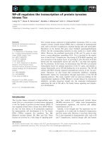
TLR4 is involved in CSM-induced IL-8 production
Recently, we demonstrated that CSM-induced IL-8 pro-
duction by MDMs could be inhibited by neutralizing anti-
bodies against TLR4 [8]. To support these effects of CSM
in detail, we investigated in TLR4 stably transfected and
null HEK 293 cell lines. TLR4 stably transfected HEK 293
cells were stimulated with CSM (0.06 OD) or LPS (100
ng/ml) as a positive control. As depicted in Fig. 1 CSM and
LPS induced IL-8 release only in TLR4 stably transfected
HEK293 cells but not in LacZ HEK 293 cell line.
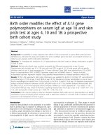
CSM modulates expression of TLR4
In both, MDMs and TLR4 stably transfected HEK 293
cells, CSM induced a concentration-dependent decrease
in surface expression of TLR4 (Fig. 2A and 2B). LPS as a
positive control induced a more pronounced decline in
TLR4 surface expressions in HEK293 cells than in MDMs.
Next, we investigated whether the surface suppression of
TLR4 was due to the internalization/shedding of recep-
tors. Therefore, intracellular level of TLR4 expression was
studied. As shown in Fig. 2C, CSM at the same time
points, intracellular levels of TLR4 in MDM was increased.
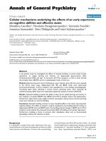
To further study the effects of CSM on modulation of
TLR4 expressions, mRNA levels of TLR4 was studied by
using PCR. MDMs were incubated with CSM (0.03, 0.06
and 0.12 OD) for 4 h and RT-PCR was performed by using
the human TLR4 and GAPDH primers as a reference gene.
CSM upregulated the expression of mRNATLR4 in MDMs
(Fig. 3A) and pre-incubation with NAC suppressed this
effect. Pre-incubation of MDMs with a neutralizing anti-
body against TLR4 (20 μg/ml) decreased the mRNA levels
of TLR4 enhancement to CSM (about 50%) while no inhi-
bition was observed when cells were pre-incubated with
the control antibody (Fig. 3B). Similar to CSM, LPS as a
positive control enhanced the TLR4 mRNA expression.
Next, in order to investigate the involvement of ROS by
CSM, MDMs were pre-treated with the antioxidant NAC
(10 mM) for 30 min and then incubated with CSM (0.03,
0.06 and 0.12 OD) for 4 h. NAC suppressed the upregula-
tion of TLR4 mRNA-induced by CSM compared to control
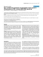
(Fig. 3A). Moreover, NAC suppressed the expression of IL-
8 at mRNA and protein levels (Fig. 4A and 4B).
CSM induces the generation of ROS by MDMs
Further, we directly measured ROS production by using a
fluorescence probe (H2DCFDA). As demonstrated in Fig.
TLR4 involved in CSM-induced IL-8 productionFigure 1
TLR4 involved in CSM-induced IL-8 production. TLR4
stably transfected HEK293 cells or 293-LacZ HEK-293 cells
(2 × 10
6
/ml) were stimulated with CSM (0.06 OD) and LPS
(100 ng/ml) for overnight. Supernatant were analyzed for IL-8
production by ELISA. Assays were performed in duplicate a
minimum of three times. Values are expressed as mean +/-
S.E. (n = 3). * signifies (**P = 0.01) of observed effect vs. con-
trol.
Journal of Inflammation 2009, 6:12 />Page 5 of 9
(page number not for citation purposes)
5, exposure of MDMs to CSM (0.03 and 0.06 OD) induces
a dose-dependent oxidation of the fluorescence probe
which indicates intracellular ROS production by CSM
(oxidative activity). ROS production by CSM was com-
pletely blocked when the cells were pre-incubated with
NAC (10 mM).
ROS generation by CSM, enhanced phosphorylation of
IRAK and induces I
κ
B-
α
degradation
It has been show that that IRAK phosphorylation is the
first step after MyD88 recruitment which finally leads to
degradation of the IκB-α and activation of NF-κB [8].
Stimulation of the MDMs with CSM for 30 min induced
the phosphorylation of IRAK which was abolished by
adding NAC (Fig. 6A). Moreover, CSM and LPS (as a con-
trol) degradated IκB-α and preincubation of MDMs with
NAC suppressed the degradation of IκB-α induced by
CSM (Fig. 6D).
Next, to confirm specific effects of CSM on TLR4 signaling,
the phosphorylation of IRAK in TLR4 stably transfected
HEK cells and null cells were studied. CSM induced phos-
phorylation of IRAK in TLR4 stably transfected HEK cells
but not in null cells (Fig. 6C).
Modulation of TLR4 expression by CSMFigure 2
Modulation of TLR4 expression by CSM. TLR4 stably transfected HEK293 cell (A) or MDMs (B) were treated with CSM
(0.03 and 0.06 OD) for 3 h and then incubated with PE conjugated anti-TLR4 or isotype control antibody as described in mate-
rials and methods. FACS analysis of a representative of at least 3 experiments showing the mean fluorescence intensity (MFI)
difference of each group. Values are expressed as mean +/- S.E.M (n = 3). *p = 0.05,***p = 0.001 significantly different com-
pared to control. C) MDMs were stimulated with CSM (0.06 OD) or LPS (100 ng/ml) for 3 h and then intracellular levels of
TLR4 were measured as described in material and methods. Values are expressed as mean +/- S.E.M (n = 3). *p = 0.05, signifi-
cantly different compared to control.
Journal of Inflammation 2009, 6:12 />Page 6 of 9
(page number not for citation purposes)
CSM modulates GSH levels
We measured the levels of GSH in MDMs after CSM stim-
ulation at various time points. CSM time-dependently
decreased GSH concentrations for 5 h and after long time
exposure this effects was restored (Fig. 7). Preincubation
of cells with NAC (10 mM) and DMSO (2%) for 20 min-
utes restored/attenuated the loss of intracellular GSH lev-
els at all time points. The period and concentration of
NAC and DMSO was chosen on the basis of previous stud-
ies with these agents [18,33].
Discussion
TLRs are found on the cell surface and in endosomes of
many different cell types. To date, 13 TLRs have been
identified in mice and humans with corresponding syn-
thetic or naturally occurring ligands. One of them is TLR4
which recognizes lipopolysaccharides (LPS) from gram
negative bacteria [13].
We have demonstrated earlier that CSM induces IL-8 pro-
duction via TLR4 in MDM. Interestingly; this effect was
not due to contamination of LPS [8]. In the current study
these pervious observations were extended in more
details.
First, as supportive evidence, we employed the HEK293
cells as stably transfected TLR4 and LACz HEK293 as a
control cell lines. Only in TLR4 stably transfected HEK
cells, CSM induced the production of IL-8. Moreover,
CSM regulates expression of TLR4 via ROSFigure 3
CSM regulates expression of TLR4 via ROS. (A) MDMs
(5 × 10
6
cells) were stimulated by CSM (0.03, 0.06 and 0.12
OD) for 4 h with and without pretreatment with NAC (10
mM) for 30 min. RNA was extracted and TLR4 and GAPDH
gene expression were quantified by real-time PCR. Results
are expressed as copies of TLR4 vs. copies of GAPDH gene.
(B) MDMs were preincubated with naturalizing anti-TLR4 or
isotype control antibodies for 30 min and then stimulated
with CSM (0.06 OD) for 4 h or LPS (100 ng/ml) and mRNA
levels of TLR4 was determined by real-time PCR method.
Values are expressed as mean +/- S.E.M (n = 3).*P <
0.05,***p = 0.001 significantly different compared to control
and # P < 0.05 significantly different compared to CSM stim-
ulated (n = 3).
IL-8 expression is ROS dependent after CSM exposureFigure 4
IL-8 expression is ROS dependent after CSM expo-
sure. MDMs (5 × 10
6
cells/ml) were pretreated with NAC
(10 mM) for 30 min and then stimulated by CSM (0.03, 0.06
and 0.12 OD) for 4 h. RNA was extracted and mRNA levels
of IL-8 were quantified by real-time PCR (A). Results are
expressed as copies of IL-8 vs. copies of GAPDH mRNA. (B)
MDMs (1 × 10
6
cells/ml) were pretreated with NAC (10 mM)
for 30 min and then stimulated by CSM (0.06 OD) for 16 h
Supernatants were collected after 16 h incubation and IL-8
production was quantified using ELISA methods. *P < 0.05 vs
baseline # P < 0.05 vs CSM stimulated (n = 3).
Journal of Inflammation 2009, 6:12 />Page 7 of 9
(page number not for citation purposes)
CSM regulates surface and intracellular TLR4 expression
in MDMs.
Interestingly, CSM induced the internalization of TLR4
receptor. TLR4, in the lung, not only could recognize
microbial components but also could sense either exoge-
nous oxidants like electrophilic compounds and free rad-
icals present in CSM or endogenous oxidants [34-36].
Activation of TLRs can lead to inflammatory response by
signaling through NF-κB, the best characterized regulator
of TLR signaling [16]. Cigarette smoke is a source of
potent reactive oxygen and nitrogen species which partic-
ipate in intracellular signaling and NF-κB activation [8].
In addition, several studies have revealed the importance
of oxidative stress in the IL-8 productions [37,38]. Thus,
we studied the role of ROS on CSM-induced increase in
mRNA TLR4 activation of MDMs. It was found that NAC
abrogated the expression of TLR4 expression. Further-
more, NAC interfered with CSM-induced IL-8 production
through a mechanism that is associated with increased
ROS production and GSH depletion.
GSH levels decreased dose- and time- dependently and
pre-treatment of the cells with antioxidants NAC and
DMSO prevented the CSM-induced decrease in GSH lev-
els in MDMs. Since NAC is able to scavenge a wide range
of oxidants (hypochlorous acid, hydrogen peroxide,
superoxide and hydroxyl radical) it revealed a better anti-
oxidant effect compare to DMSO which reacts with the
hydroxyl radical [22]. By using a direct approach to meas-
ure ROS production, CSM dose dependently increases
intracellular ROS generation by MDMs. These findings
may suggest that CSM induces its effect by intracellular
ROS generation and direct electrophilic ability to decrease
intracellular GSH.
Despite of the decreased surface expression of TLR4 after
CSM, a delayed up-regulation might be induced by a pro-
tective mechanism like the enhancement of GSH. Surface
attenuation of TLR4 receptor may be explained by an
internalization/shedding of the receptor complex or by
changes in the structure of the receptor to cross-link with
other TLR4 molecule since recent evidence indicates that
cross-linking is necessary for signal transduction [39].
Cross-linking of receptors or receptor clustering by thiol-
reactive mercury or ultraviolet radiation have been docu-
mented which activates downstream signaling [40,41].
The downregulation of TLR4 receptor presented here is in
CSM induces generation of ROS in MDMsFigure 5
CSM induces generation of ROS in MDMs. MDMs were
pretreated with NAC (10 mM) for 30 min and then stimu-
lated with CSM (0.03 and 0.06 OD) for 1 h. Intracellular ROS
concentration was measured by incubation of cells with
H2DCFDA as a probe for 30 min at 37 oC. Then after wash-
ing, the density of flurochrom as indicator for generation of
ROS was determined by FACS analysis. The results were
expressed as fold increase over control cells. Data represent
means ± SEM of triplicate experiments (n = 3). * p < 0.05
versus unstimulated control; # p < 0.05 versus CSM.
CSM regulates phosphorylation of IRAK and degradation of IκB-α by MDMs and phosphorylation of IRAK in HEK cellsFigure 6
CSM regulates phosphorylation of IRAK and degra-
dation of IκB-α by MDMs and phosphorylation of
IRAK in HEK cells. MDMs (3 × 10
6
cells) were pretreated
with NAC (10 mM) for 30 min and then stimulated with CSM
(0.06 OD) and LPS (100 ng/ml) for 30 min as described at
material and methods section. The expression of phospho
IRAK (A) and IκB-α degradation (B) were determined by
whole lysates of cells by Western blot analysis. Representa-
tive results of three independent experiments and β-actin
(C) served as loading controls from cytoplasm. D) TLR4 sta-
bly transfected HEK293 cells or 293-LacZ HEK-293 cells (3 ×
10
6
cells) were stimulated with CSM for 30 min as described
at material and methods section. The expression of phospho
IRAK were determined by whole lysates of cells by Western
blot analysis. Representative results of three independent
experiments and β-actin served as loading controls from
cytoplasm.
Journal of Inflammation 2009, 6:12 />Page 8 of 9
(page number not for citation purposes)
contrast to the result from experiments with RAW 264.7
cells exposed to hydrogen peroxide (H2O2)[34]. It is not
clear whether this discrepancy reflects genetic differences
between human and mice [42], cell differences or the type
of oxidant.
Next, the TLR4 expression at mRNA levels was studied. We
and found that CSM increases mRNA levels of TLR4.
Upregulation of mRNA level inside cells could lead to
upregulation of intracellular protein levels of TLR4 which
is reflected by increased intracellular expression.
The antioxidant NAC prevented the upregulation of TLR4
mRNA which indicates a role of oxidative stress induced
by CSM. NAC prevents the oxidative stress via counteract-
ing with electrophilic group of ROS (direct effect) or stim-
ulating the synthesis of the cellular GSH levels and
therefore protecting the cells against oxidants (indirect
effect) by modulating the redox signaling pathways
[22,23]. Thus these results indicate that CSM by inducing
ROS generation, may modulates the expression of TLR4.
TLRs ligations lead to recruitment of many proteins to the
cytoplasmic domain of the receptor like adapter mole-
cules MyD88. MyD88 recruits and promotes the interac-
tion between IL-1R-associated kinases (IRAK)-4 and
IRAK-1, resulting in the phosphorylation and activation
of IRAK-1 by IRAK-4 [43,44]. Subsequently, dissociation
of IRAK1 from the receptor complex and association with
the signal transducer tumor necrosis factor receptor-asso-
ciated factor 6 (TRAF6) occur. The subsequent down-
stream signaling leads to the degradation of the IκB-α and
activation of NF-κB [45-47]. CSM induced the phosphor-
ylation of IRAK-1 and degradates IκB-α [8]. In this study
by using NAC, we have demonstrated that ROS play an
important role in CSM-induced TLR4 associated intracel-
lular signaling. Interestingly, we have found that CSM spe-
cifically induced phosphorylation of IRAK-1 in stably
transfected TLR4 HEK cells but not in null TLR4 cells.
In conclusion, these results indicate that CSM induces a
ROS mediated signal transduction pathway via TLR4 in
MDMs. Induction of oxidative stress plays an important
role in the regulation of TLR4 and the production of IL-8.
Abbreviations
COPD: Chronic Obstructive Pulmonary Disease; TLR4:
Toll-like receptor-4; ROS: reactive oxygen Species; CSM:
Cigarette Smoke Medium; CS: Cigarette smoke; IL-8:
interleukin-8; NAC: N-acetyl-L-cysteine; OD: Optical
Density; TNF-α: Tumor necrosis factor-α; GSH: Glutath-
ione; CS: Cigarette smoke; MDMs: monocyte-derived
macrophages; LPS: Lipopolysaccharide.
Competing interests
The authors declare that they have no competing interests.
Authors' contributions
HS and EM equally conceived of the study, and partici-
pated in the design of the study and performed immu-
noassays, FACS analysis, statistical analysis, and wrote the
first draft and final version of the manuscript. KK, AK IR
and FN participated in designing the experiments and
took part in critical revision of the manuscript. FN partic-
ipated in the design and coordination of the study. GF
conceived of the study, and participated in the design of
the study and supervised the project. All authors read and
approved the final manuscript.
Acknowledgements
This study was performed within the framework of Dutch Top Institute
Pharma (project number D1.101). IR was supported by NIH-R01-
HL085613, NIEHS-ES01247 and NIEHS Toxicology Training grant ES07026.
References
1. Fels AO, Cohn ZA: The alveolar macrophage. J Appl Physiol 1986,
60:353-369.
2. Jonsson S, Musher DM, Goree A, Lawrence EC: Human alveolar
lining material and antibacterial defenses. Am Rev Respir Dis
1986, 133:136-140.
3. Barnes PJ: Alveolar macrophages as orchestrators of COPD.
Copd 2004, 1:59-70.
4. Shapiro SD: The macrophage in chronic obstructive pulmo-
nary disease. Am J Respir Crit Care Med 1999, 160:S29-32.
5. Pryor WA, Stone K: Oxidants in cigarette smoke. Radicals,
hydrogen peroxide, peroxynitrate, and peroxynitrite. Ann N
Y Acad Sci 1993, 686:12-27.
6. Rahman I, MacNee W: Role of oxidants/antioxidants in smok-
ing-induced lung diseases. Free Radic Biol Med 1996, 21:669-681.
7. Rahman I, Adcock IM: Oxidative stress and redox regulation of
lung inflammation in COPD. Eur Respir J 2006, 28:219-242.
Antioxidant prevents intracellular GSH depletion-induced by CSMFigure 7
Antioxidant prevents intracellular GSH depletion-
induced by CSM. MDMs (5 × 10
6
cells) were pretreated
with NAC (10 mM) and DMSO (2%) for 30 min and then
stimulated with CSM (0.06 OD) at various time points (1,
2.5, 5, and 24 h). Intracellular GSH contents were measured
by cellular lysate as described at "material and methods" sec-
tion and expressed as mean ± SEM of medium-treated cells. *
p < 0.05 versus un-stimulated control; # p < 0.05 versus.
Journal of Inflammation 2009, 6:12 />Page 9 of 9
(page number not for citation purposes)
8. Karimi K, Sarir H, Mortaz E, Smit JJ, Hosseini H, de Kimpe S, Nijkamp
F, Folkerts G: Toll-like receptor-4 mediates cigarette smoke-
induced cytokine production by human macrophages. Respir
Res 2006, 7:66.
9. Moodie FM, Marwick JA, Anderson CS, Szulakowski P, Biswas SK,
Bauter MR, Kilty I, Rahman I: Oxidative stress and cigarette
smoke alter chromatin remodeling but differentially regu-
late NF-kappaB activation and proinflammatory cytokine
release in alveolar epithelial cells. Faseb J 2004, 18:1897-1899.
10. Medzhitov R, Janeway C Jr: Innate immunity. N Engl J Med 2000,
343:338-344.
11. .
12. Iwasaki A, Medzhitov R: Toll-like receptor control of the adap-
tive immune responses. Nat Immunol 2004, 5:987-995.
13. Chow JC, Young DW, Golenbock DT, Christ WJ, Gusovsky F: Toll-
like Receptor-4 Mediates Lipopolysaccharide-induced Signal
Transduction. J Biol Chem 1999, 274:10689-10692.
14. Akira S: Toll-like receptor signaling. J Biol Chem 2003,
278:38105-38108.
15. Barton GM, Medzhitov R: Toll-like receptor signaling pathways.
Science 2003, 300:1524-1525.
16. Akira S, Takeda K: Toll-like receptor signalling. Nat Rev Immunol
2004, 4:499-511.
17. Ryan KA, Smith MF Jr, Sanders MK, Ernst PB: Reactive Oxygen and
Nitrogen Species Differentially Regulate Toll-Like Receptor
4-Mediated Activation of NF-κB and Interleukin-8 Expres-
sion. Infect Immun 2004, 72:2123-2130.
18. Asehnoune K, Strassheim D, Mitra S, Kim JY, Abraham E: Involve-
ment of Reactive Oxygen Species in Toll-Like Receptor 4-
Dependent Activation of NF-κB. J Immunol 2004,
172:2522-2529.
19. Rahman I, MacNee W: Lung glutathione and oxidative stress:
implications in cigarette smoke-induced airway disease. Am
J Physiol 1999, 277(6 Pt 1):
L1067-L1088.
20. Long-term oral acetylcysteine in chronic bronchitis. a dou-
ble-blind controlled study. Eur J Respir Dis Suppl 1980,
111:93-108.
21. Tattersall AB, Bridgman KM, Huitson A: Acetylcysteine (Fabrol)
in chronic bronchitis–a study in general practice. J Int Med Res
1983, 11:279-284.
22. Aruoma OI, Halliwell B, Hoey BM, Butler J: The antioxidant action
of N-acetylcysteine: its reaction with hydrogen peroxide,
hydroxyl radical, superoxide, and hypochlorous acid. Free
Radic Biol Med 1989, 6:593-597.
23. Moldeus P, Cotgreave IA, Berggren M: Lung protection by a thiol-
containing antioxidant: N-acetylcysteine. Respiration. 1986,
50(Suppl 1):31-42.
24. Basu S, Fenton MJ: Toll-like receptors: function and roles in
lung disease. Am J Physiol Lung Cell Mol Physiol 2004, 286:L887-892.
25. Zhang X, Shan P, Jiang G, Cohn L, Lee PJ: Toll-like receptor 4 defi-
ciency causes pulmonary emphysema. J Clin Invest 2006,
116:3050-3059.
26. Zhang X, Shan P, Qureshi S, Homer R, Medzhitov R, Noble PW, Lee
PJ: Cutting edge: TLR4 deficiency confers susceptibility to
lethal oxidant lung injury. J Immunol 2005, 175:4834-4838.
27. Matsuzawa A, Saegusa K, Noguchi T, Sadamitsu C, Nishitoh H, Nagai
S, Koyasu S, Matsumoto K, Takeda K, Ichijo H: ROS-dependent
activation of the TRAF6-ASK1-p38 pathway is selectively
required for TLR4-mediated innate immunity. Nat Immunol
2005, 6:587-592.
28. Delneste Y, Charbonnier P, Herbault N, Magistrelli G, Caron G, Bon-
nefoy JY, Jeannin P: Interferon-gamma switches monocyte dif-
ferentiation from dendritic cells to macrophages. Blood 2003,
101:143-150.
29. Sarir H, Mortaz E, Karimi K, Johnson M, Nijkamp FP, Folkerts G:
Combination of fluticasone propionate and salmeterol
potentiates the suppression of cigarette smoke-induced IL-8
production by macrophages. Eur J Pharmacol 2007,
571:55-61.
30. Bihl F, Salez L, Beaubier M, Torres D, Lariviere L, Laroche L, Bene-
detto A, Martel D, Lapointe J-M, Ryffel B, Malo D: Overexpression
of Toll-Like Receptor 4 Amplifies the Host Response to
Lipopolysaccharide and Provides a Survival Advantage in
Transgenic Mice. J Immunol 2003, 170:6141-6150.
31. Tietze F: Enzymic method for quantitative determination of
nanogram amounts of total and oxidized glutathione: appli-
cations to mammalian blood and other tissues. Anal Biochem
1969, 27:502-522.
32. Rahman I, Kode A, Biswas SK: Assay for quantitative determina-
tion of glutathione and glutathione disulfide levels using
enzymatic recycling method. Nat Protoc 2006, 1:3159-3165.
33. Vlahopoulos S, Boldogh I, Casola A, Brasier AR: Nuclear factor-
kappaB-dependent induction of interleukin-8 gene expres-
sion by tumor necrosis factor alpha: evidence for an antioxi-
dant sensitive activating pathway distinct from nuclear
translocation. Blood 1999, 94:1878-1889.
34. Powers KA, Szaszi K, Khadaroo RG, Tawadros PS, Marshall JC, Kapus
A, Rotstein OD: Oxidative stress generated by hemorrhagic
shock recruits Toll-like receptor 4 to the plasma membrane
in macrophages. J Exp Med 2006, 203:1951-1961.
35. Maes T, Bracke KR, Vermaelen KY, Demedts IK, Joos GF, Pauwels
RA, Brusselle GG: Murine TLR4 Is Implicated in Cigarette
Smoke-Induced Pulmonary Inflammation. Int Arch Allergy
Immunol 2006, 141:354-368.
36. Williams AS, Leung S-Y, Nath P, Khorasani NM, Bhavsar P, Issa R,
Mitchell JA, Adcock IM, Chung KF: Role of TLR2, TLR4 and
MyD88 in ozone-induced airway hyperresponsiveness and
neutrophilia. J Appl Physiol 2007, 103:1189-95.
37. Rahman I, Gilmour PS, Jimenez LA, Biswas SK, Antonicelli F, Aruoma
OI: Ergothioneine inhibits oxidative stress- and TNF-alpha-
induced NF-kappa B activation and interleukin-8 release in
alveolar epithelial cells. Biochem Biophys Res Commun 2003,
302:860-864.
38. Roebuck KA: Oxidant stress regulation of IL-8 and ICAM-1
gene expression: differential activation and binding of the
transcription factors AP-1 and NF-kappaB (Review). Int J Mol
Med 1999,
4:223-230.
39. Weber AN, Tauszig-Delamasure S, Hoffmann JA, Lelievre E, Gascan
H, Ray KP, Morse MA, Imler JL, Gay NJ: Binding of the Drosophila
cytokine Spatzle to Toll is direct and establishes signaling.
Nat Immunol 2003, 4:794-800.
40. Nakashima I, Pu MY, Nishizaki A, Rosila I, Ma L, Katano Y, Ohkusu K,
Rahman SM, Isobe K, Hamaguchi M, et al.: Redox mechanism as
alternative to ligand binding for receptor activation deliver-
ing disregulated cellular signals. J Immunol 1994, 152:1064-1071.
41. Rosette C, Karin M: Ultraviolet light and osmotic stress: acti-
vation of the JNK cascade through multiple growth factor
and cytokine receptors. Science 1996, 274:1194-1197.
42. Rehli M: Of mice and men: species variations of Toll-like
receptor expression. Trends Immunol 2002, 23:375-378.
43. Li S, Strelow A, Fontana EJ, Wesche H: IRAK-4: a novel member
of the IRAK family with the properties of an IRAK-kinase.
Proc Natl Acad Sci USA 2002, 99:5567-5572.
44. Suzuki N, Suzuki S, Yeh WC: IRAK-4 as the central TIR signaling
mediator in innate immunity. Trends Immunol 2002, 23:503-506.
45. Burns K, Janssens S, Brissoni B, Olivos N, Beyaert R, Tschopp J: Inhi-
bition of interleukin 1 receptor/Toll-like receptor signaling
through the alternatively spliced, short form of MyD88 is due
to its failure to recruit IRAK-4. J Exp Med 2003, 197:263-268.
46. Kanakaraj P, Schafer PH, Cavender DE, Wu Y, Ngo K, Grealish PF,
Wadsworth SA, Peterson PA, Siekierka JJ, Harris CA, Fung-Leung
WP: Interleukin (IL)-1 receptor-associated kinase (IRAK)
requirement for optimal induction of multiple IL-1 signaling
pathways and IL-6 production. J Exp Med 1998, 187:2073-2079.
47. Nomura F, Akashi S, Sakao Y, Sato S, Kawai T, Matsumoto M, Nakan-
ishi K, Kimoto M, Miyake K, Takeda K, Akira S: Cutting Edge:
Endotoxin Tolerance in Mouse Peritoneal Macrophages Cor-
relates with Down-Regulation of Surface Toll-Like Receptor
4 Expression. J Immunol 2000, 164:3476-3479.