Báo cáo y học: "TNF-α and IL-10 downregulation and marked oxidative stress in Neuromyelitis Optica" pptx
Bạn đang xem bản rút gọn của tài liệu. Xem và tải ngay bản đầy đủ của tài liệu tại đây (349.08 KB, 9 trang )
BioMed Central
Page 1 of 9
(page number not for citation purposes)
Journal of Inflammation
Open Access
Research
TNF-
α
and IL-10 downregulation and marked oxidative stress in
Neuromyelitis Optica
Giselle Pentón-Rol*
†1
, Majel Cervantes-Llanos
†1
, Gregorio Martínez-
Sánchez
2
, José A Cabrera-Gómez
3
, Carmen M Valenzuela-Silva
1
,
Omar Ramírez-Nuñez
2
, Mayté Casanova-Orta
2
, María A Robinson-
Agramonte
3
, Ileana Lopategui-Cabezas
4
and Pedro A López-Saura
1
Address:
1
Clinical Trials Division, Center for Biological Research, Havana, Cuba,
2
Center for Research and Biological Evaluations, Institute of
Pharmacy and Food Sciences, University of Havana, Havana, Cuba,
3
International Center of Neurological Restoration, Havana, Cuba and
4
Higher
Institute of Medical Sciences "Victoria de Girón", Havana, Cuba
Email: Giselle Pentón-Rol* - ; Majel Cervantes-Llanos - ; Gregorio Martínez-
Sánchez - ; José A Cabrera-Gómez - ; Carmen M Valenzuela-
Silva - ; Omar Ramírez-Nuñez - ; Mayté Casanova-Orta - ;
María A Robinson-Agramonte - ; Ileana Lopategui-Cabezas - ; Pedro A López-
Saura -
* Corresponding author †Equal contributors
Abstract
Background: Neuromyelitis optica is a central nervous system demyelinating and inflammatory syndrome. The
objective of this study is to identify cytokines related to the cellular immune response as well as blood brain
barrier integrity and oxidative stress.
Methods: We performed a molecular characterization of cellular immune response and oxidative stress in serum
from relapsing-NMO (R-NMO) patients and established the correlations between the clinical measurements and
molecular parameters using the Bayesian approach.
Serum samples from 11 patients with R-NMO diagnosed according to Wingerchuk criteria and matched in terms
of age, gender and ethnicity with the healthy controls were analyzed. The levels of TNF-
α
, IFN-
γ
, IL-10, MMP-9,
TIMP-1 and oxidative stress markers: malondialdehyde, advanced oxidation protein products, peroxidation
potential, superoxide dismutase, catalase, and total hydroperoxides were measured.
Results: We found almost undetectable levels of TNF-
α
, a decreased production of IL-10 and a significant up-
regulation of every oxidative stress biomarker studied. The insufficient production of TNF-
α
and IL-10 in R-NMO
patients, which are two important players of T cell mediated immunoregulation, suggest an effector – regulator
imbalance. The overproduction of oxygen reactive species as a consequence of the chronic inflammatory milieu
is reflected on the excess of oxidative damage mediators detected. Furthermore, Multidimensional Scaling and a
Bayesian linear regression model revealed a significant linear dependence between Expanded Disability Status
Scale Kurtzke and TIMP-1; pointing to a possible predictive or prognostic value of this clinical-molecular
relationship.
Conclusion: These results suggest that there is a breakdown in immunoregulatory mechanisms and noteworthy
pro-oxidant environment contributing to NMO pathogenesis.
Published: 2 June 2009
Journal of Inflammation 2009, 6:18 doi:10.1186/1476-9255-6-18
Received: 22 December 2008
Accepted: 2 June 2009
This article is available from: />© 2009 Pentón-Rol et al; licensee BioMed Central Ltd.
This is an Open Access article distributed under the terms of the Creative Commons Attribution License ( />),
which permits unrestricted use, distribution, and reproduction in any medium, provided the original work is properly cited.
Journal of Inflammation 2009, 6:18 />Page 2 of 9
(page number not for citation purposes)
Background
NMO is a neurological disease clinically characterized by
severe optic neuritis and transverse myelitis, where the
clinical monophasic and relapsing forms are distin-
guished. Recently recognized as a distinct clinical entity
from Multiple Sclerosis (MS)[1], NMO patients show B
cell autoimmunity; while NMO-IgG, an antibody that tar-
gets aquaporin-4, was found in the serum of 70% of the
patients [2].
Plasmapheresis and immunosuppressive therapies are
beneficial but they do not eliminate the disease, thereby
suggesting other pathogenic mechanisms such as cellular
immune response and oxidative stress (OS).
Since immunoregulation highly contributes to homeosta-
sis, its loss, and the consequent rupture of the balance
between the effector and regulator environments, could
be one of the etio-pathogenic mechanisms of NMO.
Oxidative stress is defined as an imbalance in the genera-
tion of reactive oxygen species (ROS) and plays a major
role in disease pathogenesis. The immune release of ROS
has been involved in demyelination and axonal damage;
while the weak cellular antioxidant defense systems in the
CNS and its vulnerability to ROS may increase damage[3].
Here we study the relevance of the cellular immune
response and OS in NMO. Serum levels of TNF-
α
, IFN-
γ
,
IL-10, MMP-9 metalloproteinase, its inhibitor TIMP-1 and
OS markers: malondialdehyde, advanced oxidation pro-
tein products, peroxidation potential, superoxide dis-
mutase, catalase, and total hydroperoxides from R-NMO
patients and healthy controls (HC) were measured. We
also explored the correlation between clinical measure-
ments and molecular parameters for associations that
may predict or act as prognostic factors of NMO.
Methods
Patients
Serum samples from 11 patients with R-NMO diagnosed
according to Wingerchuk et al. [4] were analyzed in the
present study. Eligibility criteria included no evidence of
relapse and no corticosteroids, immunomodulatory or
immunosuppressive treatment for at least 30 days previ-
ous sample collection.
Serum samples from apparently healthy donors were used
as controls. HC were matched in terms of age, gender and
ethnicity with the group of patients enrolled in the study.
After collection, specimens were stored at -70°C until use.
Written informed consent of each subject was obtained
prior to sample collection. The study protocol was
approved by the institutional review board according to
the guidelines of the national legislation and the Code of
Ethical Principles for Medical Research Involving Human
Subjects of the World Medical Association.
ELISA
Quantification of serum levels of cytokines, chemokine,
matrix metalloproteinase and its inhibitor were assessed
using commercially available quantitative sandwich
ELISA kits according to instructions of the manufacturers.
Cytokines included hu-IFN-
γ
(High-Sensitivity Assay, Cat-
alog Number RPN2787, Biotrak, GE Healthcare UK Ltd
Amersham Place, England), hu-TNF-
α
(Quantikine
®
HS
High Sensitivity, Catalog Number HSTA00C, R&D Sys-
tems, Minneapolis, MN), hu-IL-10 (Quantikine
®
HS High
Sensitivity, Catalog Number HS100B, R&D Systems, Min-
neapolis, MN), hu-CCL2 (Quantikine
®
, Catalog Number
DCP00, R&D Systems, Minneapolis, MN), hu-MMP-9
(total) (Quantikine
®
, Catalog Number DMP900, R&D
Systems, Minneapolis, MN) and hu-TIMP-1 (Quantikine
®
,
Catalog Number DTM100, R&D Systems, Minneapolis,
MN).
In brief, microtiter strips pre-coated with monoclonal
antibodies generated against the proteins were used for
quantification. After adding standards or samples and
washing away any unbound molecule, a second enzyme-
or biotin-conjugated antibody was added to the wells.
Following washes, a chromogenic substrate for the
enzyme or streptavidin-peroxidase and then a chromoge-
nic substrate for biotin were added to the wells. A stop-
ping solution was added and the intensity of the color was
measured. The standard curve was used to extrapolate
sample protein levels.
All samples were analyzed undiluted and duplicated.
Biochemical determinations
All biochemical parameters were measured with spectro-
photometric methods. The biochemical markers deter-
mined are: advanced oxidation protein products (AOPP);
Malonyldialdehyde (MDA); peroxidation potential (PP);
superoxide dismutase (SOD); catalase (CAT); total
hydroperoxides (THP).
Advanced Oxidation Protein Products
The AOPP were measured through the oxidation of iodide
anion to diatomic iodine by AOPP [5]. and were cali-
brated with chloramine-T solutions as described else-
where [6]. AOPP concentrations were expressed as
micromoles per liter of chloramine-T equivalents.
Malondialdehyde
Concentrations of MDA were analyzed using the LPO-586
kit obtained from Calbiochem (La Jolla, CA). In the assay,
the production of a stable chromophore after 40 min of
incubation at 45°C was measured at 586 nm. Freshly pre-
Journal of Inflammation 2009, 6:18 />Page 3 of 9
(page number not for citation purposes)
pared solutions of malondialdehyde bis [-dimethyl
acetal] (Sigma) was used and assayed as the standard
under identical conditions [7].
Peroxidation Potential
The PP was measured inducing lipid peroxidation by add-
ing Cu
+
(2 mM) to the serum (incubated for 24 h at
37°C), to determine the balance between pro-oxidant-
antioxidant factors. The difference between MDA concen-
trations, measured at 0 and 24 h after induction, was cal-
culated for each sample[8].
Superoxide Dismutase
Total SOD activity was measured using pyrogallol as the
substrate[9]. This method follows the superoxide driven
auto-oxidation of pyrogallol at pH 8.2 in the presence of
ethylenediaminetetraacetic acid (EDTA). The standard
assay mixture contained 1 mM EDTA in 50 mM
Tris(hydroxymethyl)-aminomethan-HCl buffer (pH 8.2)
with or without the sample. The reaction was started by
the addition of pyrogallol (final concentration 0.124
mM) and the oxidation of pyrogallol was followed for 1
min at 420 nm. The percentage of inhibition of the auto-
oxidation of pyrogallol by the SOD present in the serum
sample was determined, and standard curves were estab-
lished using known amounts of purified SOD (Sigma)
under identical conditions. One unit of SOD activity was
defined as the amount that reduced the absorbance
change by 50%, and results were expressed as U·mL·
-
1
min·
-1
.
Catalase
CAT activity was measured by following the decomposi-
tion of hydrogen peroxide at 240 nm at 10 second inter-
vals for one min[10].
Total Hydroperoxides
Quantification of THP was measured by Bioxytech H
2
O
2
-
560 Cat. 21024 kit, (Oxis internacional Inc. Portland,
USA). The assay is based on the oxidation of ferrous ions
to ferric ions by hydroperoxides under acidic conditions.
The ferric ions bind with the indicator dye xylenol orange
(3,3'-bis(N,N-di(carboxymethyl)-aminomethyl)-o-
cresolsulfone-phatein, sodium salt) to form a stable col-
oured complex, which can be measured at 560 nm.
Statistical analysis
To analyze immunoregulatory and oxidative stress molec-
ular markers we estimated the mean difference between
patients and the controls and tested the hypothesis H
0
: D
∈ [d1, d2] vs. H
1
: D<d1 ó D>d2 (D: difference of the
means) with a Bayesian approach, estimating the Bayes
factor and the probability that H
0
is true. To detect simi-
larities or dissimilarities (distances) between immunoreg-
ulatory and oxidative stress molecular markers with
respect to clinical stage (EDSS) and number of relapses,
we used the Multidimensional Scaling as a multivariate
exploratory technique and then we considered a bayesian
linear regression on the expectation of "y = EDSS" assum-
ing "non-informative" distributions a priori for the param-
eters of the model.
Results
Serum levels of Th1 (TNF-
α
, IFN-
γ
)/Th2-regulatory (IL-
10) cytokines were evaluated in R-NMO patients and HC.
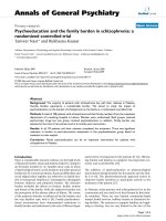
NMO patients had a TNF-
α
level lower than HC (Figure
1A). The confidence interval for the difference (CI 95%)
"HC-NMO" was (CI: 8.116; 19.050), indicating a differ-
ence greater than 8 units in HC with a high probability
(0.976). Interestingly, the levels of this cytokine were uni-
formly close to zero in all NMO patients. No significant
differences were observed in IFN-
γ
(Figure 1B) levels and
the measurements resulted widely dispersed in both
groups.
Concerning IL-10, we obtained a significant down-regula-
tion in R-NMO patients compared to controls. (CI: 1.476;
7.856), indicating a difference greater than 1.5 units in
HC with a high probability (0.956) (Figure 1C).
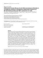
Lipid peroxidation, which may cause oligodendrocytic
damage, was assessed through MDA concentration and PP
evaluation. In fact, our results indicated a significant up-
regulation of the PP with a probability for the difference
to be over 0 of 0.990 (Figure 2A) and therefore of the
MDA with a probability for the difference to be over 0 of
0.986 (Figure 2B) in R-NMO patients compared to HC.
Proteins are major targets for radicals and other oxidants
when these are formed in both intra- and extracellular
environments in vivo. Damaged proteins may be highly
sensitive protein-based biomarkers of oxidative processes
in mammalian systems. Oxidized proteins are often func-
tionally inactive and their unfolding is associated with
enhanced susceptibility to proteinases. Here, we found
that the AOPP and THP were also up-regulated in R-NMO
patients (the probability that the difference be over 0 is
0.970 for AOPP and 0.976 for THP) (Figures 2C and 2D).
In our results we found that SOD and CAT enzymes were
up-regulated in R-NMO patients compared to HC (proba-
bility for the difference to be over 0 of 0.987 for SOD and
0.974 for CAT) suggesting the activation of a detoxifica-
tion feedback mechanism in turn to abolish the excess of
superoxide radicals as a result of the CNS inflammatory
process (Figures 2E and 2F). However, CAT/SOD ratio was
down-regulated in NMO patients compared to HC (Figure
2G).
Journal of Inflammation 2009, 6:18 />Page 4 of 9
(page number not for citation purposes)
We evaluated the serum levels of the metalloproteinase
MMP-9 and its inhibitor TIMP-1 from R-NMO patients
and HC (data not shown) and significant variations were
not found.
Furthermore, correlations between MMP-9, TIMP-1 and
MMP-9/TIMP-1 and EDSS scale and number of relapses in
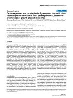
NMO patients were determined. Figure 3A shows the final
configuration to MDS viewing the distance (based on two
dimensions) between EDSS and molecular parameters in
patients. We can observe that EDSS is as close to TIMP-1
and MMP-9 as is the relation MMP-9/TIMP-1 with a high
degree of correspondence between the distances among
points implied by the MDS map and the matrix input, by
a stress of 0.0086.
In order to establish the specific relation between these
variables, we adjusted a Bayesian linear model:
where τ is the precision of the normal distribution
τ
=
σ
-2
with minimally informative distributions a priori for the
parameters of the model:
EDSS
TIMP
i
i
~ Normal
i
μτ
μαβ
,
*( )
()
=+ −
⎧
⎨
⎪
⎩
⎪
1
αβ
τ
,~ Normal
~ amma 10 10
-3 -3
010
6
,
,
−
()
()
⎧
⎨
⎪
⎩
⎪
i
G
Serum levels of cytokines in NMO patients and HC measured by ELISAFigure 1
Serum levels of cytokines in NMO patients and HC measured by ELISA. (A) TNF-
α
; (B) IFN-
γ
and (C) IL-10. CI:
Confidence interval for the difference "HC-NMO".
Journal of Inflammation 2009, 6:18 />Page 5 of 9
(page number not for citation purposes)
Serum levels of oxidative stress markersFigure 2
Serum levels of oxidative stress markers. (A) PP; (B) MDA; (C) AOPP; (D) THP; (E) SOD; (F) CAT and (G) CAT/
SOD ratio. CI: Confidence interval for the difference "HC-NMO".
Journal of Inflammation 2009, 6:18 />Page 6 of 9
(page number not for citation purposes)
Correlations between clinical and molecular parametersFigure 3
Correlations between clinical and molecular parameters. (A) Final configuration to Multidimensional Scaling (MDS).
(B) Adjusted Bayesian linear model between EDSS and TIMP-1. Stress: Degree of correspondence between the distances
among points implied by MDS map and the matrix of the observed data.
Journal of Inflammation 2009, 6:18 />Page 7 of 9
(page number not for citation purposes)
There is a significant linear dependence between EDSS
and TIMP-1 as can be observed in the graphic of density
function (Figure 3B), the confidence interval does not
contain 0, thereby an increase of the TIMP-1 values, repre-
sents an increase of the EDSS scale.
A similar analysis was done for the EDSS-oxidative stress
markers and the number of relapses with all molecular
parameters. We did not find any correlation between clin-
ical and molecular markers (data not shown).
Discussion
One theory of immune regulation involves homeostasis
between Th1 and Th2 activity. Overactivation of either
pattern can cause disease and either pathway can down-
regulate the other. But the hypothesis has major inconsist-
encies, human cytokine activities rarely fall into exclusive
pro-Th1 or -Th2 patterns. The regulatory T cells, likely
influence immunity in a manner comparable to Th1 and
Th2 cells. Many diseases previously classified as Th1 or
Th2 dominant fail to meet the set criteria [11], therefore
the most important element to considered is the effector
(Th1 or Th2) – regulator (T regulatory cells) equilibrium
which is lost in autoimmune diseases.
Some cytokines, such as TNF-
α
, IL-1 and IFN-
γ
, are well
known for their promotion of inflammatory responses.
However, these cytokines also have immunosuppressive
functions and their subsequent expressions also assist in
repair or recovery processes, suggesting a dual role for
some pro-inflammatory cytokines[12].
TNF-
α
is considered an inflammatory cytokine that
induces pleiotropic responses which comprise apoptosis
in some cells and proliferation in others. Reports indicate
either exacerbation or amelioration of pathological condi-
tions in the brain during TNF-
α
treatment, including
experimentally induced brain trauma [13] and a murine
model of multiple sclerosis [14,15]. No clinical efficacy or
worsening of the symptoms has been reported in some
Multiple Sclerosis patients treated with TNF-
α
inhibitors
[16]. In an animal model of demyelination/remyelina-
tion, the lack of TNF-
α
and TNFR2 provoked a significant
reduction in oligodendrocytes leading to a delay in the
generation of these cells, showing an unexpected role for
TNF-
α
and TNFR2 in repair of the CNS [17]. In humans,
beneficial effects of anti-TNF-
α
treatments in patients with
autoimmune diseases are proved; though surprisingly a
side effect of this treatment is the induction or exacerba-
tion of humoral autoimmunity disorders[18]. Although
controversial, human and murine studies suggest a link
between reduced TNF-
α
production and the development
of humoral autoimmunity, due to the prevention of CTL
induction and the subsequent control of autoreactive B
cells [19]. Recently, 15 cases of anti-TNF-
α
-associated
optic neuropathy were reported [20], the effects of anti-
TNF-
α
therapy may be related to changes in the balance of
immunologic homeostasis [21].
According to our results, the TNF-
α
levels from NMO
patients are practically undetectable evidencing that this
cytokine is important to protect the CNS from the damage
caused by the cascade of immune and inflammatory
events that characterize this disease and consequently
with reports that demonstrate neuroprotective [22,23]
neurotrophic [24] and immunomodulatory actions of
TNF-
α
.
Additionally, our results show a down-regulation of IL-10,
a Th2 and regulatory cytokine, suggesting not a specific
Th1/Th2 pattern but the imbalance of effector – regulator
mechanisms is implied. Low serum levels of IL-10 have
been reported in MS patients compared to HC, which
have been found to be more drastic in patients with the
progressive form of the disease [25] and treatments result-
ing in the up-regulation reduce the disease burden [26].
IL-10 levels of MS patients in remission were significantly
higher than those in the active phase [27] indicating that
in MS and probably in NMO, the regulatory cytokine, IL-
10, is impaired.
Regulatory mechanisms are essential in preventing
autoimmune disorders. Regulatory cytokines therapy is a
tempting strategy for reestablishing the immune balance
and thus preventing or reversing these disorders. Evi-
dently, the down-regulation of TNF-
α
and IL-10 cytokines
in NMO demonstrate a remarkable imbalance possibly
responsible for the several events of the NMO pathogene-
sis. Our results are consistent to those obtained by others
for MS [27] suggesting that it is possible to control NMO
restoring the regulatory cytokines balance.
The role of OS in the progressive demyelinating NMO dis-
ease has not been studied. It is seductive to speculate that
free radical oxygen chemistry contributes to pathogenesis
in this condition [28].
Such oxidative stress can damage the lipids, proteins and
nucleic acids of cells and mitochondria, potentially caus-
ing cell death. Oligodendrocytes are more sensitive to OS
[29] in vitro than are astrocytes and microglia, seemingly
due to a diminished capacity for antioxidant defense and
the presence of raised risk factors, including high iron
content. OS might therefore result in vivo in selective oli-
godendrocyte death, and thereby demyelination. The ROS
may also damage the myelin sheath, promoting its attack
by macrophages [30].
Damage can occur directly by lipid peroxidation and indi-
rectly by the activation of proteases and phospholipase
Journal of Inflammation 2009, 6:18 />Page 8 of 9
(page number not for citation purposes)
A2. Evidence for the existence of OS within inflammatory
demyelinating lesions includes the presence of both lipid
and protein peroxides.
Protein oxidation and enhanced proteolytic degradation,
therefore, have been suggested to cause a net increase in
ROS scavenging capacity. However, certain oxidized pro-
teins are poorly handled by cells, and together with possi-
ble alterations in the rate of production of oxidized
proteins, may contribute to the observed accumulation
and damaging actions of oxidized proteins during several
pathologies [31] such as shown by our results in NMO.
High activity of SOD in NMO patients may respond to an
over generation of O
2
•-
. If excess O
2
•-
is present, it can react
with norepinephrine, dopamine and serotonin [32], to
initiate their oxidation, which then continues with the
production of more ROS, quinones, etc. Damage to brain
readily releases iron (and copper) ions in forms capable of
catalyzing free radical reactions. Catalytic iron released by
the brain damage can persist because CSF has little or no
iron-binding capacity [33].
There is a high activity of SOD in patients without the pro-
portional increment of CAT to establish an appropriate
balance CAT/SOD; in addition, the apparently normal
level of THP, probably indicate an iron overload. In this
condition the over production of H
2
O
2
can occur. Neuro-
nal damage can involve direct effects of H
2
O
2
in inactivat-
ing sensitive enzymes as well as its reaction with iron ions
to form •OH [33].
Matrix metalloproteinases are a family of enzymes found
in the extracellular matrix. The control of the secretion of
these proteases as well as the balance between MMP secre-
tion and the secretion of their natural inhibitors TIMPs,
has an important relevance in the pathogenesis of demy-
elinating diseases [34]. MMP-9/TIMP-1 ratio may be
viewed as a reliable marker and may be predictive of MRI
activity in relapsing-remitting MS [35].
In a comparative study on the MMP-9 and TIMP-1 levels
in CSF from MS and NMO patients reported that the levels
of MMP-9 and TIMP-1 in NMO were similar to HC resem-
bling our results [36]. We also found that there is a specific
relationship between MMP-9, TIMP-1 and MMP-9/TIMP-
1 molecular parameters and EDSS clinical scale in a MDS
map and a significant linear dependence between EDSS
and TIMP-1. This result may suggest the relevance of these
molecular parameters as a prognostic factor of the clinical
measurement in the same way that it could have a predic-
tive value for the MRI activity.
Conclusion
Well designed clinical studies using pharmaceutical prod-
ucts directed towards the restoration of immuno-regula-
tory mechanisms, combined with an antioxidant or iron-
chelating agents are needed to assess whether they could
be beneficial for NMO treatment.
Competing interests
The authors declare that they have no competing interests.
Authors' contributions
GP-R, MC-Ll and GM-S participated in the conception and
design of experiments, acquisition and analysis of data,
interpretation of results and drafting the manuscript; IL-C
performed the ELISA assays. OR-N and MO-G carried out
the oxidative stress biochemical assays; JAC-G identified
and classified the NMO patients; MAR-A participated in
the NMO-IgG determinations; CV-S performed statistical
analysis. PAL-S contributed in the drafting of the manu-
script and revising it critically.
References
1. Argyriou AA, Makris N: Neuromyelitis optica: a distinct demy-
elinating disease of the central nervous system. Acta Neurol
Scand 2008, 118:209-217.
2. Lennon VA, Wingerchuk DM, Kryzer TJ, Pittock SJ, Lucchinetti CF,
Fujihara K, Nakashima I, Weinshenker BG: A serum autoantibody
marker of neuromyelitis optica: distinction from multiple
sclerosis. Lancet 2004, 364:2106-2112.
3. Gilgun-Sherki Y, Melamed E, Offen D: The role of oxidative stress
in the pathogenesis of multiple sclerosis: the need for effec-
tive antioxidant therapy. J Neurol. 2004, 251(3):261-268.
4. Wingerchuk DM, Lennon VA, Pittock SJ, Lucchinetti CF, Weinsh-
enker BG: Revised diagnostic criteria for neuromyelitis
optica. Neurology 2006, 66:1485-1489.
5. Witko-Sarsat V, Friedlander M, Nguyen Khoa T, Capeillère-Blandin C,
Nguyen AT, Canteloup S, Dayer JM, Jungers P, Drüeke T, Descamps-
Latscha B: Advanced oxidation protein products as novel
mediators of inflammation and monocyte activation in
chronic renal failure. J Immunol 1998, 161:2524-2532.
6. Witko VA, Nguyen T, Descamps-Latscha B: Microtiter plate assay
for phagocyte-derived taurine-chloramines. J Clin Lab Anal
1992, 6:47.
7. Esterbauer H, Cheeseman KH: Determination of aldehydic lipid
peroxidation product: malonaldehyde and 4-hydroxynone-
nal. Method in Enzymology 1990, 186:407-421.
8. Özdemirler G, Mehmetçik G, Oztezcan S, Toker G, Sivas A, Uysal M:
Peroxidation potential and antioxidant activity of serum in
patients with diabetes mellitus and myocard infarction.
Horm Metab Res 1995, 27:194-196.
9. Shukla GS, Hussain T, Chandra SV: Possible role of superoxide
dismutase activity and lipid peroxide levels in cadmium neu-
rotoxicity: in vivo and in vitro studies in growing rats. Life Sci
1987, 41(19):2215-2221.
10. BOEHRINGER MANNHEIM: Biochemica Information. A
revised biochemical reference source. In Enzymes for routine 1st
edition. Germany: Boehringer Mannheim; 1987:15-16.
11. Kidd P: Th1/Th2 balance: the hypothesis, its limitations, and
implications for health and disease. Altern Med Rev 2003,
8:223-246.
12. Correale J, Villa A: The neuroprotective role of inflammation
in nervous system injuries. J Neurol 2004, 251:1304-1316.
13. Shohami E, Ginis I, Hallenbeck JM: Dual role of tumor necrosis
factor
α
in brain injury. Cytokine Growth Factor Rev 1999,
10(2):119-130.
14. Liu J, Marino MW, Wong G, Grail D, Dunn A, Bettadapura J, Slavin AJ,
Old L, Bernard CC: TNF is a potent anti-inflammatory
Publish with BioMed Central and every
scientist can read your work free of charge
"BioMed Central will be the most significant development for
disseminating the results of biomedical research in our lifetime."
Sir Paul Nurse, Cancer Research UK
Your research papers will be:
available free of charge to the entire biomedical community
peer reviewed and published immediately upon acceptance
cited in PubMed and archived on PubMed Central
yours — you keep the copyright
Submit your manuscript here:
/>BioMedcentral
Journal of Inflammation 2009, 6:18 />Page 9 of 9
(page number not for citation purposes)
cytokine in autoimmune-mediated demyelination. Nat Med
1998, 4:78-83.
15. Kassiotis G, Kollias G: Uncoupling the pro-inflammatory from
the immunosuppressive properties of tumor necrosis factor
(TNF) at the p55 TNF receptor level. Implications for patho-
genesis and therapy of autoimmune demyelination. J Exp Med
2001, 193:427-434.
16. The Lenercept Group: TNF neutralization in MS: results of a
randomized, placebo-controlled multicenter study. Neurology
1999, 53:457-465.
17. Arnett HA, Mason J, Marino M, Suzuki K, Matsushima GK, Ting JP:
TNF alpha promotes proliferation of oligodendrocyte pro-
genitors and remyelination. Nat Neurosci 2001, 4:1116-1122.
18. Charles PJ, Smeenk RJ, De Jong J, Feldmann M, Maini RN: Assess-
ment of antibodies to double-stranded DNA induced in
rheumatoid arthritis patients following treatment with inf-
liximab, a monoclonal antibody to tumor necrosis factor
alpha: findings in open-label and randomized placebo-con-
trolled trials. Arthritis Rheum 2000, 43:2383-2390.
19. Via CS, Shustov A, Rus V, Lang T, Nguyen P, Finkelman FD: In vivo
neutralization of TNF-alpha promotes humoral autoimmu-
nity by preventing the induction of CTL. J Immunol 2001,
167:6821-6826.
20. Journal compilation: Royal Australian and Zealand College of
Ophthalmologists. Letters to the Editor 2007.
21. Robinson WH, Genovese MC, Moreland LW: Demyelinating and
neurologic events reported in association with tumor necro-
sis factor alpha antagonism: by what mechanisms could
tumor necrosis factor alpha antagonism improve rheuma-
toid arthritis but exacerbate multiple sclerosis? Arthritis Rheum
2001, 44:1977-1983.
22. Turrin NP, Rivest S: Tumor necrosis factor alpha but not inter-
leukin 1 beta mediates neuroprotection in response to acute
nitric oxide excitotoxicity. J Neurosci 2006, 26:143-151.
23. Lastres-Becker I, Cartmell T, Molina-Holgado F: Endotoxin precon-
ditioning protects neurones from in vitro ischemia: role of
endogenous IL-1beta and TNF-alpha. J Neuroimmunol 2006,
173:108-116.
24. Pickering M, Cumiskey D, O'Connor JJ: Actions of TNF-alpha on
glutamatergic synaptic transmission in the central nervous
system. Exp Physiol 2005, 90:663-670.
25. Salmaggi A, Dufour A, Eoli M, Corsini E, La Mantia L, Massa G, Nes-
polo A, Milanese C: Low serum interleukin-10 levels in multiple
sclerosis: further evidence for decreased systemic immuno-
suppression? J Neurol 1996, 243:13-17.
26. Ozenci V, Kouwenhoven M, Huang YM, Xiao B, Kivisäkk P, Fredrik-
son S, Link H: Multiple sclerosis: levels of interleukin-10-
secreting blood mononuclear cells are low in untreated
patients but augmented during interferon-beta-1b treat-
ment. Scand J Immunol 1999, 49:554-561.
27. Perrella O, Sbreglia C, Perrella M, Spetrini G, Gorga F, Pezzella M,
Perrella A, Atripaldi L, Carrieri P: Interleukin-10 and tumor
necrosis factor-alpha: model of immunomodulation in multi-
ple sclerosis. Neurol Res 2006, 28:193-195.
28. Calabrese V, Lodi R, Tonon C: Oxidative stress, mitochondrial
dysfunction and cellular stress response in Friedreich's
ataxia. J Neurol Sci 2005, 233:145-162.
29. Kanwar JR: Anti-inflammatory immunotherapy for multiple
sclerosis/experimental autoimmune encephalomyelitis
(EAE) disease. Curr Med Chem 2005, 12:2947-2962.
30. Smith KJ, Kapoor R, Felts PA: Demyelination: the role of reac-
tive oxygen and nitrogen species. Brain Pathol 1999, 9:69-92.
31. Martínez-Sánchez G, Giuliani A, Pérez-Davison G: Oxidized pro-
teins and their contribution to redox homeostasis. Redox
Report 2005, 10:174-184.
32. Wrona MZ, Dryhurst G: Oxidation of serotonin by superoxide
radical: implications to neurodegenerative brain disorders.
Chem Res Toxicol 1998, 11:639-650.
33. Halliwell B: Oxidative stress and neurodegeneration: where
are we now? J Neurochem 2006, 97:1634-1658.
34. Cross AK, Woodroofe MN: Chemokine modulation of matrix
metalloproteinase and TIMP production in adult rat brain
microglia and a human microglial cell line in vitro. Glia 1999,
28:183-189.
35. Avolio C, Filippi M, Tortorella C, Rocca MA, Ruggieri M, Agosta F,
Tomassini V, Pozzilli C, Stecchi S, Giaquinto P, Livrea P, Trojano M:
Serum MMP-9/TIMP-1 and MMP-2/TIMP-2 ratios in multiple
sclerosis: relationships with different magnetic resonance
imaging measures of disease activity during IFN-beta-1a
treatment. Mult Scler 2005, 11:441-446.
36. Mandler RN, Dencoff JD, Midani F, Ford CC, Ahmed W, Rosenberg
GA: Matrix metalloproteinases and tissue inhibitors of met-
alloproteinases in cerebrospinal fluid differ in multiple scle-
rosis and Devic's neuromyelitis optica. Brain 2001,
124:493-498.