Báo cáo y học: "Cigarette smoke regulates VEGFR2-mediated survival signaling in rat lungs" pot
Bạn đang xem bản rút gọn của tài liệu. Xem và tải ngay bản đầy đủ của tài liệu tại đây (966.96 KB, 9 trang )
RESEA R C H Open Access
Cigarette smoke regulates VEGFR2-mediated
survival signaling in rat lungs
John A Marwick
1,2
, Indika Edirisinghe
4
, Gnanapragasam Arunachalam
4
, Christopher S Stevenson
1,2
,
William MacNee
3
, Paul A Kirkham
1,2*
, Irfan Rahman
4*
Abstract
Background: Vascular endothelial growth factor (VEGF) and VEGF receptor 2 (VEGFR2)-mediated survival signaling
is critical to endothelial cell survival, maintenance of the vasculature and alveolar structure and regeneration of
lung tissue. Reduced VEGF and VEGFR2 expression in emphysematous lungs has been linked to increased
endothelial cell death and vascular regression. Previously, we have shown that CS down-regulated the VEGFR2 and
its downstream signaling in mouse lungs. However, the VEGFR2-mediated survival signaling in response to
oxidants/cigarette smoke (CS) is not known. We hypothesized that CS exposure leads to disruption of VEGFR2-
mediated endothelial survival signaling in rat lungs.
Methods: Adult male Sprague-Dawley rats were exposed CS for 3 days, 8 weeks and 6 months to investigate the
effect of CS on VEGFR2-mediated survival signaling by measuring the Akt/PI3-kinase/eNOS downstream signaling in
rat lungs.
Results and Discussion: We show that CS disrupts VEGFR2/PI3-kinase association leading to decreased Akt and
eNOS phosphorylation. This may further alter the phosphorylation of the pro-apoptotic protein Bad and increase
the Bad/Bcl-xl association. However, this was not associated with a significant lung cell death as evidenced by
active caspase-3 levels. These data suggest that although CS altered the VEGFR2-mediated survival signaling in the
rat lungs, but it was not sufficient to cause lung cell death.
Conclusion: The rat lungs exposed to CS in acute, sub-chronic and chronic levels may be representative of
smokers where survival signaling is altered but was not associated with lung cell death whereas emphysema is
known to be associated with lung cell apoptosis.
Introduction
Maintenance of the microvasculature in the lung is criti-
cal for gas exchange, the integrity of the alveolar struc-
ture and tissue repair [1]. Cigarette smoke (CS)-induced
emphysema is characterized by enlargement of the air-
spaces and a loss of alveolar structure [2,3] . Endothelial
cell death and the regression of lung parenchyma, capil-
lary density seen in emphysema may be linked to this
loss of the alveolar structure [4,5].
Vascular e ndothelial growth factor (VEGF) plays vital
role in development and maintenance of vasculature
and tissue regeneration [6]. VEGF signaling on
endothelial cells is involved in several key processes dur-
ing wound healing including degradation of the extracel-
lular matrix of existing vessels, migration and
proliferation of capillary endothelial cells, formation of
new capillaries and restitution of the air-blood barrier in
the alveoli [1, 7]. Targeted disruption of VEGF gene in
mice impairs blood vessel formation, growth retardation
and premature death [8]. Furthermore, deletion or inhi-
bition of VE GF in specific tissues in ad ult mice has
shown noticeable effects, mainly significant reduction in
capillary density with tissue cell apoptosis [9].
VEGF signaling through VEGF receptor 2 or kinase
insert domain receptor (a type III receptor tyrosine
kinase) or protein-tyrosine kinase receptor FLk-1
(VEGFR2) is key in endothelial survival and th e mainte-
nance of the vasculature [10,11]).). VEGFR2 inhibition
leading to endothelial cell death has been linked to both
* Correspondence: ;
edu
1
National Heart and Lung Institute, Imperial College London, UK
4
Department of Environmental Medicine, Lung Biology and Disease Program,
University of Rochester Medical Centre, Rochester, NY, USA
Marwick et al. Journal of Inflammation 2010, 7:11
/>© 2010 Marwick et a l; licensee BioMed Central Ltd. This is an Open Access article distributed under the terms of the Creative Commons
Attribution License ( which permits unrestricted use, distribution, and repro duct ion in
any medium, pro vided the original work is properly cited.
lung vascular regression and alterations in alveolar
structure [12,13]) VEGF/VEGFR2-mediated endothelial
survival signals is predominantly mediated through
phosphatidylinositol-3-OH kinase (PI-3K) and its down-
stream target of the serine-theronine kinase Akt [10].
Akt is a general mediator of growth factor-induced sur-
vival and has shown to suppress the apoptotic death in
vitro induced by a variety of stimuli, including growth
factor withdrawal, cell-cycle discordance, loss of cell
adhesion and DNA damage [14-17]. VEGF-mediated
survival signaling is mediated through the upregulation
of anti-apoptotic proteins such as Bcl-2 and A1 [18],
and IAP (inhibitors of apoptosis proteins), survivin and
IXAP (X-chromosome-linked IAP) [19], which may inhi-
bit upstream caspases and terminal effecter caspases
respectively.
Bad is an pro-apoptotic member of the Bcl-2 family
proteins that can displace Bax binding to Bcl-2 and Bcl-
xl, results in cell death [20,21]. Survival factor IL-3 can
inhibit the apoptotic activity of Bad by activating intra-
cellular signaling pathways that results in phosphoryla-
tion of Bad (Ser112 and Ser136) [22]. This further leads
to binding of Bad to 14-3-3 proteins and inhibition of
Bad binding to Bcl-2 and Bcl-xl [22]. Akt has been
shown to promote cell survival via its ability to phos-
phorylate Bad at Ser136 residue [23].
VEGF and VEGFR2-mediated downstream signaling
activates eNOS [24], and release nitric oxide (NO) [25].
The mechanism of cell survival by NO can be directly
linked to increased neovascularisation and cell migration
[26] or by increasing Bcl-2 expression [27]. Previously,
we have demonstrated that CS-induced oxidative stress
impairs VEGF-mediated VEGFR2 phosphorylation and
VEGFR2 expression i n both endothelial cells and mouse
lung [28], and in emphysematous lungs o f both smokers
and non-smokers [4,29]. However, the mechanism of
CS-induced VEGFR2-mediated impaired Akt and its
downstream signaling leading t o apoptotic cell death in
lung has not been studied. Therefore, we hypothesized
that CS regulates VEGFR2-mediated survival signaling
via Akt-dependent pa thways in rat lung. To test this
hypothesis, rats were exposed to CS for different time
points (3 days, 8 weeks and 6 months) and VEGFR2/
PI3-kinase association, Akt, eNOS, Bad phosphorylation
and active caspase levels were determined.
Materials and methods
Animals
Adult male Sprague-Dawley rats (323 ± 2.5 g) (Charles
River, Margate, UK) w ere divided into 6 exposure
groups: (a) 3 day sham exposed (n = 6), (b) acute 3 day
CS exposed (n = 6), (c) 8 week sham exposed (n = 6),
(d) sub-chronic 8 week CS exposed (n = 6), (e) 6
mont hs sham exposed (n = 6) and (f) chronic 6 months
CS exposed (n = 6). The rats were exposed to whole
body CS generated from 2R4F research cigarettes (Uni-
versity of Kentucky, Lexington, Kentucky, USA, total
particulate matter (TPM) concentration 27.1 ± 0.8 mg
per cigarette) in 7 L smoking chambers at 4 c igarettes
per day, Monday to Friday. To ensure a consistent expo-
sure across exposed animals, cotinine levels were mea-
sured. Plasma cotinine levels were 2.66 ± 0.12 μMafter
1 hr exposure (cotinine was not detectable in the plasma
fromair-exposedanimals)and0.51±0.07μM after 24
hr. There was no progressive increase in cotinine levels
over 1 week of exposures. Carboxyhemoglobin level was
measured immediately after the animals were removed
from the chambers. A peak level of 42 ± 4.0 μMwas
reached after the 4
th
cigarette, which quickly decreases
after the exposure was stopped. Sham exposed animals
where exposed to medical grade air under the same
conditions as CS exposed animals. Rats were sacrificed 2
hr post-last exposure by intra-peritoneal injection of 200
mg sodium pentobarbital.
Tissue processing
The lungs were excised from rats, the right lobe tied off
and then snap frozen in liquid nitrogen for immunoblot-
ting and immunoprecipitation experiments. The left lobe
was inflated with 5 ml of 10% neutral buffered forma lin
and then immersed in NBF to complete fixation for 24
hr. The left lobe was then sliced tangentially into 6
slices, which were processed as two tissue blocks. Sec-
tions ( 3 μm) of the 4 central lung slices were cut using
a Leica rotary microtome. The sections were mounted
on to Polysine slides (Surgip ath Europe Ltd, Cambridge,
UK) and dried overnight at 37°C.
Immunohistochemistry
Briefly, lung sections we re dewaxed in xylene, rehy-
drated and endogenous peroxidase inhibited with 0.5%
hydrogen peroxide in methanol for 10 minutes. Sections
were stained with anti-active caspase 3 (Abcam, Cam-
bridge,UK),overnightat4°C. Immunodetection was
preformed using biotinylated rabbit anti-mouse IgG
antibody/reagent (Dako Cytomation, Cambridgeshire,
UK), SABC reagent (Dako Cyto mation, Cambridgeshire,
UK), and 3,3’ -diaminobenzidine (DAB) (Sigma, Dorset,
UK). The nuclei were counterstained with Cole’s haema-
toxylin solution. Tonsil was used as a positive control
and for negative controls the primary antibody was
omitted from one section of each of the samples. Two
fields to the right of the large airway in two pieces of
the left lobe were counted (i.e. 4 fields, total area
approximately 6.5 mm
2
). When there was a difference
of more than 5 cells/mm
2
between the average counts
of 2 and 4 fields, an extra field was counted in piece 3
(total area of approximately 8.3 mm
2
).
Marwick et al. Journal of Inflammation 2010, 7:11
/>Page 2 of 9
Whole cell lung homogenate
Rat lung tissue (0.1 g) were homogenized in 1 ml of ice-
cold lysis buffer containing 1% Nonident 40 (NP-40),
0.1% SDS, 0.01 M deoxycholic acid and a complete pro-
tease cocktail inhibitor with EDTA (Roche, East Sussex,
UK) and incubated on ice for 45 min. The samples were
then centrifuged at 13,000 rpm for 25 min at 4°C and
the supernatant aliquoted and stored at -80°C.
Western Blotting
Lung tissue homogenate samples were separated on SDS
polyacrylamide gel. Separated proteins were electro-
blotted onto nitrocellulose membranes (Schleicher and
Schuell, Dassel, Germany) and blocked for 1 hr at room
temperature with 5% nonfat dry milk. The membranes
were incubated with anti-VEGFR2 (Santa Cruz, Santa
Cruz, CA, USA), PI-3K (Upstate, Milton Keynes, UK),
anti-Akt, anti-phosphoacetylated-Akt, anti-Bad, anti-
phosphoacetylated Bad (Ser1 36), anti-Bcl-2, anti- Bcl-xl,
anti-phosphorylated eNOS ( Ser1177), anti-eNOS (Cell
Signaling) and anti-b-actin (Santa Cruz Biotechnology).
Immunoprecipitation
Lung homogenate (200 μgofprotein)inafinalvolume
of 100 μl lysis buffer w as pre-cleared with Protein-A-
agarose beads (Calbiochem, Merck Biosciences, Notting-
ham, UK) for 30 minutes at 4°C with constant agitation.
The samples were then incubated wit h the antibody for
1 hr at 4°C with constant agitation. Protein-A-agarose
beads were then added and left at 4°C overnight with
constant agitation. The samples were then centrifuged,
the supernatant discarded and the beads were washed in
lysis buffer and heated with sample loading buffer. The
samples were then run on Western blots using SDS-
PAGE. Blots were probed with anti-PI-3K (Upstate) and
anti-Bad (Cell Signaling) a nd stripped, reprobed with
anti-Bcl-xl (Cell Signaling) or anti-VEGFR2 (Santa Cruz)
as loading controls.
Protein Assay
Protein level was measured with a bicinchoninic acid kit
(Bio-Rad Laboratories Inc., Hercules, California, USA).
Protein standards were obtained by dilution of a stock
solution of BSA. Linear regression was used to deter-
mine the actual protein concentration of the samples.
RNA isolation and reverse-transcriptase PCR
Lung tissue (0.1 g) was homogenized in 1 ml of Trizol
(Invitrogen Life Technologies, Paisley, UK) and left at
room temperature for 15 minutes. RNA was extracted
according the manufacturer’s instructions. The RNA was
the aliquoted and stored at -80°C until further use . RNA
was quantified by a spectrophotometer at 260 nm. Protein
contamination was estimated at 280 nm and a ratio of <
1.6 was accepted. RNA (2 μg) was reverse transcribed in
to cDNA using oligo-dT MMLV-Reverse Transcriptase in
a20μl final volume. RT-PCR was then preformed using 5
μg of cDNA (primers see below) using 1× PCR buffer (50
mM KCl, 10 mM Tris-HCl pH 9.0, 1.5 mM MgCl
2
and
0.1% Triton X-100), 400 μM dNTP, 1 mM MgCl
2
,50pM
of each 5’ and 3’ primer and 2 U of Taq DNA polymerase
(Promega). RT-PCR amplification was carried out using
rat Bcl-2 up 5’-CCG GGA GAT CGT GAT GAA GTA-3’;
Bcl-2 rev 5’-CAT ATT TGT TTG GGG CAT GTC T-3’
and rat GAPDH).(36) as a loading control using previously
described RT-PCR parameters [bcl2: 94°C for 45 sec; 58°C
for 45 sec; 72°C for 60 sec for 25 cycles and then final 72°
C for 10 min (product size 508 bp) and GAPDH: 95°C for
30 sec; 60°C for 60 sec; 60°C for 2 mins for 25 cycles and
then 68°C for 7 mins (product size 520 bp)] [29]. RT-PCR
reactions were carried out using a thermocycler and the
PCR products were electorophoresed on a 1.5% agarose
gel containing ethidium bromide (EtBr). The bands were
visualised and quantified by densitometry using a UV
Grab-IT software package.
Statistical analysis
All the data are expressed as Mean ± SEM. Statistical
analysis of significance was preformed using Minitab
software. The da ta were normally distributed and values
obtained in the different groups of rats were compared
using one-way analysis of variance (ANOVA) using
Tukey’s post-hoc test. Statistical significance was consid-
ered at P < 0.05, P < 0.01 and P < 0.001 levels.
Results
Cigarette smoke regulates VEGFR-2-PI3K interaction in rat
lung
Our previous studies have shown that CS reduced
VEGFR2 phosphorylation and its total levels in mouse
and rat lungs [28,29]. One of the key initiators of
VEGFR2-mediated endothelial survival signaling is PI-
3K. It is known that PI-3K interacts with VEGFR2
directly by the p85 catalytic subunit [11]. We therefore
investigated the effect of CS on VEGFR2-PI-3K interac-
tion in rat lungs (Fig. 1). No significant alteration was
observed on PI-3K (p85 subunit) interaction with
VEGFR2 after acute or sub-chronic CS exposure com-
pared to sham-exposed animals. However, significant (P
< 0.01) reduction was observed after 6 months of CS
exposure suggesting that chronic CS exposure reduced
VEGFR2-PI-3K interaction in rat lungs.
Cigarette smoke reduced Akt phosphorylation
Previous study have shown that activation of Akt is
pivotal in VEGF/VEGFR2-mediated endothelial cell sur-
vival [10]. We assess ed the effect of CS on Akt phos-
phorylation by immunoblotting in rat lung . We fo und
Marwick et al. Journal of Inflammation 2010, 7:11
/>Page 3 of 9
that Akt phosphorylation was significantly (P < 0.001)
reduced after sub-chronic and chronic CS exposure
compared to sham exposed animals (Fig. 2). However,
CS exposure did not alter the total Akt level in acute,
sub-chronicandchronicexposuretimepoints.This
data suggested that sub-chronic and chronic CS expo-
sure impaired Akt survival signaling but not with acute
CS exposure in rat lungs.
Chronic CS exposure led to decreased Bad
phosphorylation
Bad can inhibit anti-apoptotic signals of Bcl-2 and Bcl-
xl, resulting in apoptotic cell death. Phosphorylation of
Bad leads its inactivation and inhibition of Bad binding
to Bcl-2 and Bcl-xl [22]. We therefore determined the
effect of CS on Bad phosphorylation by immunoblotting.
Phosphorylation of B ad (Ser136) was significantly (P <
0.01) reduced in chronic CS exposure compared to
sham exposed groups (Fig. 3). However, acute and sub
chronic CS exposures did not show any effect on B ad
phosphorylation. The alterations of Bad phosphorylation
inchronicCSexposurewascorrelated with decreased
phosphorylation of Akt in rat lungs suggesting that CS
exposure alters Akt-mediated survival signal by modula -
tion on Bad (Ser136) phosphorylation.
Cigarette smoke increased the Bcl-xl-Bad association
It has been shown that interaction of Bad with Bcl-xl
inhibit its anti-apopto tic effect and activates apoptotic
events [20]. Hence the interaction of Bad with Bcl-xl
was assessed by immunoprecipitation and immunoblot-
ting. We found that acute and chronic CS exposures
significantly (P < 0.01) increased th e Bad/Bcl-xl binding
compared with sham exposed animals (Fig. 4). These
data indicate that increased association of B ad/Bcl-xl
may lead to increased apoptotic cell death in rat lungs.
Cigarette smoke had no affect on Bcl-2 mRNA expression
It has been shown that VEGF/VEGFR2-mediated survi-
val signal may be mediated through sustained upregula-
tion of Bcl2 expression [18]. CS had no effect on
expression of Bcl-2 mRNA as measured by RT-PCR in
both acute and chronic time points compared with
sham operated animals (Fig. 5). This data suggests that
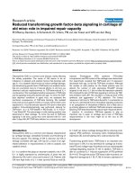
Figure 1 VEGFR2-PI-3K association in rat lungs exposed to CS.
(A) A representative immunoblot picture of immunoprecipitated
VEGFR2 probed for the p85 catalytic subunit of PI-3K after 3 days, 8
weeks and 6 months of CS exposure in rat lungs. The interaction of
the p85 subunit of PI-3K was unaltered after 3 days and 8 weeks of
CS exposure but was significantly increased after 6 months of CS
exposure compared to sham-exposed animals (n = 6). (B)
Histograms represent the Mean ± SE of percentage of VEGFR2/PI-3K
association. ** p < 0.01 compared to sham-exposed animals.
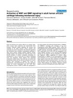
Figure 2 Effect of CS on Akt phosphoryaltion.(A)A
representative immunoblot picture of Akt phosphorylation after 3
days, 8 weeks and 6 months of CS exposure in rat lungs. Akt
phosphorylation was significantly reduced both at 8 weeks and 6
months, but not after 3 days of CS exposure, compared to sham-
exposed animals (n = 6). (B) Histograms represent the Mean ± SE of
percentage of Akt phosphorylation. *** p < 0.001 compared to
sham-exposed animals.
Marwick et al. Journal of Inflammation 2010, 7:11
/>Page 4 of 9
CS does not affect Bcl-2 mRNA expression in response
to either acute or chronic exposures in rat lungs.
Cigarette smoke reduced the eNOS level and its
phosphorylation
VEGF-induced VEGFR2 phosphorylation and downstream
signaling leading to activation of eNOS, a key enzyme
linked to endothelial survival and function. Therefore, the
effect of CS on phosphorylated and total eNOS levels was
ass essed by immunoblotting. CS significantly (P < 0.001)
reduced the level of phophorylated and total eNOS com-
pared to sham-ex posed rat lungs after sub-chronic expo-
sure without any significant change aft er acute exposure
(Fig. 6). We expe ct a similar reduction in total and phos-
phorylation eNOS after chronic CS exposure compared to
sham-exposed rat lungs. These data suggested that chronic
CS exposure impairs the activation of eNOS in rat lungs
which may have further implication on decreased NO pro-
duction and endothelial dysfunction.
Cigarette smoke exposure had no effect on activation of
caspase 3 or lung cell death
Increased endothelial cell death was observed in emphy-
sematous lungs of smokers indicate that apoptotic cell
death may play a role in pathogenesis of COPD [4]. To
investigate the effect of CS on lung cell death, expres-
sion of active caspase 3 was assessed by immunohisto-
chemmistry. There was no difference in active caspase 3
expression between CS exposed and sham-exposed ani-
mals after either acute or chronic exposures (Fig. 7).
These data suggested that CS exposure was not asso-
ciated with lung cell death.
Discussion
VEGFR2-mediated Akt survival signaling has been
shown to be critical in endothelial cell survival [10]. Pre-
vious studies have shown that emphysema patients have
decreased VEGF and VEGFR2 expression along with
increased endothelial cell death [4,29]. Moreover, inhibi-
tion of VEGFR2 has also showed increased lung
endothelial cell death in rats [12]) VEGFR2 activates
Akt by interacting with PI-3K through its p85 subunits
[10,11]. In our data we observed significant alteration in
VEGFR2/PI-3K interaction after chronic CS exposure in
ratlungs.ThissupportsourconceptthatchronicCS
exposure in rat lungs reduces the interaction of both PI-
3K and VEGFR2 thus further leads to alteration of Akt-
mediated downstream survival signaling.
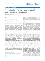
Figure 3 Effect of CS on Bad phosphorylation.(A)A
representative immunoblot picture of Bad phosphorylation (Ser136)
after 3 days, 8 weeks and 6 months of CS exposure in rat lung. Bad
phosphorylation (Ser136) was unaltered after 3 days and 8 weeks
but significantly decreased after 6 months of CS exposure
compared to sham-exposed animals (n = 6). (B) Histograms
represent the Mean ± SE of percentage of phosphorylated Bad
levels. ** p < 0.01 compared sham-exposed animals.
Figure 4 Bad-Bcl-x l interaction in CS exposed rat lungs.(A)A
representative immunoblot picture of immunoprecipitated Bcl-xl
probed for Bad after 3 days, 8 weeks and 6 months of CS exposed
rat lungs. Bad interaction with Bcl-xl was significantly increased at 3
days, 8 weeks and 6 months CS exposure compared to sham-
exposed animals (n = 6). (B) Histograms represent the Mean ± SE of
percentage of Bad interaction with Bcl-xl. ** p < 0.01 compared to
sham-exposed animals.
Marwick et al. Journal of Inflammation 2010, 7:11
/>Page 5 of 9
Our previous studies showed that CS significantly
decreases the VEGFR2 and Akt levels i n mouse and rat
lungs [28,29], and this is likely to be linked to VEGFR2-
mediated survival signaling. In present study, we show
that Akt phosphorylatio n was significantly reduced after
sub-chronic and chronic CS exposures compared to
sham-exposed animals, which was directly correlated
with decreased PI-3K/VEGFR2 interaction on CS
exposed animals. T his altered VEGFR2/PI-3K associa-
tion and impaired Akt phosphorylation may further
leads to modifications on its downstream targets via Bad
phosphoryation. The reason for CS-mediated reduction
of VEGFR2 is not known but our recent study suggested
that VEGFR2 is post-translationally modified by ROS/
RNS present or derived by CS [30].
The pro-apoptotic Bad is the primary target o f Akt and
Akt phosphorylates Bad and rendering it inactive for
apoptotic signal [20]. In this study, we show that after 6
months of CS exposure, there was a significant decrease
in Bad phosphoryation (Ser136) in lungs as compared to
sham-exposed animals. These data are consistent with
the reduction of Akt phosphorylation seen in the CS
exposed animals, and indicates that decreased phosphor-
ylation of Bad may further increase its association with
Bcl-xl. The lack of any change seen after acute and sub-
chronic CS exposure may be due to a cross-talk with
other receptors and signaling pathways, compensating
for any decrease in Akt activation or the decrease seen in
Akt activation was not sufficient to impact on Bad
Ser136 phosphorylation levels. However, further studies
are required to clarify the role played by Bad phosphory-
lation in response to CS exposure in lung cell death.
Bcl-xl is an anti-apoptotic pro tein that prom otes cell
survival by inhibiting caspase-mediated apoptotic cell
death [20]. Heterodimerization of Bad with Bcl-xl pre-
vents anti-apoptotic effect of Bcl-xl [31]. Bcl-xl can also
binds directly to the outer membrane of the mitochon-
dria, formi ng a pore to allow anionic metabolit e
exchange across the membrane and promoting cell sur-
vival during apoptotic signaling [32]. Native Bad can
bind to Bcl-xl, displacing Bax and preventing the Bcl-xl
binding to the mitochondria membrane [20]). Hence, we
studied the Bcl-xl/Bad interactions in acute , sub-chronic
and chronic CS exposed ani mals. We found that Bcl-xl/
Bad interaction was significantly elevated after acute and
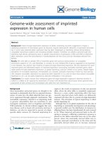
Figure 5 Bcl-2 mRNA levels in CS exposed rat lung.(A)A
representative RT-PCR picture of Bcl-2 mRNA levels after 3 days, 8
weeks and 6 months of CS exposure. Bcl-2 mRNA expression remains
unaltered at all time point compared to sham-exposed animals (n =
6). (B) Histograms represent the Mean ± SE of percentage of Bcl-2
mRNA expression levels.
Figure 6 Phosphorylated and total eNOS levels in CS exposed
rat lung.(A) A representative immunoblot picture of
phosphorylated and total eNOS after 3 days and 8 weeks of CS
exposure in rat lungs. Phophorylated and total eNOS levels were
significantly reduced in 8 weeks, but not after 3 days of CS
exposure compared to sham-exposed animals (n = 6). (B)
Histograms represent the Mean ± SE of percentage of eNOS
phosphorylation. *** p < 0.001 compared to sham-exposed animals.
Marwick et al. Journal of Inflammation 2010, 7:11
/>Page 6 of 9
chronic CS exposures in rat lungs. However, there was
no alteration in Bad phosphorylation (Ser136) after
acute and sub-chronic CS exposures indicating that this
elevated Bad-Bcl-xl interaction may be independent of
Bad phosphorylation at this site. Bad is also phosph ory-
lated by protein kinase C at Ser155. Phosphorylation of
Bad a t Ser155 is also thought to play an important role
in prevention of dimerization of Bad to Bcl-xl due to its
position on the BH3 domain [33]. Therefore, further
studies are required to assess the role of phosphorylated
and total levels Bad in apoptotic-mediated cell death.
VEGF-mediated VEGFR2 phosphorylation and its
downstream signaling via Akt induced the a ctivation of
eNOS in endothelium (24). It has been shown that
eNOS inhibits apoptosis and increase cell survival
through Bcl-2-dependent pathway [27]. Since eNOS is
also an essential mediato r of VEGFR2-mediated
endothelial survival, we determined whether CS exp o-
sure had any effect on phosphorylate d and total eNOS
levels. Our data showed that chronic CS exposure
decreased the phosphorylated and total eNOS levels in
rat lungs. These data are in agreement with reduction in
Akt phosphorylation in response to CS. Recently, we
have shown that CS impairs the VEGF induced
VEGFR2-mediated eNOS phosphorylation levels in
human microvascular endothelial cells [30 ]. Upon acti-
vation by its upstream kinases eNOS release NO in
endothelial cells, which is known to mediates cell survi-
val and resistance to apoptosis [25]. Hence this data
further support our concept that CS decreases VEGFR2-
mediated downstream signaling thus leading to dimin-
ished NO production and cell survival.
Caspases, in particular caspase 3, are important mar-
ker of cell undergoing apoptotic cell death. CS had no
effect on the level of active caspase 3 in lung cells. This
data corroborates with previous studies demonstrating
that lung cell death events were not significant between
smokers and non-smokers though CS causes destruction
of alveolar wall and reduction in vascular density only in
emphysematous lungs [4]. We have recent ly reported
that emphysema-like changes were observed after 8
months of CS exposure in rat model [34]. It is possible
that 6 months of CS exposure may explain the n otice-
able changes and different events of apoptotic cell
death. Taken together, our data indicate that CS expo-
sure alters VEGFR2-mediated survival signaling in rat
lungs. However, despite the reduced VEGFR2-PI3-K
association, Akt activation, Bad phosphorylation and
increased Bad/Bcl-xl interacti on, the studied time points
were unable to explain the CS induced global lung cell
Figure 7 Effect of CS smoke on active caspase 3 activation in rat lungs.(A) Representative pictures show the IHC staining of active caspase
3 at 3 days, 8 weeks and 6 months CS smoke-exposed rat lungs. Arrows represent cells positively stained for active caspase 3. (B) Graph
representing positive cell counts in rat lungs. There was no difference in the number of positively stained cells between sham-exposed and CS-
exposed animals after either 3 days, 8 weeks or 6 months of CS exposure in rat lungs (n = 6).
Marwick et al. Journal of Inflammation 2010, 7:11
/>Page 7 of 9
death. It is important to note that impaired VEGFR2-
mediated survival signaling pathway may have further
implication on CS-mediated endothelial cell apoptosis.
Further investigations are required to substantiate the
VEGFR2-mediated c ell survival signaling mechanism in
CS-induced emphysematous lungs. In conclusion, CS
downregulates VEGFR2-medi ated cell survival signaling
pathways in rat lungs in vivo (Fig 8), however, these
alterations were unable to induce apoptotic-mediated
cell death. These findings suggest that in human lungs,
as it is possible that CS exposure does not cause lung
cell apoptosis unless lungs have unde rgone airspace
enlargement. As emphysematous lungs, but not smo-
kers’ lungs, display reduced endothelial cell survival and
vascular regres sion; the lungs of the rat exposed to
chronic cigarette smoke may be representative of
smokers whereas emphysema is known to be associated
with lung cell apoptosis (airspace enlargement).
Abbreviations
CS: cigarette smoke; COPD: chronic obstructive pulmonary disease; eNOS:
endothelial nitric oxide synthase; VEGF: vascular endothelial growth factor;
VEGFR2: VEGF receptor 2.
Acknowledgements
J.A. Marwick was co-sponsored by a Medical Research Council CASE award
Ph.D studentship with Novartis Institute for Biomedical Research. This study
was supported by the Environmental Health Sciences Centre ES01247.
Author details
1
National Heart and Lung Institute, Imperial College London, UK.
2
Respiratory
Disease Area, Novartis Institute for Biomedical Research, Horsham, UK.
3
Edinburgh Lung and the Environment Group Initiative Colt Labora tories,
MRC Centre for Inflammation Research, University of Edinburgh, Edinburgh,
UK.
4
Department of Environmental Medicine, Lung Biology and Disease
Program, University of Rochester Medical Centre, Rochester, NY, USA.
Authors’ contributions
JAM, IE and GA contributed in the study design and planning, and
performed the experiments. JAM, IE and GA performed immunoassays,
immunoblottings and cell counts. IE and GA performed chronic smoke
inhalation experiments. JAM, IE and GA performed the statistical analysis.
JAM wrote the first draft of the manuscript. IE and GA revised the
subsequent drafts. CSS performed the cigarette smoke in vivo inhalation
experiments along with some studies by IE and GA. WM, PAK and IR
supervised the study and contributed in data discussions and correcting the
drafts. IR conceived the study, contributed in the study design, planning and
revised the manuscript as well as handled the publication process with PAK.
CSS and WM participated in designing the experiments and coordinated in
completing the study. All authors read and approved the final manuscript.
Competing interests
The authors declare that they have no competing interests.
Received: 17 December 2009
Accepted: 13 February 2010 Published: 13 February 2010
References
1. Ferrara N, Davis-Smyth T: The biology of vascular endothelial growth
factor. Endocr Rev 1997, 18:4-25.
2. Kim WD, Eidelman DH, Izquierdo JL, Ghezzo H, Saetta MP, Cosio MG:
Centrilobular and panlobula r emphysema i n smokers. Two distinct
morphologic and functional entities. Am Rev Respir Dis 1991, 144:1385-1390.
3. Shapiro SD: Vascular atrophy and VEGFR-2 signaling: old theories of
pulmonary emphysema meet new data. J Clin Invest 2000, 106:1309-1310.
4. Kasahara Y, Tuder RM, Cool CD, Lynch DA, Flores SC, Voelkel NF:
Endothelial cell death and decreased expression of vascular endothelial
growth factor and vascular endothelial growth factor receptor 2 in
emphysema. Am J Respir Crit Care Med 2001, 163:737-744.
5. Voelkel NF, Cool CD: Pulmonary vascular involvement in chronic
obstructive pulmonary disease. Eur Respir J Suppl 2003, 46:28s-32s.
6. Dvorak HF, Brown LF, Detmar M, Dvorak AM: Vascular permeability factor/
vascular endothelial growth factor, microvascular hyperpermeability, and
angiogenesis. Am J Pathol 1995, 146:1029-1039.
7. Folkman J: Angiogenesis and angiogenesis inhibition: an overview. Exs
1997, 79:1-8.
8. Carmeliet P, Ferreira V, Breier G, Pollefeyt S, Kieckens L, Gertsenstein M,
Fahrig M, Vandenhoeck A, Harpal K, Eberhardt C, et al: Abnormal blood
vessel development and lethality in embryos lacking a single VEGF
allele. Nature 1996, 380:435-439.
9. Tang K, Breen EC, Gerber HP, Ferrara NM, Wagner PD: Capillary regression
in vascular endothelial growth factor-deficient skeletal muscle. Physiol
Genomics 2004, 18:63-69.
10. Gerber HP, McMurtrey A, Kowalski J, Yan M, Keyt BA, Dixit V, Ferrara N:
Vascular endothelial growth factor regulates endothelial cell survival
Figure 8 Hypothesized mechanism of CS-impaired VEGFR2-
mediated survival signaling. CS decreases VEGFR2 levels thereby
alters the survival signaling via PI-3K. Downregulation of VEGFR2-PI-3
K-Akt pathways will lead to reduction of eNOS and NO
bioavailability as well as reduced survival signaling via increasing the
Bad-Bcl-xL interaction.
Marwick et al. Journal of Inflammation 2010, 7:11
/>Page 8 of 9
through the phosphatidylinositol 3’-kinase/Akt signal transduction
pathway. Requirement for Flk-1/KDR activation. J Biol Chem 1998,
273:30336-30343.
11. Thakker GD, Hajjar DP, Muller WA, Rosengart TK: The role of
phosphatidylinositol 3-kinase in vascular endothelial growth factor
signaling. J Biol Chem 1999, 274:10002-10007.
12. Kasahara Y, Tuder RM, Taraseviciene-Stewart L, Le Cras TD, Abman S,
Hirth PK, Waltenberger J, Voelkel NF: Inhibition of VEGF receptors causes
lung cell apoptosis and emphysema. J Clin Invest 2000, 106:1311-1319.
13. Taraseviciene-Stewart L, Kasahara Y, Alger L, Hirth P, Mc Mahon G,
Waltenberger J, Voelkel NF, Tuder RM: Inhibition of the VEGF receptor 2
combined with chronic hypoxia causes cell death-dependent pulmonary
endothelial cell proliferation and severe pulmonary hypertension. FASEB
J 2001, 15:427-438.
14. Ahmed NN, Grimes HL, Bellacosa A, Chan TO, Tsichlis PN: Transduction of
interleukin-2 antiapoptotic and proliferative signals via Akt protein
kinase. Proc Natl Acad Sci USA 1997, 94:3627-3632.
15. Kennedy SG, Wagner AJ, Conzen SD, Jordan J, Bellacosa A, Tsichlis PN,
Hay N: The PI 3-kinase/Akt signaling pathway delivers an anti-apoptotic
signal. Genes Dev 1997, 11:701-713.
16. Kulik G, Klippel A, Weber MJ: Antiapoptotic signaling by the insulin-like
growth factor I receptor, phosphatidylinositol 3-kinase, and Akt. Mol Cell
Biol 1997, 17:1595-1606.
17. Fujio Y, Walsh K: Akt mediates cytoprotection of endothelial cells by
vascular endothelial growth factor in an anchorage-dependent manner.
J Biol Chem 1999, 274:16349-16354.
18. Nor JE, Christensen J, Mooney DJ, Polverini PJ: Vascular endothelial growth
factor (VEGF)-mediated angiogenesis is associated with enhanced
endothelial cell survival and induction of Bcl-2 expression. Am J Pathol
1999, 154:375-384.
19. Tran J, Rak J, Sheehan C, Saibil SD, LaCasse E, Korneluk RG, Kerbel RS:
Marked induction of the IAP family antiapoptotic proteins survivin and
XIAP by VEGF in vascular endothelial cells. Biochem Biophys Res Commun
1999, 264:781-788.
20. Adams JM, Cory S: The Bcl-2 protein family: arbiters of cell survival.
Science 1998, 281:1322-1326.
21. Gross A, McDonnell JM, Korsmeyer SJ: Bcl-2 family members and the
mitochondria in apoptosis. Genes Dev 1999, 13:1899-1911.
22. Zha J, Harada H, Yang E, Jockel J, Korsmeyer SJ: Serine phosphorylation of
death agonist BAD in response to survival factor results in binding to
14-3-3 not BCL-X(L). Cell 1996, 87:619-628.
23. Datta SR, Dudek H, Tao X, Masters S, Fu H, Gotoh Y, Greenberg ME: Akt
phosphorylation of BAD couples survival signals to the cell-intrinsic
death machinery.
Cell 1997, 91:231-241.
24. Shen BQ, Lee DY, Zioncheck TF: Vascular endothelial growth factor
governs endothelial nitric-oxide synthase expression via a KDR/Flk-1
receptor and a protein kinase C signaling pathway. J Biol Chem 1999,
274:33057-33063.
25. Kang-Decker N, Cao S, Chatterjee S, Yao J, Egan LJ, Semela D,
Mukhopadhyay D, Shah V: Nitric oxide promotes endothelial cell survival
signaling through S-nitrosylation and activation of dynamin-2. J Cell Sci
2007, 120:492-501.
26. Ying L, Hofseth LJ: An emerging role for endothelial nitric oxide synthase
in chronic inflammation and cancer. Cancer Res 2007, 67:1407-1410.
27. Dodd F, Limoges M, Boudreau RT, Rowden G, Murphy PR, Too CK: L-
arginine inhibits apoptosis via a NO-dependent mechanism in Nb2
lymphoma cells. J Cell Biochem 2000, 77:624-634.
28. Edirisinghe I, Yang SR, Yao H, Rajendrasozhan S, Caito S, Adenuga D,
Wong C, Rahman A, Phipps RP, Jin ZG, et al: VEGFR-2 inhibition augments
cigarette smoke-induced oxidative stress and inflammatory responses
leading to endothelial dysfunction. FASEB J 2008, 22:2297-2310.
29. Marwick JA, Stevenson CS, Giddings J, MacNee W, Butler K, Rahman I,
Kirkham PA: Cigarette smoke disrupts VEGF165-VEGFR-2 receptor
signaling complex in rat lungs and patients with COPD: morphological
impact of VEGFR-2 inhibition. Am J Physiol Lung Cell Mol Physiol 2006, 290:
L897-908.
30. Edirisinghe I, Arunachalam G, Wong C, Yao H, Rahman A, Phipps RP, Jin ZG,
Rahman I: Cigarette smoke-induced oxidative/nitrosative stress impairs
VEGF- and fluid shear stress-mediated signaling in endothelial cells.
Antioxid Redox Signal 2009.
31. Minn AJ, Kettlun CS, Liang H, Kelekar A, Heiden Vander MG, Chang BS,
Fesik SW, Fill M, Thompson CB: Bcl-xL regulates apoptosis by
heterodimerization-dependent and -independent mechanisms. EMBO J
1999, 18:632-643.
32. Heiden Vander MG, Li XX, Gottleib E, Hill RB, Thompson CB, Colombini M:
Bcl-xL promotes the open configuration of the voltage-dependent anion
channel and metabolite passage through the outer mitochondrial
membrane. J Biol Chem 2001, 276:19414-19419.
33. Datta SR, Katsov A, Hu L, Petros A, Fesik SW, Yaffe MB, Greenberg ME: 14-3-
3 proteins and survival kinases cooperate to inactivate BAD by BH3
domain phosphorylation. Mol Cell 2000, 6:41-51.
34. Stevenson CS, Docx C, Webster R, Battram C, Hynx D, Giddings J,
Cooper PR, Chakravarty P, Rahman I, Marwick JA, et al: Comprehensive
gene expression profiling of rat lung reveals distinct acute and chronic
responses to cigarette smoke inhalation. Am J Physiol Lung Cell Mol
Physiol 2007, 293:L1183-1193.
doi:10.1186/1476-9255-7-11
Cite this article as: Marwick et al.: Cigarette smoke regulates VEGFR2-
mediated survival signaling in rat lungs. Journal of Inflammation 2010
7:11.
Submit your next manuscript to BioMed Central
and take full advantage of:
• Convenient online submission
• Thorough peer review
• No space constraints or color figure charges
• Immediate publication on acceptance
• Inclusion in PubMed, CAS, Scopus and Google Scholar
• Research which is freely available for redistribution
Submit your manuscript at
www.biomedcentral.com/submit
Marwick et al. Journal of Inflammation 2010, 7:11
/>Page 9 of 9