Báo cáo y học: "IGF-1 regulates cAMP levels in astrocytes through a β2-adrenergic receptor-dependant mechanism" pot
Bạn đang xem bản rút gọn của tài liệu. Xem và tải ngay bản đầy đủ của tài liệu tại đây (239.64 KB, 4 trang )
Int. J. Med. Sci. 2008, 5
240
International Journal of Medical Sciences
ISSN 1449-1907 www.medsci.org 2008 5(5):240-243
© Ivyspring International Publisher. All rights reserved
Short Research Communication
IGF-1 regulates cAMP levels in astrocytes through a β
2
-adrenergic recep-
tor-dependant mechanism
Daniel Chesik, Nadine Wilczak and Jacques De Keyser
Department of Neurology, University Medical Center Groningen, Hanzeplein 1, 9713 GZ Groningen, the Netherlands
Correspondence to: Daniel Chesik, Department of Neurology, University Medical Center Groningen, Hanzeplein 1, 9713 GZ Groningen,
The Netherlands. Tel.: 0031-50-3637719; Fax: 0031-50-3611707; e-mail:
Received: 2008.06.10; Accepted: 2008.08.04; Published: 2008.08.06
We have recently demonstrated that neonatal astrocytes derived from mice lacking beta-2 adrenergic receptors
(β
2
AR) possess higher proliferation rates, as compared to wild-type cells, an attribute that was shown to involve
insulin-like growth factor (IGF) signaling. In the present study, we demonstrate that basal cAMP levels in β
2
AR
knockout astrocytes were significantly lower than in wild type cells. Furthermore, treatment with IGF-1 reduced
intracellular cAMP levels in wild type astrocytes, yet had no effects on cAMP levels in β
2
AR deficient astrocytes.
Our data suggests that IGF-1 treatment influences cAMP production through a β
2
AR-dependant mechanism in
astrocytes. A deficit of β
2
AR on astrocytes, as previously reported in multiple sclerosis, may influence cell pro-
liferation, an action which could have implications in processes involved in astrogliosis.
Key words: beta adrenergic receptors, insulin-like growth factor, cyclic adenosine monophosphate, astrocytes, multiple scle-
rosis
Introduction
Beta-adrenergic receptors (βARs) are members of
the superfamily of G-protein coupled receptors
(GPCRs) and involved in fundamental processes such
as cell growth, differentiation, and metabolism. They
are stimulated by catecholamines, epinephrine and
norepinephrine (NE) and play important roles in car-
diovascular, respiratory, metabolic, reproductive and
central nervous system (CNS) functions [1]. One of the
major cellular signaling pathways of the βARs is me-
diated by the G-protein G
s
α leading to activation of
adenylyl cyclase and increases in the second messen-
ger cyclic adenosine monophosphate (cAMP), an acti-
vator of cAMP-dependent protein kinase A (PKA).
The type 1 insulin-like growth factor receptor
(IGF-1R) is activated by its ligands, insulin-like growth
factor-1 and -2 (IGF-1 and -2), which results in intrinsic
tyrosine kinase receptor activity and the transduction
of intracellular signaling pathways, including MAPK
and PI3K pathways [2]. It is becoming evident that
signaling pathways induced by receptor tyrosine
kinases (RTK) may interact with GPCR pathways at a
variety of intracellular levels, including direct recep-
tor-receptor interactions [3,4]. For example, insulin and
IGF-1 have been shown to stimulate insulin and type 1
IGF receptor-catalyzed phosphorylation of the β
2
AR,
which has been shown to result in loss of receptor
function and its activation of adenylyl cyclase [5,6].
Multiple sclerosis (MS) is an inflammatory de-
myelinating disease of the CNS characterized by infil-
tration of macrophages and T-cells into brain paren-
chyma. This is accompanied by cytokine and
chemokine expression and release. Astrocytes respond
to this insult with onset of cellular reactivity, which is
particularly prominent in MS and ultimately leads to
the formation of chronic lesions [7,8]. IGF is essential
for proper CNS development and a potent stimulator
of myelin synthesis and, therefore, possesses thera-
peutic potential for remyelination strategies in MS.
However, due to its mitogenic capacity on astrocytes,
treatment based on enhancing IGF-1 signaling could
augment the process of astrogliosis and further exac-
erbate astrogliotic scaring, a mechanism which is
thought to impede remyelination processes. Investiga-
tions in our laboratories have demonstrated a defi-
ciency of the β
2
AR on astrocytes in lesions and normal
appearing white matter of MS patients, whereas these
receptors were present on neurons [9]. Astrocytic
β
2
ARs are known to engage in a variety of cellular
functions, such as regulation of immune-inflammatory
responses, glutamate uptake, and energy metabolism
[10-13]. Many of these functions operate via the G
s
α -
Int. J. Med. Sci. 2008, 5
241
adenylyl cyclase pathway, which enhances cAMP,
leading to PKA activation and further phosphorylation
of down stream targets. Because of the importance of
both the β
2
adrenergic and type 1 IGF receptor signal-
ing in CNS and the potential role of a deficit of the
β
2
AR on astrocytes in MS lesions, we investigated the
influence of IGF-1, a known modulator of β
2
AR func-
tions and a potential candidate for therapeutic pur-
poses in MS, on cAMP production in astrocytes de-
rived from mice deficient in β
2
ARs.
Materials and methods
Materials
Tissue culture plasticware was obtained from
Nalge Nunc International (Roskilde, Denmark). All
other cell culture materials were purchased from Gibco
BRL (UK). For immunohistochemistry, primary anti-
bodies used were rabbit anti-GFAP (Sigma; St Louis,
MO, USA) and mouse anti-S-100β (Swant, Bellinzona,
Switzerland). Secondary antibodies for immunofluo-
rescent stainings were, Alexa-fluor 488
goat-anti-mouse-IgG (FITC-conjugated) and Al-
exa-fluor 568-goat-anti-rabbit (TRITC-conjugated)
(Sigma). Anti-fading fluorescent mounting medium
was from DAKO (Ca., USA). Human recombinant
IGF-1 and NE were purchased from Sigma.
Generation of Knockout Mice
Mice lacking functional β
2
-adrenergic receptors
have been generated previously and the strategy for
disrupting this gene has been described [14]. The
β
2
AR-deficient mouse (β
2
AR -/-) was generated by
homologous recombination resulting in the insertion
of a neomycin resistance gene cassette into the fourth
transmembrane domain of the β
2
-AR gene. These mice
are viable, fertile and showed no overt phenotypic
abnormalities. All mice were maintained under speci-
fied pathogen-free conditions and animal studies were
in accordance with the University and government
authority’s guidelines. Using PCR techniques, mRNA
for the β
2
AR gene could be detected in β
2
AR +/+, but
was absent in β
2
AR -/- mice astrocytes (data not
shown).
Cell cultures
Astrocyte cultures from wild-type (WT) and
β
2
AR knockout (KO) mice were prepared according to
a shake-off protocol described previously [15]. Cere-
bral hemispheres of 1 day-old mouse pups were freed
from the meninges and mechanically disrupted using a
Pasteur pipette. After centrifugation (10 min, 300*g)
single cell suspensions were transferred to culture
flasks (1 brain/flask) and cultivated for 5 days in
growth medium (DMEM containing; 10% FCS, 5
μg/ml pyruvate, 2 mmol/l glutamine, 50 U/ml peni-
cillin, and 50 μg/ml streptomycin and 1000mg glu-
cose/liter). Growth medium was replaced with fresh
medium twice a week. 7 to 12 days after plating, O2A
precursor cells and microglia were removed by shak-
ing-off overnight at 250 rpm and 37°C. Two shake-off
procedures were performed followed by trypsinisation
of the astrocytic monolayer. The suspended cells were
filtrated through 100 µm mesh nylon membranes,
centrifuged (300*g, 15 min), counted and plated into
poly-L-lysine- (PLL)-coated culture 10cm2 dishes
(1,000,000 cells/dish) or 12-well multiwell dishes
(50,000 cells/well), for mRNA isolation or immuno-
cytochemistry, respectively. 2 h after plating, growth
medium was removed and cells rinsed with phosphate
buffered saline (PBS) followed by addition of a
chemically-defined, insulin-free medium (CDM:
DMEM containing 5 μg/ml pyruvate, 2 mmol/l
glutamine, 50 U/ml penicillin, 50 μg/ml streptomycin,
5 μg/ml transferrin, and 5 ng/ml selenite) or supple-
mented with 10% FCS for further cultivation. Purity of
cultures were examined by staining with the astrocytic
markers S-100β and found to be more than 95% pure
(data not shown).
cAMP assay
Astrocytes were cultured in 96-well plates at a
density of 20,000 cells / well, incubated in CDM for 48
hrs and treated with IGF-1 (100ng/ml) or NE (10
-5
M)
prior to cell lysis. The cAMP Biotrak competitive en-
zyme immunoassay system kit from Amersham Bio-
science (Buckinghamshire, UK) was utilized for cAMP
measurement and applied according to manufacturer’s
suggestions. Samples were subjected to cAMP extrac-
tion and incubated with anti-cAMP antiserum, which
was immobilized onto secondary antibody pre-
coated-microplates. Peroxidase-labelled cAMP conju-
gate was applied for assay competition. Following
substrate conversion, absorption was spectropho-
tometrically measured at 450nm. A cAMP standard
was provided for quantitative calculation of cAMP
concentrations of astrocyte samples per well.
Statistical analysis
All experiments were performed a minimum of 3
times each, in triplet. For any given experiment, each
data bar represents the mean +/- SEM of values ob-
tained in separate experiments. Statistical significance
was determined by one-way Anova analysis. Values of
P<0.05 were considered significant.
Results
Previous examination of isolated astrocyte cul-
tures by light microscopy revealed contrasting prop-
erties regarding proliferation rates of astrocytes de-
Int. J. Med. Sci. 2008, 5
242
rived from WT mice, as compared to those derived
from mice with a β
2
AR deletion. While cell morphol-
ogy remained unaltered, KO astrocytes grew more
rapidly, a characteristic which was visible and quanti-
fiable after 3 days in culture [16].
The effects of cAMP on cell proliferation and dif-
ferentiation are well documented and was our motive
for evaluation of cAMP levels in WT and KO astro-
cytes. In untreated cells, basal cAMP levels demon-
strated 47.3% lower concentrations in KO astrocytes as
compared to WT cells (p<0.05; figure 1). NE, a
non-specific agonist which interacts with both alpha-
and beta-adrenergic receptor sites and generally
regulates cAMP concentrations. Treatment of astro-
cytes with NE (10
-5
M) for 15 minutes, prior to har-
vesting of cell lysates, resulted in enhanced levels of
cAMP in both WT and KO cells by 115% and 135.3%,
as compared to untreated cells, respectively. Despite
the absence of β
2
ARs in KO cells, NE increased cAMP
levels, which is likely a result of stimulation of other
adrenergic receptors that signal via Gs and activate
adenylyl cyclase. NE-induced increase in cAMP pro-
duction reached a maximum after 15-minute treatment
and declined after a 30-minute exposure to NE in both
WT and KO cells. In response to IGF-1 (50ng/ml; 1
minute treatment), cAMP levels were reduced in WT
cells by 50% and remained reduced after a 30-minute
exposure to IGF-1 (figure 1; p<0.05, as compared to
untreated WT cells). β
2
AR deficient astrocytes demon-
strated no changes in cAMP levels in response to IGF-1
treatment.
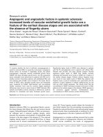
Figure 1: Intracellular cAMP levels in astrocytes. Astrocytes
were plated in 96-well plates (20,000 cells/well) and cultivated
in CDM containing 0% FCS. Basal levels of untreated cells
demonstrate 47.3% lower cAMP levels in KO astrocytes, as
compared to WT cells. Treatment with NE for 15 minutes in-
creased cAMP levels in both WT and KO astrocytes by 115.1%
and 135.3%, respectively. NE-induced cAMP concentration
was reduced in both WT and KO after 30 minutes treatment.
Treatment with IGF-1 for 1 minute reduced cAMP levels by
50% in WT cells only, an effect that was still observed after 5,
10 and 30 minute treatments. β
2
AR deficient astrocytes dem-
onstrated no changes in cAMP levels in response to IGF-1
treatment. Data represents mean +SEM. *, p<0.05 compared to
untreated WT cells; #, p<0.05 compared to untreated KO cells.
Discussion
cAMP is involved in cellular prolifera-
tion-differentiation processes as demonstrated on as-
trocytes with dibutyryl cAMP (dBcAMP), which initi-
ates a more differentiated status of astrocyte with re-
duced proliferative capacity [17]. dBcAMP has also
been shown to inhibit IGF-1-induced mitogenesis in
cells by inhibiting Raf-1 kinase activity, an effect that
has been attributed to phosphorylation of Raf-1 by
protein kinase A [17-19]. Interestingly, we have shown
that cultured β
2
AR KO astrocytes, which proliferate
more rapidly than wild type cells [16], display reduced
basal cAMP levels. This reduction in basal cAMP in
β
2
AR deficient cells might be accounted for by the ab-
sence of constitutive activity, which is known for the
β
2
AR. In addition to agonist-induced activation, β
2
AR
can undergo spontaneous conformational changes,
resulting in ligand-independent activation [20].
In addition to G
s
activation and consequent
cAMP production, the β
2
AR can activate G
i
proteins, a
property that is unique amongst the beta-adrenergic
receptors. Activation of G
i
results in enhanced sur-
vival, an effect which is thought to be mediated by
elevated activity of Akt/PKB [21]. An absence of β
2
AR
signaling in astrocytes and consequent reduction of
Akt/PKB activation might be compensated by in-
creased IGF signaling as demonstrated previously [16].
This compensatory mechanism might explain en-
hanced astrocytic proliferation.
We have demonstrated that treatment with IGF-1
reduced cAMP levels in WT astrocytes, yet had no
effect on KO cells. Although IGF-1 had a clear effect on
cAMP production in WT cells, the mechanism by
which IGF receptor signaling is involved in this regu-
lation is uncertain. As mentioned above, IGF-1 can
catalyze the phosphorylation of the β
2
AR, which re-
sults in loss of function of this receptor and inhibition
of adenylyl cyclase G
s
activation [6,22]. Although we
demonstrate that IGF-1 has the potential to regulate
cAMP levels in astrocytes in a β
2
AR-dependent man-
ner, we do not know whether this occurs through di-
rect phosphorylation and subsequent desensitization
of the β
2
AR by the IGF-1R, a notion which remains
speculative.
We have previously reported a loss of β
2
AR on
astrocytes in cerebral white matter of patients with MS,
Int. J. Med. Sci. 2008, 5
243
a deficit which may contribute to pathology of this
disease by several possible mechanisms such as im-
paired astrocytic glycogenolysis. Insufficient glyco-
genolysis could decrease energy supplies to axons and
may contribute to axonal degeneration [23]. In this
context, cAMP physiological effects include crucial
roles in regulating energy metabolism, such as lipoly-
sis, gluconeogenesis, and glycogenolysis [24]. Loss of
β
2
AR on astrocytes in MS might also contribute to en-
hanced astrogliosis and cellular reactivity, a hallmark
trait in MS lesions. Regulation of cell growth by the
β
2
AR, a receptor which is involved in processes of
proliferation and differentiation, has been implicated
in astrocytes in vitro [16].
In summary, β
2
AR deficient astrocytes have
lower basal cAMP levels as compared to wild type
cells. This reduction of cAMP levels may be involved
in the increased cell proliferation as reported previ-
ously. We show here that stimulation with IGF-1 re-
duces cAMP levels in astrocytes in a β
2
AR-dependent
manner, a reduction of which may promote some of
the mitogenic properties of IGF signaling.
Acknowledgments
We would like to thank Prof. Brian Kobilka
(Stanford University School of Medicine) and Prof.
Lutz Hein (University of Wuerzburg) for kindly pro-
viding us with β
2
-AR -/- mice.
Conflict of Interest
The authors have declared that no conflict of in-
terest exists.
References
1. Gutkind JS. Cell growth control by G protein-coupled receptors:
from signal transduction to signal integration. Oncogene 1998;
17: 1331–1342.
2. Hung KS, Tsai SH, Lee TC, Lin JW, Chang CK, Chiu WT. Gene
transfer of insulin-like growth factor-1 providing neuroprotec-
tion after spinal cord injury in rats. J Neurosurg Spine 2007; 6:
35-46.
3. Daub H, Weiss FU, Wallasch C, Ullrich A. Role of transactivation
of the EGF receptor in signalling by G-protein coupled receptors.
Nature 1996; 379:557–560.
4. Dalle S, Imamura T, Rose DW, Worrall DS, Ugi S, Hupfeld CJ, et
al. Insulin induces heterologous desensitization of
G-protein-coupled receptor and insulin-like growth factor I
signaling by down-regulating b-arrestin-1. Mol Cell Biol 2002;
22:6272–6285.
5. Hadcock JR, Port JD, Gelman MS, Malbon CC. Crosstalk be-
tween tyrosine kinase and G-protein-linked receptors. Phos-
phorylation of β2-adrenergic receptors in response to insulin. J
Biol Chem 1992; 267:26017–26022.
6. Karoor V, Malbon CC. Insulin-like growth factor receptor-1
stimulates phosphorylation of the b2-adrenergic receptor in vivo
on sites distinct from those phosphorylated in response to insu-
lin. J Biol Chem 1996; 271:29347–29352.
7. Eng LF, Ghirnikar RS. GFAP and astrogliosis. Brain Pathol 1994;
4:229–237.
8. Fawcett JW, Asher RA. The glial scar and central nervous system
repair. Brain Res Bull. 1999; 49:377–391.
9. De Keyser J, Wilczak N, Leta R, Streetland C. Astrocytes in mul-
tiple sclerosis lack β2 adrenergic receptors. Neurology 1999;
53:1628–1633.
10. Hertz L, Chen Y, Gibbs ME, Zang P, Peng L. Astrocytic adreno-
ceptors: a major drug target in neurological and psychiatric dis-
orders? Curr Drug Targets CNS Neurol Disord 2004; 3:239-67.
11. Fahrig T. Receptor subtype involved and mechanism of norepi-
nephrine-induced stimulation of glutamate uptake into primary
cultures of rat brain astrocytes. Glia 1993; 7:212–218.
12. Fillenz M, Lowry JP, Boutelle MG, Fray AE. The role of astro-
cytes and noradrenaline in neuronal glucose metabolism. Acta
Physiol Scand 1999; 167:275–284.
13. Feinstein DL, Heneka MT, Gavrilyuk V, Dello Russo C,
Weinberg G, Galea E. Noradrenergic regulation of inflammatory
gene expression in brain. Neurochem Int 2002; 41:357–365.
14. Chruscinski AJ, Rohrer DK, Schauble E, Desai KH, Bernstein D,
Kobilka BK. Targeted disruption of the b2 adrenergic receptor
gene. J Biol Chem 1999; 274:16694–16700.
15. McCarthy KD, de Vellis J. Preparation of separate astroglial and
oligodendroglial cell cultures from rat cerebral tissue. J Cell Biol
1980; 85:890–902.
16. Chesik D, Glazenburg L, De Keyser J, Wilczak N. Enhanced
proliferation of astrocytes from beta(2)-adrenergic receptor
knockout mice is influenced by the IGF system. J Neurochem
2007; 100:1555-64.
17. Kurino M, Fukunaga K, Ushio Y, Miyamoto E. Cyclic AMP
inhibits activation of mitogen-activated protein kinase and cell
proliferation in response to growth factors in cultured rat corti-
cal astrocytes. J Neurochem 1996; 67:2246–2255.
18. Howe LR, Leevers SJ, Gomez N, Nakielny S, Cohen P, Marshall
CJ. Activation of the MAP kinase pathway by the protein kinase
raf. Cell 1992; 71:335–342.
19. Valverde AM, Teruel T, Lorenzo M, Benito M. Involvement of
Raf-1 kinase and protein kinase Cf in insulin-like growth factor
I-induced brown adipocyte mitogenic signaling cascades: inhi-
bition by cyclic adenosine 3`,5`-monophosphate. Endocrinology
1996; 137:3832–3841.
20. Ballesteros JA, Jensen AD, Liapakis G, Rasmussen SG, Shi L,
Gether U, et al. Activation of the beta 2-adrenergic receptor in-
volves disruption of an ionic lock between the cytoplasmic ends
of transmembrane segments 3 and 6. J Biol Chem 2001;
276:29171-7.
21. Zhu WZ, Zheng M, Koch WJ, Lefkowitz RJ, Kobilka BK, Xiao RP.
Dual modulation of cell survival and cell death by
beta(2)-adrenergic signaling in adult mouse cardiac myocytes.
Proc Natl Acad Sci U S A 2001; 98:1607-12.
22. Baltensperger K, Karoor V, Paul H, Ruoho A, Czech MP, Malbon
CC. The beta-adrenergic receptor is a substrate for the insulin
receptor tyrosine kinase. J Biol Chem 1996; 271:1061-4.
23. De Keyser J, Zeinstra E, Mostert J, Wilczak N. β2-Adrenoceptor
involvement in inflammatory demyelination and axonal degen-
eration in multiple sclerosis. Trends Pharmacol Sci 2004; 25:
67–71.
24. Collins S, Surwit RS. The β-adrenergic recept
ors and the control
of adipose tissue metabolism and thermogenesis. Recent Prog
Horm Res 2001; 56:309–328.