Handbook of ECOTOXICOLOGY - Section 5 (end) pot
Bạn đang xem bản rút gọn của tài liệu. Xem và tải ngay bản đầy đủ của tài liệu tại đây (3.69 MB, 218 trang )
© 2003 by CRC Press LLC
SECTION V
Special Issues in Ecotoxicology
39 Endocrine Disrupting Chemicals and Endocrine Active Agents Timothy S. Gross,
Beverly S. Arnold, María S. Sepúlveda, and Kelly McDonald
40 A Review of the Role of Contaminants in Amphibian Declines
Donald W. Sparling
41 Genetic Effects of Contaminant Exposure and Potential Impacts on Animal
Populations Lee R. Shugart, Christopher W. Theodorakis, Amy M. Bickham, and
John W. Bickham
42 The Role of Ecotoxicology in Industrial Ecology and Natural Capitalism
John Cairns, Jr
43 Indirect Effects of Pesticides on Farmland Wildlife Nick Sotherton and
John Holland
44 Trace Element and Nutrition Interactions in Fish and Wildlife Steven J. Hamilton
and David J. Hoffman
45 Animal Species Endangerment: The Role of Environmental Pollution Oliver H. Pattee,
Valerie L. Fellows, and Dixie L. Bounds
© 2003 by CRC Press LLC
CHAPTER 39
Endocrine Disrupting Chemicals and
Endocrine Active Agents
Timothy S. Gross, Beverly S. Arnold, María S. Sepúlveda, and Kelly McDonald
CONTENTS
39.1 Introduction and Historical Background
39.1.1 General and Comparative Endocrinology
39.1.2 Mechanisms of Endocrine Modulation
39.2 Screening and Monitoring for Endocrine Disrupting Chemicals
39.2.1 In Vitro Assays
39.2.2 In Vivo Assays
39.3 EDC Effects: Evidence for Specific Chemicals and Chemical Classes
39.3.1 Polycyclic Aromatic Hydrocarbons (PAHs)
39.3.2 Polychlorinated and Polybrominated Biphenyls (PCBs and PBBs)
39.3.3 Polychlorinated Dibenzo-p-Dioxins (PCDDs) and Polychlorinated
Dibenzo-p-Furans (PCDFs)
39.3.4 Organochlorine Pesticides and Fungicides
39.3.4.1 Cyclodienes
39.3.4.2 Chlordecones (Kepone and Mirex)
39.3.4.3 Dichlorodiphenylethanes
39.3.4.4 Hexachlorocyclohexane
39.3.4.5 Vinclozolin
39.3.5 Non-Organochlorine Pesticides
39.3.5.1 Organophosphate Pesticides (OPs)
39.3.5.2 Carbamate Pesticides
39.3.5.3 Organometal Pesticides
39.3.5.4 Triazine Pesticides
39.3.6 Complex Environmental Mixtures
39.3.6.1 Pulp- and Paper-Mill Effluents
39.3.6.2 Sewage-Treatment Effluents
39.3.7 Metals
39.3.7.1 Mercury (Hg)
39.3.7.2 Other Metals
39.4 Summary and Conclusions
References
© 2003 by CRC Press LLC
39.1 INTRODUCTION AND HISTORICAL BACKGROUND
It has been established that a wide variety of anthropogenic (man-made) chemicals in the
environment are capable of modulating and adversely affecting or disrupting endocrine function in
vertebrate organisms.
1–13
Th
e physiological effects of exposure to these chemicals have been termed
“endocrine disruption” and the active compounds labeled as “endocrine-disrupting chemicals”
(EDCs) or “endocrine-active-agents.” Endocrine disruption has been defined by the U.S. Environ
-
mental Protection Agency (EPA)
12
as the actio
n of “an exogenous agent that interferes with the
production, release, transport, metabolism, binding, action, or elimination of natural hormones in
the body responsible for the maintenance of homeostasis and the regulation of developmental
processes.” This definition was further expanded by the U.S. EPA Endocrine Disruption Screening
and Testing Advisory Committee (EDSTAC)
14
to indicate that these effects are “adverse” and may
involve a wide assortment of endocrine-mediated functions and potential receptor-mediated events.
Indeed, effects may involve the steroid receptor superfamily, including the sex steroids, thyroid
hormones, and adrenal hormones, as well as hypothalamic-pituitary and other protein hormones.
The physiological processes regulated by the endocrine system are diverse and numerous.
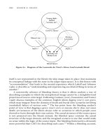
Likewise, the mechanisms of action and effects of potential EDCs are equally diverse (see Figure
39.1). Receptor-mediated events involve EDCs acting as hormone mimics (agonists or antagonists)
and adversely impacting hormone synthesis, catabolism, secretion, transport, and signal transduc
-
tion. Examples of nonreceptor-mediated modes of EDC action include altered enzyme function
and selective toxicities for endocrine-active or target tissues, whereas altered gene expression and
induction of oxidative stress are types of receptor mediated events. EDCs may also act by altering
developmental processes, often producing multigenerational effects.
Endocrine-active anthropogenic chemicals are also numerous and diverse (see References 1–13).
Evidence
for endocrine-disrupting effects due to these chemicals comes from a diverse array of
Figure 39.1 Schematic representation of the hypothalamic-pituitary-gonad-liver axis of teleost fishes. Asterisks
denote areas at which EDCs can exert their effects. In general, this model is also applicable for
other oviparous vertebrates. Abbreviations: GnRH (gonadotropin releasing hormone); GTH (gonad-
otropin hormone); GSI (gonadosomatic index); SHBG (serum binding hormone globulin); VTG
(vitellogenin); T (testosterone); E
2
(17β estradiol); 11KT (11-ketotestosterone).
Hypothalamus
GnRH Dopamine
GTH IIGTH I
+
_
Pituitary
GTH
***
Steroid Pathway
Cholesterol
***
T
E
2
11-KT
SHBG
***
E
2
Spermiation
Ovulation
***
Fry
Development
***
VTG
***
External and Internal
Stimuli
Secondary Sex
Characteristics
***
E
2
Sex Steroids
Estrogens ( )
Androgens (T, 11-KT)
***
Hepatic Metabolism
***
Sperm/Eggs
(gamete size, morphology,
and number)
***
Gonad (GSI)
***
oocyte
growth
***
Liver
DNA
VTG
***
***
******
© 2003 by CRC Press LLC
reports involving multiple vertebrate taxonomic groups, limited invertebrate taxa, and results from
both in vitro and in vivo studies. Reported effects of EDCs have included effects at multiple levels
of biological organization including molecular, biochemical, cellular, tissue, and organismal. How
-
ever, few reports have documented effects at the population level and higher. In addition, most
studies have focused upon reproductive effects; however, effects on growth, metabolism, and thyroid
and immune function have also been noted. This chapter summarizes the current evidence for the
endocrine-disrupting effects of specific chemicals and chemical classes in vertebrate wildlife with
a discussion on potential mechanisms/modes of action.
39.1.1 General and Comparative Endocrinology
To fully understand the mechanisms by which anthropogenic or natural EDCs may modulate
endocrine function, normal functioning of the endocrine system must be understood. Indeed, an
assessment of the risk of potential EDC exposures and effects requires critical information from a
variety of disciplines, including endocrinology, and an understanding of the variation among and
within vertebrate classes. The following section is a brief overview of vertebrate endocrinology
and the hormones that may be involved in endocrine modulation or disruption.
The endocrine system is a collection of hormone-secreting cells, tissues, and ductless glands
(e.g., pituitary, thyroid, adrenal, and gonads) that play an important role in growth, development,
reproduction, and homeostasis. Tissues of the endocrine system synthesize and secrete hormones
that influence virtually every stage of the life cycle of an organism, from gametogenesis and
fertilization, through development into a sexually mature organism and senescence. Endocrinology
is the study of tissues that secrete hormones into the blood and the subsequent effects hormones
have on target tissues. Hormones are released into the extracellular environments and affect neigh
-
boring cells (paracrine control), the emitting cell (autocrine control), or other target tissues (endo-
crine control). Some nerve cells also release hormones into the blood (neuroendocrine control) or
into extracellular fluid for communication with other nerve cells or nonnerve cells (neurotransmis
-
sion). Pheromones are hormones secreted into the external environment for communication with
other individuals or species. In addition, there are several hormones that act through more than one
of these chemical-signaling modes.
Figure 39.1 summarizes the hypothalamic-pituitary-gonadal axis for fish as an example of the
endocrine system, its diverse control over reproductive and developmental processes, and sites at
which EDCs may exert endocrine-disrupting effects. In general, this model is also applicable to
other oviparous vertebrate species including birds, amphibians, and reptiles.
The vertebrate hypothalamus and the pituitary gland (or hypophysis) have an essential role in
regulating endocrine and nonendocrine target tissues.
15–17
The hypothalamus and pituitary are func-
tionally and anatomically linked, forming the hypothalamic-pituitary axis. In mammals, the pituitary
is composed of four anatomically and functionally distinct regions: the adenohypophysial pars distalis
and pars intermedia, and the neurohypophysial median eminence and pars nervosa. In fish, the pars
distalis is additionally separated into two regions that contain different cell types and produce different
hormones.
18
The pituitary gland of amphibians, birds, and reptiles is similar to the mammalian
pituitary gland.
16
Indeed, the basic arrangement of the hypothalamic-pituitary axis is essentially the
same in all vertebrate groups, with the exception of teleost fishes, which lack a median eminence.
16
The hypothalamus directly controls pituitary hormone secretion via the production and release
of a number of peptide and nonpeptide hormones. These pituitary-tropic hormones are generally
categorized as releasing hormones (RH) or release-inhibiting hormones (RIH), depending on their
function. Hypothalamic hormones include corticotropin-releasing hormone (CRH), thyrotropin-
releasing hormone (TRH), gonadotropin-releasing hormone (GnRH), growth-hormone-releasing
hormone (GHRH), growth-hormone release-inhibiting hormone (GHRIH, somatostatin), and pro
-
lactin release-inhibiting hormone (PRIH). Other hypothalamic hormones also include critical neuro-
transmitters such as catecholamine and dopamine.
19
© 2003 by CRC Press LLC
The principal neurohypophysial (neuropituitary) hormones in mammals are arginine vasopressin
and oxytocin. Birds, reptiles, and amphibians have structurally-related peptides: mesotocin and
arginine vasotocin,
20
while fish in general have arginine vasotocin and isotocin or mesotocin,
depending on the species.
16
These hormones are critical for milk secretion, oviductal and uterine
contraction, renal water absorption, and vaso-constriction and dilation. In all vertebrates, these
neurohypophysial hormones are produced in the hypothalamus and are transported to the pituitary,
where they are stored until release into the bloodstream.
Hormones produced by the mammalian adenohypophysis are the pituitary-derived tropic hor-
mones including growth hormone (GH), adrenocorticotropin (ACTH), melanotropin (MSH), thy-
roid-stimulating hormone (TSH), prolactin (PRL), and the gonadotropins — follicle-stimulating
hormone (FSH), and luteinizing hormone (LH). Secretions of ACTH, TSH, and the gonadotropins
(FSH and LH) are each regulated by negative feedback. Although structurally related counterparts
for the adenohypophysial hormones have been identified in fish, amphibians, birds, and reptiles,
16
there are important differences in hormone actions across vertebrate groups. For instance, PRL is
associated with reproduction and lactation in mammals but is an important osmoregulatory hormone
in fish.
21
Although FSH and LH function similarly in mammalian and avian reproduction, reptiles
do not synthesize an LH-like gonadotropin and instead utilize FSH to regulate gonadotropin-related
functions.
15
In fish and amphibians, two different gonadotropins, GTH-I and GTH-II, have been
identified that act similarly to mammalian FSH and LH, respectively.
17
GH generally regulates
body and tissue growth; however, in nonmammalian vertebrates, it is also involved in osmoregu
-
lation. In mammals and birds, ACTH is responsible for stimulating the production of corticosteroids
by the adrenal gland, which in turn plays a role in metabolism, ion regulation, and stress responses.
The role of ACTH in fish and amphibians is less clear, however, and MSH may have similar
properties in these taxonomic groups. Indeed, similarities in hormone structure may not necessarily
represent similar hormone function in nonmammalian vertebrates.
GH is important for bone growth and as an anabolic hormone during development.
22
It has
direct effects on a wide variety of tissues as well as indirect effects that are modulated by growth
factors such as insulin-like growth factor-I (IGF-I).
22
In conjunction with thyroid hormones, GH is
necessary for the development of a wide number of tissues ranging from cardiac
23
and skeletal
muscle,
24
to bone
25
and brain development.
26
In nonmammalian species, GH probably functions in
a similar manner; however, less is known about growth hormone in fish, amphibians, and reptiles.
The adrenal glands, thyroid gland, and gonads are all directly regulated by the pituitary gland.
16
Thyroid hormones, which are produced by thyroid glands, and steroids produced by the adrenal
cortex and gonads can indirectly inhibit their own secretion by inhibiting the release of pituitary
and hypothalamic hormones (negative feedback). In response to TSH, the thyroid gland produces
two hormones, triiodothyronine (T
3
) and tetraiodothyronine (T
4
). In mammals, T
3
and T
4
have
important effects on metabolism and development.
16
Thyroid hormones also play an essential role
in fish and amphibian metamorphosis. Indeed, thyroid hormones determine the timing of develop
-
mental processes, and metamorphosis is almost entirely controlled directly by thyroid hor-
mones.
16,27–29
Some metamorphic processes that are under the control of thyroid hormones include
the migration of the eye and dorsal fin growth in fish,
30,31
amphibian tail and gut resorption,
27,32,33
restructuring of the amphibian head,
34,35
amphibian limb development,
36
and amphibian gill resorp-
tion.
37
Thyroid hormones also have important roles during fish smoltification.
38–40
The mammalian adrenal gland produces two important steroid hormones — aldosterone and
corticosterone. Aldosterone plays an important role in the maintenance of sodium concentrations,
and corticosterone is primarily involved in regulating blood glucose.
16
Adrenal steroids function
similarly in birds but very differently in other nonmammalian vertebrates. In amphibians, aldos
-
terone and corticosterone are equally effective as regulators of blood glucose, whereas in fish and
reptiles, corticosterone serves to regulate blood glucose and sodium. While adrenal hormones have
critical roles in all vertebrates, characterizations of their functions in nonmammalian vertebrates
are limited, and interspecies differences have not been thoroughly evaluated.
© 2003 by CRC Press LLC
In all vertebrate classes, gonadal function is dependent upon the hypothalamic-pituitary axis
through the production of GnRH and gonadotropins.
16
In mammals, the gonadotropins include FSH
and LH, which control different gonadal events. In females, FSH promotes ovarian follicular growth,
and LH induces ovulation. Both gonadotropins are also required for normal estrogen synthesis: LH
stimulates the synthesis of androgens, and FSH stimulates aromatization of androgens to estrogen.
In males, FSH promotes spermatogenesis, and LH promotes steroidogenesis and spermiation. The
mammalian gonad also produces the peptide hormone inhibin, which feeds back to inhibit FSH
production. In both males and females, the pulsatile release of GnRH is regulated by the feedback
of high circulating levels of androgens and estrogens. In birds, gonadotropins function in a similar
manner; however, reptiles do not synthesize an LH-like gonadotropin and utilize FSH to regulate
all gonadotropin-related functions.
15
In fish and amphibians, two different gonadotropins — GTH-
I and GTH-II — have been identified, and they act similarly to mammalian FSH and LH, respec
-
tively. GTH-I is involved in gonadal development, gamete production, and vitellogenesis, a process
that involves the hepatic synthesis of yolk protein precursors, vitellogenin (VTG), under the stimulus
of estrogens.
16,17
GTH-II stimulates the final stages of oocyte maturation as well as ovulation in
females and spermiation in males.
In general, gonadotropins exert effects on vertebrate gonads by binding to specific receptors.
The primary gonadal response to gonadotropins is the synthesis and secretion of assorted sex
steroids. In all vertebrates, the primary reproductive sex steroids include androgens [e.g., testoster
-
one (T), 11-ketotestosterone (11KT), androstenedione (A), dihydrotestosterone (DHT)], estrogens
[estradiol (E
2
), estrone (E
1
), estriol (E
3
)], and progestins [progesterone (P
4
), dihydroxyprogesterone
(DHP)]. Gonadal steroid hormones are involved in every aspect of reproduction, from sex deter
-
mination to the control of courtship behaviors and the development of secondary sex characteristics.
Sex steroids also play an important role in brain development. For example, in mammals, E
2
and DHT are involved in normal sexual differentiation of the brain.
41–43
Although reproductive
function is regulated and modulated by sex steroids in all vertebrates,
16,28,44–47
there are distinct
functional differences that must be noted. Indeed, functional differences in sex steroids are most
evident for fish, amphibians, and reptiles, with significant differences also existing within each of
these taxonomic classes.
17
For instance, the primary androgen for spermatogenesis in mammals,
birds, and reptiles is T, but in many fish and some amphibians the critical androgen for spermato
-
genesis is 11KT. Preliminary results from our laboratory would suggest that 11KT might not be
the predominant androgen in live-bearing fish (such as mosquito fish Gambusia holbrooki). E
2
is
the sex steroid responsible for oocyte growth and maturation in all vertebrates; however, it also
regulates and induces the synthesis of VTG in oviporous vertebrate species.
16,17,48,49
Progestins are critical to pregnancy in mammals but function in reptiles and birds in post-
ovulatory events such as the regulation of eggshell deposition. In fish, progestins are responsible
for final egg maturation prior to oviposition. Gonadal sex steroids can also have dramatic effects
on sex differentiation in fish, amphibians, and reptiles, effects that are not observed in birds or
mammals.
17,28,50
When applied early during development, sex steroids can cause sex reversal in
fish, amphibians, and reptiles. Therefore, the genetic sex of the individual can be different from
the phenotypic sex. Finally, the effects of sex steroids on gonadal differentiation and sex reversal
vary dramatically between species and across developmental stages, and therefore these differences
need to be noted and considered in any study of potential EDC effects in vertebrate wildlife.
39.1.2 Mechanisms of Endocrine Modulation
There is significant evidence to suggest that a wide variety of anthropogenic chemical contam-
inants in the environment can disrupt or modulate endocrine function in a wide variety of vertebrate
and some invertebrate organisms. However, information regarding the mechanisms that lead to
these endocrine modifications is limited. It is, nonetheless, critical that mechanisms and modes of
action for EDCs and endocrine-active agents be understood. Mechanisms of action are generally
© 2003 by CRC Press LLC
difficult to elucidate and are complicated by multiple factors including chemical properties, routes,
timing, and lengths of exposure, as well as endocrine-system and species- and tissue-specific
physiological differences. Furthermore, the integration of the nervous, endocrine, reproductive,
hepatic, and other target systems, as well as multiple feedback regulatory pathways, adds to the
complexity of understanding EDC mechanisms (see, for example, Figure 39.1).
Potential mechanisms of action for EDCs are diverse. EDCs may interrupt multiple pathways
along the hypothalmic-pituitary–target-tissue axis, potentially disturbing the normal synthesis,
transport, release, binding, action, biotransformation, or elimination of natural hormones in the
body. EDCs may alter the hypothalamic-pituitary axis, which can have widespread effects through
the disruption of endocrine functions downstream of the hypothalamus. There is increasing evidence
that EDCs may disrupt endocrine function by influencing the regulation/release of the pituitary-
tropic hormones. Indeed, polychlorinated biphenyls (PCBs) have been shown to interfere with the
neurotransmitters that control GnRH secretion, resulting in decreased GnRH production as well as
subsequent reductions in gonad size and plasma concentrations of sex steroids.
51
In mammals,
neonatal exposure to diethylstilbestrol (DES) or dichlorodiphenyltrichloroethane (DDT) results in
both reduced GnRH and LH production.
51
These results demonstrate that interference at one site
along the hypothalmic-pituitary axis can affect multiple downstream events. Furthermore, the
hypothalamus and pituitary are regulated by the feedback of hormones from several other endocrine-
active tissues; therefore, alterations in different hormone concentrations can also affect hypothalmic
and pituitary function.
EDCs can exert effects and disrupt the function of other endocrine tissues and hormones
downstream of the hypothalamus and pituitary. Hormones are synthesized by specific endocrine
tissues, secreted into the bloodstream, and transported by binding proteins to target tissues to interact
with receptors, elicit responses, and be metabolized or degraded. EDCs can block or enhance the
function of hormones by interfering with any one or several of these critical steps. For instance,
EDCs may interfere with hormone synthesis, thereby altering endocrine activity by directly affecting
the availability of specific hormones or critical precursors.
28,52
Failure to synthesize appropriate
hormones can result from either an alteration in the biosynthetic enzymes and in the availability
of precursor molecules. The initial, as well as rate-limiting, step in the biosynthesis of hormones
may often be affected. EDCs can inhibit the uptake of critical precursors and the subsequent
conversion to hormone products.
53–55
EDCs can alter the rate at which hormones are metabolized. The cytochrome P450 (CYP450)
monooxygenases constitute a super family of enzymes that play essential roles in both the synthesis
(steroidogenesis) and metabolism of steroid hormones. Many of these enzymes appear to be sensitive
to EDCs.
52,56–59
EDCs can affect the number or activity of specific monooxygenases, thereby
affecting the rate of hormone metabolism and clearance. Since specific CYP450 enzymes — like
CYP1A — are also responsible for metabolizing foreign compounds — like EDCs — EDC stim
-
ulation of CYP1A and other monooxygenases that hydroxylate them prior to their elimination may
in turn contribute to increased clearance of sex hormones by inducing other monooxygenase
activities.
60
EDCs have also been reported to increase the activity of several other microsomal
enzymes including aminopyrine demethylase, glucuronyl transferase, and p-nitroreductase.
61,62
Some
EDCs may also induce hormone-like effects due to alternating rates of degradation. For example,
many synthetic hormones, such as ethynyl estradiol (EE
2
), a synthetic estrogen used in birth control
pills, are not degraded readily by the enzymes that normally metabolize the endogenous hormones.
63
EDCs can also interfere with the binding of hormones to transport proteins, preventing their delivery
to target tissues.
64,65
The absence of available binding proteins may result in both faster uptake or
increased degradation of free-circulating hormones.
66–68
For example, the sex-hormone-binding
globulin (SHBG) has high affinity for both T and E
2
, which is necessary to prevent degradation and
clearance of these hormones as well as enable their transport to target tissues.
69
EDCs, which mimic
estrogens or androgens, may bind to these globulin proteins and displace the endogenous sex steroids,
© 2003 by CRC Press LLC
thereby increasing the elimination rates for endogenous hormones. Although several studies suggest
that globulins may also facilitate the transport of EDCs to target tissues,
69
the greater binding affinity
of globulins for endogenous hormones probably limits this process.
70
EDCs may bind to hormone receptors and either activate (agonize)
71–73
or inhibit (antagonize)
74
receptor function. Indeed, many studies have focused on EDCs as hormone-mimics and the potential
for these compounds to interact with hormone-specific receptors. Potential EDCs have been eval
-
uated extensively for their ability to bind to the estrogen receptor (ER). Estrogens normally bind
to the ER located in the nucleus of target cells. The E
2
-bound ER has a high affinity for DNA
sequences called estrogen response elements (ERE). After binding the ERE, the ER-DNA complex
interacts with various transcription factors, chromosomal proteins, and regulatory factors in order
to induce or inhibit the transcription of specific genes and enable endocrine-specific response.
EDCs can block or enhance the function of a hormone or endocrine target tissue by interfering
with any one or several of these critical steps. Although the potential estrogenic activities of EDCs
have overshadowed studies of other receptor-mediated EDC activities, EDCs that act as androgens
or antiandrogens via interaction with the androgen receptor (AR) have also been noted.
74–76
Unlike the ER, which has an E
2
specific response element, the response element for the AR is
shared with other steroid receptors including the glucocorticoid (GR), progesterone (P
4
), and
mineralocorticoid (MR) receptors. Therefore, EDCs that have androgenic activities may exert
broader effects than those attributed to a simple androgen mimic. EDCs may also interact with a
wider variety of receptors important for endocrine function. For example, some EDCs (e.g., 2,3,7,8-
tetrachlorodibenzo-dioxin [TCDD] and other planar hydrocarbons) are reported to have antiestro
-
genic activities by interacting with the aryl hydrocarbon receptor (AhR) rather than by competitively
binding to the ER. The AhR is an intracellular receptor that is expressed by many different cell
types and that functions as a transcription factor.
77,78
EDC interactions with the AhR may interfere
with estrogen responses in a number of ways: by reducing E
2
binding to the ER,
79
by blocking the
binding of the ER to the ERE,
80
by impairing nuclear translocation,
81
or by suppressing gene
transcription.
82
These examples demonstrate the varied receptor-mediated activities of EDCs.
Endocrine-disrupting effects may also occur due to direct or indirect toxicities for specific
endocrine-active or target tissues. For example, many lipophilic EDCs will accumulate primarily
in fatty tissues, such as the liver and gonads, potentially interfering with the synthesis and mobi
-
lization of lipids and thereby inhibiting specific endocrine-related functions such as vitellogenesis.
It is important to point out, however, that specific mechanisms or modes of action for most EDCs
are not well elucidated or understood. This stems from the fact that mechanisms are often difficult
to identify and are complicated by multiple factors including differences in EDC-specific properties,
routes of exposure, and vertebrate class and species differences. Nonetheless, it is critical that
mechanisms of action for EDCs and endocrine-active agents be understood in order that effects in
wildlife be prevented and that appropriate screening and testing methods be developed.
39.2 SCREENING AND MONITORING FOR ENDOCRINE DISRUPTING CHEMICALS
Analytical methods have long been used to determine concentrations of chemical residues that
persist in the environment (e.g., water, sediment) and accumulate in biota (e.g., tissue and body
burdens). Although these approaches are useful for characterizing the presence and distribution of
specific EDCs in the environment, they fail to indicate whether chemical exposures have biological
consequences. The development of EDC-specific screening and monitoring procedures aid in the
establishment of potential relationships between environmental EDC concentrations and biological
responses. In the past decade, several in vitro and in vivo assays have been proposed that can be
used to screen or monitor individual EDCs, specific EDC mixtures, or complex environmental
mixtures for potential endocrine disrupting or modulating activity.
© 2003 by CRC Press LLC
39.2.1 In Vitro Assays
Several in vitro assays have been described for evaluating potential endocrine-disrupting or
modulating activities of EDCs.
75
These assays are based on several specific mechanisms of action
for EDCs, including receptor binding, gene expression, cell proliferation, and cell differentiation.
83
Advantages of in vitro systems include low cost, high reproducibility, and the rapid analysis of large
numbers of samples. These assays are also valuable for studying mechanisms of action of com
-
pounds, screening effects of mixtures, and detecting potential interaction effects. Results from these
screening procedures can aid in the subsequent development and validation of assays. In vitro assays,
however, generally lack ecorelevance because pharmacokinetics, biotransformation, and binding to
carrier proteins may not be accurately represented. For example, some EDCs are activated or
deactivated in vivo by enzymatic conversion during metabolism, conjugation, and excretion. These
limitations must be considered when interpreting or applying results from in vitro screening tests.
Receptor-binding assays can be utilized to screen for and identify potential EDCs (which
function via receptor-mediated pathways) since they can evaluate whether specific EDCs can bind
to specific receptors. Depending on the receptor of interest, receptor-binding assays utilize either
crude cell fractions such as plasma membranes, cytosol, or the nucleus. Cell fractions may be
obtained from specific vertebrate organisms or from established cell lines, transformed cells,
84,85
or transfected cells.
86
Although in vitro receptor-binding assays are relatively simple and inexpensive
to conduct, they do not necessarily reflect binding under in vivo conditions and are of very little
use in screening for EDCs that operate by nonreceptor-mediated pathways. Finally, these assays
do not differentiate between agonist and antagonist properties.
Additional in vitro assays have utilized the ability of EDCs to induce target-cell-specific
proliferation and differentiation. For instance, MCF-7 cells, derived from human breast cancer cells,
have been widely utilized for the development of the E-screen assay, which evaluates the ability
of specific EDCs or EDC mixtures to both bind and express the ER
87
and the resultant cell
proliferation as a response.
88–94
EDCs are identified as potential E
2
agonists if there is a significant
increase in cell proliferation, which in turn is quantified by counting cell nuclei
92
or measuring
other responses such as metabolic reductions. Although the E-screen assay has been extensively
used as a screen for estrogenicity,
76,92,95
a positive response cannot be necessarily interpreted as an
indicator for the presence of E
2
agonists. In addition, ER antagonists and antiandrogens are not
detected using this assay, and thus a significant number of false negatives are common. Before a
compound is identified as an EDC, positive responses with the E-screen assay should be confirmed
by in vivo studies.
A number of additional in vitro cell-based expression assays have also been developed to
measure receptor-dependent biological responses. Expression assays evaluate the induction or
suppression of proteins by specific genes in response to potential receptor-mediated EDCs and
mixtures. Measured protein endpoints for these receptor-specific expression assays include:
VTG,
71–73,94,96,97
sex-hormone-binding globulins,
98
luciferase,
99
galactosidase,
100
and chlorampheni-
col acetyltransferase (CAT).
86
However, these assays are general and are not limited to the action
of EDCs. Additional cell types/lines that have also been utilized for in vitro expression assays
include fish hepatocytes,
71,73,94,98
MCF-7,
95,101
HeLA,
86,98
and yeast.
101
The types of cells used in
expression assays are critical to any interpretations. Indeed, significant differences in responses
between yeast-cell-based assays and mammalian-cell assays have been reported,
98
and sensitivities
vary greatly.
100
Nonetheless, expression assays have several advantages as compared to other in
vitro screening assays. Unlike receptor-binding or cell-proliferation assays, expression assays can
be used to detect both agonists and antagonists.
86,99,102
Expression assays can also evaluate potential
EDCs that influence many aspects of gene expression in addition to those that operate through
receptor-mediated functions. Nonetheless, in vitro expression assays generally have high variability
and lack ecorelevance.
© 2003 by CRC Press LLC
39.2.2 In Vivo Assays
The effects of EDCs occur at many biological levels of organization including molecular,
biochemical, organelle, cell, tissue, organism, population, community, and ecosystem. The use of a
battery of biomarkers that reflect multiple biological levels of organization would enable a more
thorough evaluation of both exposure and the potential mechanism of action. Although responses
at the population level and higher are the most biologically ecorelevant, they are rarely utilized as
biomarkers since these responses are complex, less specific, and require greater effort and time.
Indeed, most of the current biomarkers are limited to the measurement of responses at the molecular,
biochemical, cellular, and organism levels. In vivo assays for the identification of EDCs are not
mechanism-dependent and provide results that are more environmentally relevant than in vitro assays.
Indeed, in vivo assays rely upon either natural exposures or controlled exposures based on expected
or predicted environmental exposures. In vivo assays for EDCs can detect effects on endocrine
function, regardless of the mechanism of action, as well as identify a potential EDC that would not
necessarily exhibit activity in an in vitro screening assay. Most importantly, in vivo screening assays
both identify potential EDCs and enable the description and evaluation of potential effects.
In vivo assays for evaluating EDCs may involve the utilization of specific endocrine biomarkers
as a way to evaluate potential effects. Widely used endocrine-endpoint-based in vivo assays have
included the uterotropic assay, the Hershberger assay, and the thyroid-function assay. Although these
assays were not originally designed for the evaluation or identification of EDCs, they have demon
-
strated the utility of in vivo assays for the identification of potential EDCs. The uterotropic assay
utilizes prepubertal or adult ovariectomized female rats to assess uterine weight and histological
responses to potential EDCs. The Hershberger assay evaluates androgenicity using androgen-depen
-
dent tissue (e.g., prostate and seminal vesicles) responses to potential EDCs. The thyroid-gland-
function assay evaluates potential EDC exposures and the subsequent evaluation of plasma concen
-
trations of T
3
, T
4
, and TSH.
Biomarkers that detect alterations at the biochemical and molecular levels are frequently utilized
for in vivo EDC-screening assays.
103
Biochemical and molecular responses are generally the first
detectable responses to an environmental change or stressor and can serve as early indicators of
both exposure and effect. Aside from being highly sensitive changes at the molecular and biochem
-
ical level, they can sometimes be predictive of responses at higher levels of organization (tissue
and organism levels). Examples of molecular-based in vivo EDC-screening assays include receptor
analyses, transcriptional-based analyses, and differential display.
14
These assays are, in general,
based on an analysis of specific molecular parameters for tissues collected following either natural
or experimental exposures to potential EDCs. Although molecular-based in vivo assays are highly
sensitive, they are difficult to validate and often lack ecorelevance. Examples of current biochemical-
based in vivo EDC-screening assays include: measurement of VTG production
104
and systemic
hormone concentrations (e.g., plasma sex steroids, T
3
, and T
4
). In fact, systemic concentrations of
various hormones have been frequently utilized as biomarkers for EDCs in fish,
105–110
amphibi-
ans,
111,112
reptiles,
113,114
birds,
5,115–120
and mammals.
121
These procedures have broad application to
all vertebrate classes since hormones, especially the steroid and thyroid hormones, are evolutionarily
conserved across all vertebrate classes. However, it must be noted that the same hormones may
differ in function significantly between and within vertebrate classes. For example, the primary
androgen for spermatogenesis in mammals, birds, and reptiles is T, but in many fish and some
amphibians, the critical androgen for spermatogenesis is 11KT.
VTG has been utilized as a bioindicator of potential exposure and effects of estrogenic EDCs
in fish and other oviparous vertebrates.
96,122–124
This phospholipoprotein is produced by the liver
under the control of E
2
in oviparous female fish, amphibians, reptiles, and birds.
111
Oviparous
species have vitellogenic cycles that correspond to egg production. Potential EDCs, which mimic
or alter endogenous E
2
, may induce the expression of VTG. This assay has, in general, focused on
© 2003 by CRC Press LLC
responses in males, which do not exhibit clear vitellogenic cycles. However, it must be noted that
low background levels of VTG are likely to be normal in males. Thus, an identification of a potential
EDC by this method cannot be based solely on the presence of detectable VTG; it must additionally
be based on a species-specific VTG response that is significantly increased above background levels.
Additional in vivo EDC-screening assays involve endpoints based on responses at the tissue
and organism levels. Although these assays may have higher biological and ecological relevance,
they are more variable and often specific to vertebrate classes or species. Examples of screening
assays that rely on tissue-level responses include tissue somatic indices (e.g., gonadosomatic index-
GSI), tissue histopathology, altered secondary sex characteristics,
125–128
and egg- and sperm-quality
assessments.
94
In vivo assays that rely on organism responses may include assessments of egg
numbers/ovarian development,
128–132
se
xual maturity,
128
neon
atal/embryonic mortality,
129–131,133–135
reproductive impairment,
108,136
and evaluation of egg hatchabilities
129,134,135,137
and nest num-
bers.
133,134
Population and ecosystem endpoints of reproductive success may include evaluation of
pod size, age-class analyses, and population numbers.
135,138,139
Valid in vivo screening procedures should provide information about EDC exposure and be
indicative of expected or predicted physiological effects. In addition, in vivo biomarkers reflect the
complex pharmacokinetic and metabolic factors that can affect EDC uptake and metabolism. It is
important to keep in mind, however, that in vivo assays and endpoints are influenced by both
physiological and environmental variables, which make it difficult to establish clear cause-and-
effect relationships between responses and specific EDCs. Nonetheless, these assays are often the
most useful for evaluating potential EDC effects and for the identification of environmentally
relevant EDCs.
39.3 EDC EFFECTS: EVIDENCE FOR SPECIFIC CHEMICALS
AND CHEMICAL CLASSES
The previous section reviewed many of the possible mechanisms by which environmental
contaminants may alter endocrine function in fish and wildlife. The following section introduces
several classes of environmentally relevant contaminants with reported or potential endocrine-
disrupting activity in invertebrates and vertebrates. This review presents evidence for EDC effects
for several specific chemical classes: polycyclic aromatic hydrocarbons, polychlorinated and poly
-
brominated biphenyls, dioxins, organochlorine and other pesticides, complex environmental mix-
tures, and metals. For most chemicals, the specific mechanism of action is not well understood,
and chemical structure does not necessarily indicate or suggest endocrine functionality, mimicry,
or EDC activity (see Figure 39.2 as an example of chemical structures for several environmental
estrogens). In fact, direct evidence of endocrine activity is often difficult to demonstrate and thus
is generally absent. This review includes reports from a variety of laboratory and field studies that
have explored the effects of EDCs in fish and wildlife and discusses the potential or suspected
modes of action (MOAs) (see Table 39.1).
39.3.1 Polycyclic Aromatic Hydrocarbons (PAHs)
Polycyclic aromatic hydrocarbons (PAHs), whether of natural or anthroprogenic origin, are
products of incomplete combustion of organic compounds and enter aquatic environments via oil
spills, waste discharge, runoff, and dry or wet deposition. Although they are biodegraded in soils
and water within weeks to months, the metabolites are often longer lasting and more toxic.
Birds can be exposed to PAHs through ingestion of contaminated food and water, by preening
feathers, or through the skin in cases of oil spills. Petroleum hydrocarbons can also be absorbed
through the eggshell.
140
In a review by Hoffman,
141
PAHs applied to the shells of eggs caused
mortality and reduced hatchability. In studies reviewed by Fry,
142
exposure to petroleum oil increased
© 2003 by CRC Press LLC
circulating corticosterone levels and disrupted reproduction through negative feedback to the hypo-
thalamic-pituitary-gonadal system. However other studies
143,144
have shown decreased levels of
plasma corticosterone, suggesting that ingested petroleum may interfere with adrenocortical func
-
tion. Yolk formation may also be depressed after exposure to oil, resulting in a reduction in egg
numbers.
145
Exposure to as little as 0.1-mL weathered crude oil (equivalent to 2.5 mL/kg body wt.)
interferes with egg production, laying, incubation, and pair bonding. Field exposure of adult storm
petrels (Oceanodroma sp.) with dependent chicks reduced foraging and feeding of chicks, resulting
in reduced growth or death.
146
Population studies with pigeon guillemots (Cepphus columba) after the Exxon Valdez spill
indicated a decline in numbers for three consecutive years but no effects on reproduction.
147
Reproductive effects in the black oystercatcher (Haematopus bachmani) were noted.
148
There was
a decrease in nonbreeding pairs, a decrease in egg size, and higher chick mortality, all of which
directly related to the amount of oil present in the foraging territory. Birds exposed to oil may
exhibit changes in adrenal hormone synthesis and elevated hepatic mixed oxidase activity, which
may increase metabolic clearance of corticosterone.
140,149,150
In a laboratory study, female mallards
(Anas platyrhynchos) that ingested crude oil hatched fewer live ducklings per pair.
116,140
In this
study, there was evidence of suppression of follicular development, eggshell thinning, decreased
hatchability, and reduced levels of plasma E
2
, E
1
, P
4
, and LH in females. These results suggest that
the oil acts on ovarian steroidogenesis, reducing positive feedback to the pituitary and causing a
decline in LH, a delay in ovarian maturation, and reduced fertility.
Several field studies have documented altered reproductive activity in fish residing in PAH-
contaminated waters. For instance, gonadal development was impaired and E
2
concentrations were
depressed in English sole (Parophyrs vetulus) from highly contaminated areas of Puget Sound,
Washington. Reproductive impairment was statistically correlated with elevated PAH concentra
-
tions, as measured by the presence of fluorescent aromatic compounds (FACs) in the bile of
fish.
151,152
Other examples in which PAH exposure may have been related to endocrine alterations
or reproductive dysfunction include altered ovarian development in plaice (Pleuronectees platessa)
exposed to crude oil,
153
reduced GSI, increased liver size and ethoxyresorufin O-deethylase activity
(EROD) in white sucker (Catostomus commersori) residing downstream of pulp and paper mills,
154
Figure 39.2 Structures of some selected natural and environmental estrogens.
OH
OH
CCH
3
CH
3
Bisphenol A
OH
R
4-Alkyphenols
RCO
2
RCO
2
Phthalates
Cl
C CCl
3
H
Cl
o, p-DDT
,
PCBs
Cl Cl
OH
HO
17ß-estradiol
HO
ß-sitosterol
HO
HO
Diethylstilbestrol
CH
2
CH
3
CH
3
CH
2
© 2003 by CRC Press LLC
Table 39.1 Summary of Effects and Possible Modes of Action (MOAs) of Endocrine Disrupting Chemicals (EDCs) by Chemical Class and Taxa
Chemical Taxa Effects Possible MOA
Sample
Reference
PAHs Birds ↓Hatchability DNA damage
Oxidative stress
ER agonist
141
Fish ↓GSI
Impaired gonadal development
DNA damage
Oxidative stress
ER agonist
155
152
PCBs Mammals Abortions & stillbirths Antiestrogens
Act through Ah receptor
175
Birds ↓Eggshell thickness
↓Hatching success
↑Embryo mortality
Antiestrogens
Act through Ah receptor
189, 195
Amphibians and Reptiles ↓Sex hormones
↑Mortality & malformation rates
Unknown 211, 212
Fish ↓Spawning
↓Hatchability
Antiestrogens
Act through Ah receptor
214, 215
PBBs Mammals Fetotoxic and teratogenic
↑Menstrual cycles
↓Sex hormones
Unknown 234, 606, 607
Birds ↓Offspring viability
↓Hatchability
Unknown 235
Organochlorine pesticides
Cyclodienes Birds ↓Productivity E
2
agonist 288, 292–294
Reptiles Sex reversal
↓Sex hormones
E
2
agonist 113, 211
Fish ↓Fertilization
↓Maturing oocytes
Altered spermatogenesis
E
2
agonist 299, 304
302
Chlordecone and Mirex Mammals Persistent estrus and vaginal changes Weakly estrogenic 310
Birds ↓Clutch size, egg size, shell thickness, hatchability
↑Embryo malformations
309
Fish Gonadal abnormalities 323
DDT and derivatives Mammals Persistent vaginal estrus Androgen antagonist 325
Birds Eggshell thinning
Reproductive problems
Population reduction
338, 343
© 2003 by CRC Press LLC
Reptiles and Amphibians Sex reversal
↓Clutch viability sex
Altered plasma hormone levels
Abnormal gonadal morphology
Hormone mimicry
Estrogenicity
113, 129, 135
Fish ↑Oocyte atresia
↓Fecundity and fertility
↓Sex hormones
Hormone mimicry
Estrogenicity
Steroid receptors
136, 346
Hexachlorocyclohexane
lindane
Fish ↓Sex hormones 347–349
Vinclozolin Mammals Feminization of males Androgen antagonist 76
PCDDs & PCDFs
TCDD
Mammals Impairs sexual differentiation in male rats, delay in
testicular descendent and puberty
Antiestrogenic through AhR
receptor
244, 245
Birds Developmental alterations
Congenital deformities
Feminization
Antiestrogenic 247, 248
Amphibians and Reptiles Early metamorphosis
↑Frequency of deformities
alterations in sex ratios
Antiestrogenic 264
266
267
Fish Early-life-stage mortality
Impaired oocyte development
Antiestrogenic 273
Non-organochlorine
pesticides
Organophosphate
pesticides (OPs)
Mammals Depressed reproduction
Gonadotrophins
Acts at sites on hypothalamus-
pituitary-gonadal-liver axis;
Acetylcholinesterase inhibitor
357
Birds Gonadotrophins
Developmental defects
141
Amphibians and Reptiles Altered metamorphosis
↑Deformities and delayed development
371, 372
Fish Retarded ovarian growth
↓GSI
Arrested spermatogenesis
375
Carbamate pesticides Amphibians ↑Developmental deformities Acetylcholinesterase inhibitors
Acts on pituitary to alter GnRH
and GTH concentrations
396
© 2003 by CRC Press LLC
Table 39.1 Summary of Effects and Possible Modes of Action (MOAs) of Endocrine Disrupting Chemicals (EDCs) by Chemical Class and Taxa (Continued)
Chemical Taxa Effects Possible MOA
Sample
Reference
Fish ↑Histopathological alterations in gonads
↓GSI (oocyte atresia and spermatogonial necrosis)
379, 380
Organometal pesticides
(TBT)
Fish ↓Sperm counts
Delayed hatching
May inhibit aromatose 418
412
Invertebrates Masculinization of female gastropods
Imposex
Competitive inhibitor of
aromatase
Cytotoxic and genotoxic effects
402
Complex Mixtures
Pulp- and paper-mill
Effluents
Fish ↓Sex hormones
↓Gonadal development
Delayed sexual maturation
Altered secondary sex characteristic expression
Estrogenic
ER, AR, AhR agonists
433
453
Sewage-effluents Fish ↓Testicular growth and development
Altered spermatogenesis
Estrogenic
ER agonists/antagonist
27, 466
↑VTG production in males
↓Hatchlings
↑Oocyte atresia
Estrogenic
ER binding
418, 466, 481
476, 503
Metals
Methylmercury Mammals ↓Embryo survival
↓Sperm counts
Unknown 524, 528
Birds Impaired reproductive behavior
↓Hatchability and nesting success
Unknown 535, 539
Amphibians and Reptiles ↓GSI
↓Sperm bundles
543
Fish ↓GSI
↑Gonadal abnormalities
Altered gonadal steroidogenesis
Unknown 546
© 2003 by CRC Press LLC
and decreased GSI in bream (Abramis brama) inhabiting contaminated areas of the Rhine River.
155
Although PAH concentrations were abnormally high in the field studies described above, they
were only one of a group of pollutants that may have caused the observed effects. Furthermore,
several histological studies report no differences in the gonads of male and female fish from
control and PAH-contaminated sites.
156,157
A combination of field and laboratory experiments is
still necessary before the reproductive alterations observed in the wild can be clearly attributed
to PAH exposure.
Laboratory and field studies present clear evidence for the adverse affects of PAHs in fish.
Thomas et al.
158
have elucidated the impact of benzo(a)pyrene (BaP) on endocrine and reproductive
activities in female Atlantic croaker (Micropogonias undulatus). Atlantic croaker fed 0.4 mg
BaP/70g/day for 30 days during the period of ovarian recrudescence experienced impaired ovarian
growth with a concomitant reduction in plasma E
2
and T. GSI in control females increased fivefold
over the course of the study, whereas GSI of exposed females reached only 66% of controls.
In vitro production of sex steroids was not impaired by BaP, and there appeared to be a
relationship between the amount of ovarian tissue (i.e., size of ovaries) and steroidogenic capacity.
Similar results were reported in a separate study of female Atlantic croaker exposed to BaP via
injection for 30 days.
159
In this study, in addition to reduced GSI and plasma sex steroids, a
reduction in the number of hepatic ERs and plasma VTG was observed. BaP did not interfere
with the binding of E
2
to the ER under in vitro competition studies, and again there was no clear
evidence for a direct effect of BaP on steroidogenesis. In vitro competition studies using hepatic
ER from spotted seatrout supported earlier results on Atlantic croaker.
160
This is consistent with
mammalian studies, and suggests that BaP must undergo metabolic activation in order to interact
with the ER. The effects of the PAH 3-methylcholanthrene on endocrine and reproductive function
in ricefield eels (Monopterus albus) were similar to those observed for BaP-treated Atlantic
croaker. Exposure to 4 ppm 3-methylcholanthrene for 7 days resulted in reduced E
2
, T, VTG,
GSI, and altered ovarian histology.
161
PAHs are known CYP450 inducers. For example, the PAH naphthoflavone induced the expres-
sion of CYP4501A1 (the primary xenobiotic-metabolizing enzyme) and inhibited VTG synthesis
in E
2
-stimulated liver cells from rainbow trout (Oncorhynchus mykiss).
72
However, naphthoflavone
had no effect on vitellogenesis when incubated without E
2
. The degree of CYP4501A1 induction
was directly related to the extent of VTG inhibition, which suggests that naphthoflavone may be
acting as an antiestrogen via the AhR, the intracellular receptor involved in CYP4501A1 expression.
The effect of naphthoflavone on vitellogenesis in vivo appears to be more complicated. When
juvenile rainbow trout were treated with 0.5 ppm E
2
and 25 or 50 ppm of naphthoflavone, an
inhibitory effect on VTG synthesis was observed; however, lower concentrations of naphthoflavone
(5 or 12.5 ppm) appeared to potentiate E
2
-stimulated VTG production.
72
Furthermore, reduced VTG
synthesis by higher concentrations of naphthoflavone was correlated with a decrease in radiolabeled
E
2
binding to the ER. These results suggest that naphthoflavone influences VTG synthesis by
regulating ER function, although it is likely that the antiestrogenic activity of PAHs involves multiple
mechanisms. Several investigators have proposed that CYP4501A1-inducing compounds affect sex-
steroid concentrations by increasing their catabolism.
162
Evidence from a recent study,
163
however,
suggests that PAHs may also interfere with steroid biosynthesis. Incubating vitellogenic ovarian
tissue from female European flounder (Platichthys flesus) with 3 PAHs (phenanthrene, BaP, and
chrysene) decreased A and E
2
secretion. In addition, phenanthrene inhibited steroid conjugation,
and it was concluded that these PAHs inhibited key steroidogenic enzymes, including CYP450 17,
20 lyase, which is responsible for converting C21 to C19 steroids.
39.3.2 Polychlorinated and Polybrominated Biphenyls (PCBs and PBBs)
PCBs are a group of synthetic organic chemicals, formed by the chlorination of biphenyls,
which include 209 individual compounds (congeners). These substances were manufactured for a
© 2003 by CRC Press LLC
wide range of industrial applications including use as hydraulic fluids, lubricants, plasticizers, and
coolant/insulation fluids in transformers. Several chemical properties make these compounds both
highly useful and potentially hazardous. For instance, their chemical stability makes them ideal for
industrial activities involving high temperatures; however, this stability also renders them persistent
in the environment. The majority of PCBs that enter the water adsorb to organic particles and
sediments, although they are essentially nonbiodegradable in soils and sediments.
164
Furthermore,
they are hydrophobic, which makes them excellent lubricants, yet allows them to bioaccumulate
in tissues and biomagnify as they are passed along the food chain. Concentrations of PCBs in fish
at contaminated sites may range from ppb to ppm. The production of PCBs is currently banned or
highly restricted and the use of certain mixtures permitted only under tightly regulated conditions.
Nonetheless, PCBs originating from industrial wastes, accidental leaks or spills, and careless
disposal continue to be a source of pollution and environmental concern.
Many studies examining the health hazards of PCBs describe the effects of occupational
exposure in humans and the physiological responses of mammals and birds that have consumed
large quantities of contaminated fish. These studies provide strong evidence that PCB exposure
can lead to the development of cancer; disturbances of the immune, hepatic, pulmonary, and nervous
systems; and impaired reproduction and development. Many of these abnormalities are enhanced
in the offspring, even if exposure occurs prior to conception. Responses are believed to be dependent
on species, sex, age, and chemical structure.
165
Laboratory studies with mink (Mustela vison) have established an association between PCB
residues and reproductive effects in wildlife,
166
but there are no field studies linking PCBs with
reproductive effects.
167
In one study in which mink were fed meat from cows contaminated with
Aroclor 1254, concentrations as low as 0.87–1.33 ppm resulted in reproductive failure.
168
Other
feeding studies have shown impaired reproduction in mink with fat concentrations of 13.3 ppm
and reproductive failure at concentrations of 24.8 ppm.
169
Field studies with big brown (Eptisecus fuscus) and little brown bats (Myotis lucifugus)
suggested a correlation between PCB residue levels and reproductive toxicity;
170,171
however,
captive studies have not supported this link.
172
Studies with ringed seals (Pusa hispida) have found
a relationship between fat PCB concentrations and uterine-horn occlusions.
173
Later studies with
ringed and gray seals (Halichoerus grypus), however, failed to detect any relationship between
PCB levels and pregnancy or impairment of the uterine horns.
174
Other studies have linked PCBs
with abortions and premature pupping in California sea lions (Zalophus californianus),
175
tumors
and decreased fecundity in Beluga whales (Delphinapterus leucas),
176
skeletal lesions in harbor
(Phoca vitulina) and grey seals,
177,178
and immunosuppression in harbor seals.
179
In a field exper-
iment with harbor seals, animals fed PCB-contaminated fish had a significant reduction in repro-
ductive success; however, in this study it was difficult to separate out the influence of other possible
factors and contaminants.
180,181
PCBs have been associated with embryonic mortality, deformities, and low reproductive success
in many species of birds. Laboratory studies with chickens (Gallus gallus), ringed turtledoves
(Streptopelia risoria), and mallards have shown reproductive impairment following ingestion of
PCB-laden feed. In three studies, eggs of chickens that received 10–80 ppm Aroclor 1248 in the
diet exhibited reduced hatching success;
182–184
however, in another study, a diet of 20 ppm Aroclor
1254 did not affect this parameter.
185
Aroclor 1242 in the drinking water at 50 ppm produced chick
embryo mortality and teratogenesis.
186
Aroclor 1254 in the diet of ringed turtledoves has increased
embryonic mortality, decreased parental attentiveness,
187
and depleted brain dopamine and norepi-
nephrine.
188
Eggshell thickness was affected in mallard hens,
189
but another study produced no
eggshell changes.
190
Studies with screech owls (Otus asio)
191
and Atlantic puffins (Fratercula
arctica)
192
also produced no reproductive effects.
Field studies have indicated PCBs as the cause of mortality of ring-billed gulls (Larus delawa-
rensis) in southern Ontario
193
as well as the cause for increased embryo/chick mortality and reduced
hatching success.
194,195
PCBs have also been blamed for the low reproductive success and eggshell
© 2003 by CRC Press LLC
damage in Lake Michigan herring gulls (Larus argentatus).
196,197
Reproductive success of Forsters
terns (Sterna forsteri) from a Green Bay colony was 52% of that from inland colonies.
198,199
In this
study, hatchlings also weighed less, had shorter femurs, exhibited edema, and were malformed.
The toxicity was attributed to the PCB congeners 105 and 126, and results indicated PCB congener
77 as accounting for some of the toxicity in the tern eggs.
199–201
PCBs have also been implicated
as embryotoxic in eagles,
202
as producing decreased embryonic weight in black-crowned night
herons (Nycticorax nycticorax),
203
as reducing hatching success of American kestrels (Falco spar-
verius),
204
and as the cause of congenital anomalies and embryonic death in double-crested cormo-
rants (Phalacrocorax auritus).
205,206
In cormorants, however, there is discussion as to whether DDT
or PCBs are more strongly associated with nest failure.
207,208
American alligator eggs (Alligator mississippiensis) from Lake Apopka, Florida have residues
of PCBs as well as a combination of organochlorine pesticides.
209
Alligators from this site have also
been documented to have abnormally developed reproductive organs, altered serum hormone con
-
centrations, and decreased egg viability.
135,139,210
However, alligators from Lake Apopka are known
to be exposed to a complex mixture of potential EDCs, and therefore it is difficult to pinpoint which
compounds are responsible for the observed effects. In red-eared slider turtles (Trachemys scripta
elegans), males exposed to Aroclor 1242 had significantly lower T concentrations than controls.
211
The African clawed frog (Xenopus laevis) and the European common frog (Rana temporaria)
were exposed to the PCB mixture Clophen A50 or to PCB 126 for either 10 days or until
metamorphosis. Exposed frogs had increased mortality, higher malformation rates, and lower
thyroid hormone concentrations.
212
In a similar study, the same frog species were exposed to the
mixtures Clophen A50 and Aroclor 1254 or to PCB 126; effects of exposure depended on route
and time and length of exposure. This study also indicated a relationship between lowered concen
-
trations of retinoid and PCB exposure.
212
In another study, green frogs (Rana clamitans) and leopard
frog (Rana pipiens) were exposed throughout metamorphosis to PCB 126 at concentrations ranging
from 0.005 to 50 ppb. Survival of larvae decreased at the higher concentrations in both species.
213
In fish, reproductive impairment has been demonstrated under both in vivo laboratory studies
and in field studies of fish residing in PCB-contaminated waters. Several field studies have attempted
to correlate PCB tissue levels with observed reproductive alterations. For instance, PCB levels in
the liver and ovarian tissue of female English sole from Puget Sound were associated with the
spawning of fewer eggs.
214
Similarly, a negative correlation was found between egg hatchability
and total PCB concentrations in the eggs of lake trout (Salvelinus namaycush) from the Great
Lakes.
215
In a study of the reproductive success of lake trout residing in Lake Michigan, Mac and
Edsall
216
suggested that maternally derived PCBs were the cause of reduced egg hatchability and
increased fry mortality. Johnson et al.
217
reported decreased egg weight and increased oocyte atresia
in female winter flounder (Pseudopleuronectes americanus) with high tissue concentrations of
PCBs, although there was no evidence that PCBs altered GSI, plasma E
2
, or fecundity in this
species. Interestingly, English sole residing in the same location had reduced plasma E
2
concen-
trations and impaired gonadal development. It was proposed that the different migratory practices
of both fish species might have resulted in different susceptibilities to the chemicals, since these
behaviors resulted in differences in the timing and duration of exposure to the most highly con
-
taminated waters.
In the laboratory, female Japanese medaka (Oryzias latipes) had reduced GSI and were unable
to spawn following an injection of 150 ppm of PCB.
218
It was suggested that PCB exposure disrupted
E
2
metabolism since only control fish, excreted E
2
into the water 8 days after the injection. In male
goldfish (Carassius auratus), PCB exposure resulted in decreased plasma T and 11KT concentra
-
tions, while hepatic EROD activity was increased 15-fold.
219
Reduced plasma T in males and E
2
and P
4
in females, accompanied by an increase in several sex-steroid-metabolizing enzymes, was
observed in carp (Cyprinus carpio) injected with 250 ppm of the commercial PCB Aroclor 1248.
220
In another study, E
2
-treated juvenile rainbow trout fed a diet contaminated with PCBs (3, 30, or
300 ppm) for 6 months showed decreased synthesis of VTG.
221
© 2003 by CRC Press LLC
The decline in estrogens and androgens combined with the elevation of EROD (or other
metabolizing enzymes) would suggest that the reduction in sex steroids is related to an increase in
metabolism rather than a decreased synthesis. However, this is probably not the only mechanism
for PCB-induced damage since several studies also report abnormalities at the organ e.g., GSI,
testicular abnormalities) and organism levels (offspring survival, hatchability) in fish showing
normal sex-steroid and VTG concentrations.
222–224
In some of these cases, it is believed that the
reproductive abnormalities (e.g., delayed spawning, reduced hatchability) may be caused by the
accumulation of toxic levels of PCBs in the ovaries and maturing oocytes.
225
Evidence that PCBs
bind VTG suggest that lipoproteins are involved in the transport of the contaminants from extra-
gonadal tissue into the ovaries.
226
One explanation for the inconsistencies observed between studies
might be related to timing of exposure. For instance, the lack of effects of 3,′3′,4,′4′-tetrachloro
-
biphenyl (TCB) on plasma concentrations of sex steroids and VTG in female striped bass (Morone
saxatilis) and white perch (Morone americana) may have been related to the fact that the fish used
in these studies were already vitellogenic and not in a highly active stage of gonadal maturation.
222,223
The stage of gonadal maturation may also be important in males exposed to PCBs. Atlantic
cod (Gadus morhua) fed Aroclor 1254 (1–50 ppm) for 5.5 months accumulated significant levels
of the PCB in the testes and liver and exhibited considerable testicular damage including fibrosis
of lobule walls, necrosis and disintegration of lobule elements, and decreased spermatogenesis.
227
These authors suggest that the stage of gonadal maturation may be related to the degree of chemical
sensitivity since only males experiencing rapid spermatogenic proliferation or fully mature males
suffered testicular damage (i.e., sexually immature and regressed males were unaffected). In several
cases, substantial concentrations of PCBs have been detected in tissues of fish that showed no signs
of adverse reproductive effects.
228
It is not surprising that a wide range of responses has been observed in studies that have differed
with regards to species and experimental design. However, the chemical complexity of this class
of compounds is an additional factor that complicates interpretation. Slight structural differences
in the 209 possible PCB congeners, as well as different compositions of the mixtures, may result
in vastly different physiological responses.
There is considerable evidence that PCBs act at multiple sites along the hypothalamic-pituitary-
gonadal (HPG) axis,
158,229
and in vitro experiments are providing insight into the mechanisms
underlying these reproductive alterations. However, extrapolating the actual risks that PCBs impose
on the environment and biota are difficult due to the complexity and diversity of the commercial
mixtures (of congeners). In addition to understanding the interaction of the PCB mixtures with
other environmental pollutants and stressors, consideration must be given to the interaction (addi
-
tivity, synergism, antagonism) of the individual components that make up these mixtures.
PBBs, formed by the bromination of biphenyls, are similar in structure to PCBs. These
chemicals are stable and lipophilic and, therefore, present many of the same environmental hazards
as PCBs (e.g., persistence in the environment and long biological half-lives). There are 209 possible
PBB congeners, although only 45 have been actually synthesized.
230
FireMaster BP-6, used pri-
marily as a flame-retardant additive in the early 1970s, was the most widely used PBB, although
its production was discontinued in 1978.
231,232
The production and distribution of PBBs was
insufficient to result in widespread contamination of the environment; however, the accidental
contamination of cattle feed by the Michigan Chemical Company in 1973 resulted in the pollution
of many Michigan farmlands. Significant concentrations of PBBs were subsequently detected in
water and sediment samples and in tissues of fish and ducks residing downstream of the Michigan
Chemical Company.
233
Although information concerning the reproductive effects of PBBs in fish is lacking, there is
substantial evidence that these chemicals adversely affect reproductive processes in other species.
234
For instance, feeding adult female chickens a diet contaminated with 45 ppm of the commercial
PBB FireMaster FF-1 for 5 weeks resulted in impaired production and hatchability of eggs and in
reduced viability of offspring.
235,236
A variety of reproductive effects following PBB exposure have
© 2003 by CRC Press LLC
also been reported from other avian (quail) and mammalian (rodents, monkey, cow, and mink)
species.
230,234
Although PBBs have been detected in aquatic environments and are known to accu-
mulate in fish tissues,
237
there is very little information regarding the effects of these chemicals on
exposed fish. Like PCBs, PBBs are believed to be potent inducers of several monooxygenase
enzymes (including EROD), although it is not known whether this induction affects the metabolism
of circulating reproductive hormones.
39.3.3 Polychlorinated Dibenzo-p-Dioxins (PCDDs) and Polychlorinated
Dibenzo-p-Furans (PCDFs)
PCDDs and PCDFs are structurally related compounds produced during a variety of thermal
and chemical reactions including the combustion of PCBs, production of steel and other compounds,
and disposal of industrial wastes (via the interaction of chlorophenols). These compounds have
also been identified as components of bleached-pulp-mill effluents. PCDDs and PCDFs are halo
-
genated aromatic hydrocarbons with high chemical stability, low water solubility, and limited
solubility in many organic solvents. There are 75 possible PCDD congeners and 135 PCDF
congeners, although 2,3,7,8-tetrachlorodibenzo-p-dioxin, known as TCDD or dioxin, has received
the most attention. Concerns regarding TCDD stem from its wide distribution in the environment
and extreme toxicity to both humans and wildlife. Although a variety of PCDDs and PCDFs have
been detected in fish and wildlife, the 2,3,7,8-substituted congeners are believed to be the most
persistent and prevalent in tissue samples analyzed to date, with half-lives of over a year in some
fish species.
238
TCDD and related compounds have been implicated in a number of health-related
problems including neurotoxicity, hepatoxicity, cardiotoxicity, chloracne, birth defects, immuno
-
suppression, wasting syndrome, and endocrine and reproductive alterations.
240,241,269
In nonhuman
primates and rodents, although developmental effects of the immune, reproductive, and nervous
systems occur at body burdens in the range of 30–80 pptr, biochemical changes on cytokine
expression and metabolizing enzymes are seen at doses ten times lower.
240
Many of the toxic effects
associated with exposure to dioxins appear to be dependent on target tissue, species, sex, and age.
Studies on the developmental effects of TCDD in rodents have demonstrated that only transient
exposure to relatively low concentrations of TCDD during critical windows of development are
capable of eliciting irreversible disruption of organ functioning in offspring. For example, gesta
-
tional exposure of rats to low concentrations of TCDD (0.064–1.0 ppb) during a critical period of
development (day 15 of gestation) causes impaired sexual differentiation in male fetuses including
persistence of female traits; decrease in the concentration of T, in the weight of testis and epididymis,
and in the production of sperm; and altered sexual behavior during adulthood.
242–244
Similarly, Gray
et al.
245
reported a delay in testicular descendent and puberty, with a subsequent reduction in sperm
counts and fertility in adult male rodents after a single maternal dose of 1 µg TCDD/kg on day 15
of gestation. Other signs of developmental toxicity in mammals include decreased growth, structural
malformations (e.g., cleft palate and hydronephrosis), prenatal mortality, and neurobehavioral
changes (e.g., impaired learning in rhesus monkeys).
239
Laboratory studies on the effects of TCDD in birds have shown significant variation in sensitivity
across species, with over 40-fold differences on embryo mortality (reviewed in References 239 and
246). For example, doses of only 20–50 ppt of TCDD in chicken eggs cause mortality and
malformations, as opposed to 1000–10,000 ppt in eggs of ring-necked pheasants (Phasianus
colchinus) and eastern bluebirds (Sialia sialis). Chicken embryos are also much more sensitive to
the teratogenic effects of TCDD, particularly cardiovascular malformations.
In birds, there is considerable evidence indicating that embryonic exposure to dioxin and dioxin-
like compounds can induce developmental alterations. Indeed, several field studies with colonial
fish-eating birds from the Great Lakes have implicated dioxin equivalents (the aggregate of AhR-
active substances) as the causative factors for the increased incidence of developmental deformities
and embryo lethality observed in certain contaminated areas (see reviews in References 247–250).
© 2003 by CRC Press LLC
Together, these epidemiological studies have provided one of the strongest links between contam-
inant exposure and reproductive/developmental effects in wildlife.
The Great Lakes Embryo Mortality, Edema and Deformities Syndrome (GLEMEDS) was first
described in double-crested cormorants but has also been reported in other species including the great
blue heron (Ardea herodias) and the Caspian tern (Sterna caspia). The syndrome is characterized by
increased embryo mortality; growth retardation; subcutaneous, pericardial, and peritoneal edema;
congenital deformities of the bill and limbs; feminization of embryos; and abnormal parental behav
-
ior.
247
This syndrome closely resembles the “chick edema disease” observed in chickens after in ovo
exposure of hens to PCDDs and PCDFs. Embryotoxicity in piscivorous birds from the Great Lakes
has been associated with TCDD concentrations above 100 pg/g (reviewed in Reference 241).
Although a reduction in the release of pollutants to the Great Lakes has resulted in significant
population improvements for several avian species, particularly double-crested-cormorants and ring-
billed gulls (Larus delawarensis), reproductive and physiological alterations due to contaminants are
still associated with population-level effects in birds that feed on highly contaminated fish (such as
Caspian terns and bald eagles, Haliaeetus leucocephalus).
249,251
Similar reproductive and developmental effects due to PCDDs have also been reported from
free-ranging populations of great blue herons,
252,253
double-crested cormorants,
254
and wood ducks
(Aix sponsa)
255
sampled elsewhere. In addition, in ovo exposure to dioxins has been associated with
the development of asymmetric brains in wild (great blue herons, double-crested cormorants, and
bald eagles) and domestic (chickens) species.
256
The behavioral and physiological repercussions of
this gross brain deformity, however, are unknown at this time. Other sublethal effects observed in
birds exposed to dioxins include decrease bursa and spleen weights in developing embryos
257
and
altered thyroid-gland structure, circulating thyroid hormones, and vitamin A (retinoid) status
(reviewed in Reference 258). In Belgium and The Netherlands, PCDDs/PCDFs in common tern
(Sterna hirundo) were correlated with lower yolk sac retinoids and plasma-thyroid concentrations
in hatchlings and with unfavorable breeding parameters (delayed laying and smaller eggs and
chicks).
259
Similarly, cormorant (Phalacrocorax carbo) hatchlings from a PCDD/PCDF-contami-
nated site in The Netherlands had decreased plasma-thyroid concentrations and an increased in ovo
respiration rate.
260
Results from laboratory studies, however, have failed to replicate what has been
reported from wild avian species, and in ovo exposure to TCDD has caused either increases or no
changes in thyroid hormones.
120,261,262
Information on the developmental toxicity effects of TCDD in amphibians and reptiles is scarce.
Neal et al.
263
reported no effects in tadpoles and adult bullfrogs (Rana catesbeiana) after a single
injection of TCDD (500 ppb). Jung and Walker
264
exposed anuran eggs and tadpoles to TCDD for
24 h and observed that American toads (Bufo americanus) treated with at least 0.03 ppb appeared
to metamorphose earlier than controls and that metamorphosis tended to occur at larger body masses
after exposure to higher doses of dioxin. The authors concluded that anuran eggs and tadpoles
eliminate TCDD more rapidly and are 100- to 1000-fold less sensitive to its deleterious develop
-
mental effects when compared to fish. Differential sensitivity to TCDD and related compounds
could be related to differences in metabolism or to different patterns in AhR binding and signal
transduction across taxa. In this respect, there is recent information showing a high degree of amino-
acid-sequence conservation for the AhR among bird species (97% amino acid identity) but a much
lower percent identity across taxa (79 and 74% identity between the amphibian Necturus maculosus
and bird and mouse sequences, respectively).
265
In an epidemiological study, developmental abnormalities and hatch rates from eggs of the
common snapping turtle (Chelydra serpentina serpentina) were assessed in relation to over 70
PAHs, including 8 PCDDs and 14 PCDFs.
266
This study found an increase in the frequency of
deformities with increasing contaminant exposure in eggs, particularly PCDDs and PCDFs con
-
centrations. In the laboratory, American alligator eggs (embryo stages 19–22) were treated with
TCDD (at doses ranging from 0.1 to 10 ppm) and incubated at male-producing temperatures
© 2003 by CRC Press LLC
(33ºC).
267
High doses of TCDD in eggs resulted in dose-dependent alterations in sex ratios, with
a higher incidence of female hatchlings.
Results from several field and laboratory studies have established that fish, in particular early
life stages, are extremely sensitive to the effects caused by TCDD when compared to other taxa.
Effects on fry survival are significant at egg doses ranging from 50 to 5000 pg/TCDD/g, which
corresponds to concentrations of 75 to 750 pg TCDD/g in parent fish.
241
Signs of TCDD-induced
developmental toxicity resemble blue sac disease, which is an edematous syndrome characterized
by yolk sac and pericardial edema, subcutaneous hemorrhages, craniofacial malformations, retarded
growth, and death.
268
This syndrome has been well characterized in salmonids after exposure of
eggs via water, injection, or maternally derived TCDD. Studies with salmonids have established
differential sensitivity to induce sac fry mortality, with LD
50
values varying from less than 100 pg
TCDD/g egg in lake trout (the most sensitive fish species to TCDD developmental toxicity) to 200
and over 300 pg TCDD/g egg in brook trout (Salvelinus fontinalis) and in some strains of rainbow
trout, respectively.
269–271
TCDD-developmental toxicity has also been reported in several nonsalmo-
nid species including the northern pike (Esox lucius), the mumichog (Fundulus heteroclitus), the
Japanese medaka,
272
and the zebrafish (Brachydanio rerio).
273
Regardless of species or egg exposure
route, early-life-stage mortality occurs during the sac-fry stage, probably as a consequence of the
generalized edema.
Exposure of eggs to TCDD and related compounds may have been responsible for the decline
of some fish populations in the Great Lakes since concentrations of dioxin and dioxin equivalent
in eggs and fry of salmonids have fallen within the range of those known to induce blue sac disease
in the laboratory.
269
The reader is advised to refer to References 269 and 274 for comprehensive
reviews on the effects of TCDD and related compounds on fish of the Great Lakes.
Fish reproduction can also be affected after exposure to TCDD. In the laboratory, adult female
zebrafish fed 5–20 ng TCDD showed impaired oocyte development with fewer eggs produced and
lethal developmental abnormalities in offspring (e.g., malformations of notocord).
273
In a separate
study, although TCCD treatment of newly fertilized zebrafish eggs did not affect hatchability, doses
of 1.5 ng TCDD/g or more resulted in a variety of structural and physiological abnormalities in larvae.
275
The mechanisms by which TCDD and structurally related compounds cause endocrine/devel-
opmental effects are complex and not completely understood. The relative toxicity of TCDD and
other halogenated aromatics is likely dependent on their ability to bind and activate the AhR.
Although this AhR mechanism is known for its involvement in the antiestrogenic action of TCDD
as well as for its ability to induce structural malformations, its MOA in causing other reproductive
and developmental toxicity is less clear.
239
There is substantial evidence that these contaminants
induce the expression of certain genes (e.g., translation products comprising Phase I and Phase II
enzymes) while altering the transcription of others (ER).
Indeed, the antiestrogenic effects of TCDD in mice have been attributed to its ability to suppress
ER gene expression, probably through an inhibition of ER transcription after binding of the TCDD-
AhR complex to promoter regions of the ER gene (see Reference 276 for a review of mechanisms
of action of dioxins). Antiestrogenic effects of these contaminants have also been documented in
vitro using fish cell lines. Using carp hepatocytes, Smeets et al.
277
demonstrated that although low
concentrations of TCDD caused a suppression of VTG secretion and an induction of CYPIA, the
two phenomena were not correlated to each other. From these results, the authors concluded that
the antiestrogenic effects of TCDD were probably not caused by increased metabolism of E
2
due
to induction of CYP1A. Mechanistic studies using mammalian cell lines also support this theory,
80
although increased metabolism of sex steroids may provide an additional or secondary mechanism
of antiestrogenicity. In this respect, great blue heron hatchlings and adults exposed to TCDD had
increased testosterone hydroxylase activity, a result that was coupled with increased CYP1A1
activity.
278
Changes in hydroxylase activity, however, have not been associated with alterations in
circulating sex-steroid concentrations in TCDD-exposed herons.
119,120,261
An additional MOA of
© 2003 by CRC Press LLC
TCDD could be through the pituitary, disrupting normal feedback mechanisms between hormones
and LH secretion.
279
Since vitamin A and thyroid hormones are essential for normal differentiation and development
of tissues, alterations in their homeostasis might result in malformations and altered growth. In this
respect, there is evidence showing that dioxin may interfere with the metabolism and storage of
vitamin A (retinoids)
280
and of thyroid hormones
281
through the co-induction of Phase I (P450) and
Phase II (uridine diphosphate-glucoroyltransferase (T
4
-UDPGT) enzymes. In addition, hydroxy
metabolites of PCDDs and PCDFs can compete for thyroxine on the transthyretin (the thyroxine
binding prealbumin) binding site.
282
Finally, recent evidence suggests that the hemodynamic and
teratogenic effects observed in fish fry affected by blue sac disease could be due to the ability of
TCDD to induce oxidative stress and oxidative DNA damage. In the Japanese medaka, oxidative
stress to the vascular endothelium of developing embryos induces programmed cell death, or
apoptosis.
283
Apoptosis of vascular cells causes alterations in hemodynamics, leading to a gener-
alized loss of function and subsequent mortality.
39.3.4 Organochlorine Pesticides and Fungicides
Organochlorine pesticides (OCPs) comprise a large group of structurally diverse compounds
used to control agricultural pests and vectors of human disease. Many of these compounds, as well
as their metabolites, are environmentally persistent due to their chemical stability, low water
solubility, and high lipophilicity. The exact mode of neurotoxicity is not well understood, although
OCPs are believed to disrupt the balance of sodium and potassium in nerve cells. The ability of
these toxic compounds to bioaccumulate in and often harm unintended species has led to the
restricted use of most OCPs. Despite a general reduction in use, several field studies have suggested
that OCPs adversely affect endocrine function in fish, indicating that aquatic wildlife is still being
exposed to levels capable of altering endocrine and reproductive parameters. For instance, a negative
correlation was found between total OCPs and E
2
in male carp (Cyprinus carpio) in a large-scale
field effort to assess the reproductive health of fish in U.S. streams.
106
Similar results were reported
in largemouth bass (Micropterus salmoides) collected from a contaminated (with OCPs) site in
Florida.
284
In the following section, OCPs are discussed according to accepted structural classifi-
cations, although effects within the same chemical class may differ drastically. Furthermore,
Pickering et al.
285
have suggested that pesticide toxicity is species specific, and a single species
may be differentially susceptible to different pesticides. Indeed, the reported effects and mechanisms
of action may vary significantly between the various OCPs.
39.3.4.1 Cyclodienes
The chlorinated cyclodiene pesticides are lipophilic, stable solids with low solubility in water.
Although differing from the dichlorodiphenylethanes (i.e., DDT) in their mode of action, they
served a similar function in controlling a variety of insect pests. Examples of pesticides in this
class include endrin, dieldrin, chlordane, toxaphene, telodrin, isodrin, endosulfan, and heptachlor.
Consistent with the nature of organochlorine compounds, cyclodiene pesticides are persistent in
soils and sediments with a half-life of 1–14 years in soils following application.
The cyclodienes are believed to produce a wide range of toxic responses in wildlife and adverse
effects in laboratory animals. For example, rats exposed to endosulfan had alterations to the nervous,
immune, hepatic, renal, and reproductive systems.
286
Dieldrin levels of 9.4 ppm in purple gallinule (Porghyrula martinica) and of 17.5 ppm in the
common gallinule (Gallinula chloropus) showed no significant effects on percentage of eggs
hatched or in the survival of young.
287
Lockie et al.
288
reported that the proportion of successful
eyries of the golden eagle (Aquila chrysaetos) increased from 31 to 69% as the levels of dieldrin
fell from a mean of 0.86 ppm to 0.34 ppm. It has been postulated that dieldrin poisoning of adult
© 2003 by CRC Press LLC
birds is the likely mechanism for population decline of bird-of-prey populations, such as the
peregrine falcon (Falco peregrinus) and Eurasian sparrow hawk (Accipiter nisus) in Great Britain
and the peregrine falcon in the United States, rather than DDE effects on shell quality.
289–291
Screech
owls (Otus asio) with egg aldrin concentrations ranging from 0.12 to 0.46 ppm were 57% as
productive as controls, with lower clutch sizes, hatch rates, and survival.
292
Heptachlor epoxide
reduced nest success in Canada geese (Branta canadensis) when eggs contained > 10 ppm
293
and
reduced productivity in American kestrels when eggs contained > 1.5 ppm.
294
No relationship was
found between heptachlor epoxide residues and shell thickness in eggs of Swainson’s hawk (Buteo
swainsoni), and reproduction was not affected in wild prairie falcons (Falco mexicanus) and merlins
(Falco columbarius).
294,295
Chlordane fed to northern bobwhites (Colinus virginianus) in concen-
trations of 3 and 15 ppm and to mallards at 8 ppm had no effect on reproduction. Similarly,
toxaphene fed at 100 ppm to chickens had no significant effect on reproduction.
296
A 2-year study
with American black ducks fed a diet containing 1, 10, or 50 ppm toxaphene produced no repro
-
ductive effects, although duckling growth, skeletal development, and collagen was decreased in
offspring of parents fed 50 ppm.
297
Chlordane, dieldrin, and toxaphene were tested for their ability to override male-producing
incubation temperature in the red-eared slider turtle. Chlordane produced significant sex reversal
alone and when administered with E
2
.
113
In another study, treated male turtles exposed to chlordane
had significantly lower testosterone concentrations, and females had significantly lower P
4
, T, and
5-α-DHT concentrations than controls.
211
Studies involving the effects of cyclodienes on the reproductive success of fish have produced
a range of results, most likely resulting from species- and chemical-specific sensitivities as well as
differences in experimental design. For instance, toxaphene at concentrations ranging from 0.02–2.2
ppt did not affect the reproductive success of female zebrafish, as measured by total number of
eggs spawned, percentage of fertilized eggs, embryo mortality, and egg hatchability.
298
However,
in this species, oviposition appeared to be affected by toxaphene exposure in a dose-dependent
manner. Conversely, decreased fertilization has been observed in winter flounder after exposure to
0.001–0.002 ppm dieldrin,
299
reproduction of first-generation flagfish (Jordanella floridae) was
affected after exposure to 0.3 ppb endrin,
300
and sublethal concentrations of dieldrin and aldrin
were reported to induce abortion in mosquitofish.
Although less information is available regarding the effects of cyclodienes on reproductive
function in male fish, a laboratory study with tilapia (Oreochromis mossambicus) showed disrupted
nest-building and decreased reproductive activity.
301
In a study with male striped snakehead (Channa
striatus), testicular damage and disrupted spermatogenesis were observed after exposure to 0.75–1
ppm of endosulfan for 2–30 days.
302
Several reports indicate that oocyte development may be a target for cyclodiene-mediated
reproductive toxicity. An increase in oocyte atresia was observed in rosy barb (Barbus conchonius)
exposed to a low dose (46.6 pptr) of aldrin for 2–4 months.
303
Impaired oocyte development and
reduced GSI have also been observed in striped snakehead
304
and carp minnow (Rasbora danico-
nius)
305
exposed to endosulfan. Other toxic effects related to endosulfan exposure include reduction
in the percentage of maturing and mature oocytes, rupturing of ooctye walls, damage to yolk
vesicles, and multiple other histopathological changes in ovarian morphology.
304
Consistent with
the observations of oocyte damage and decreased GSI, endosulfan was shown to have an inhibitory
effect on vitellogenesis in clarias catfish (Clarias batrachus).
306
It is possible that endosulfan directly
interferes with VTG synthesis in the liver, a theory that is supported by evidence that endosulfan
alters protein synthesis in the liver of clarias catfish. Alternatively, other studies suggest that
endosulfan impairs steroidogenesis by interfering with enzymes along the steroid biosynthetic
pathway.
307
Likewise, the authors of the later study concluded that endosulfan affected VTG
synthesis by interfering with the production or activity of hormones responsible for regulating VTG
production. Multiple effects along the hypothalamus-hypophysial-ovarian axis of the Mozambique
tilapia (Sarotherodon mossambicus) were also observed following an exposure to 0.001 ppm
© 2003 by CRC Press LLC
endosulfan for 20 days.
308
In addition to reduced GSI and various histopathological abnormalities
associated with ovarian growth and oocyte maturation, degeneration of basophils and acidophils
(gonadotrops) in pituitary tissue of endosulfan-treated fish was apparent.
39.3.4.2 Chlordecones (Kepone and Mirex)
Chlordecone, also known as Kepone, and mirex are two structurally similar OCPs that were
manufactured and used primarily in the 1960s and 1970s. No longer permitted in the U.S., mirex
was used as a pesticide to control fire ants as well as a flame-retardant additive, and chlordecone
was used to control insects on a variety of crops and for household purposes. The toxicological
effects of chlordecone exposure in humans are well documented as a result of an incident known
as the “Kepone Episode,” in which many employees and residents in the vicinity of several Kepone
manufacturing companies were exposed to intoxicating concentrations of the chemical.
309
The
central nervous system, liver, and reproductive organs appeared to be most sensitive to the toxic
effects of chlordecone. Comparative studies using laboratory animals have since concluded that
the target organs as well as the excretion pathways for chlordecone are similar in humans and
rodents, although metabolic pathways differ significantly.
Reproductive impairment in a variety of mammalian and nonmammalian species has been
attributed to the estrogenic properties of chlordecone.
309,310
Chlordecone induced constant estrus in
mice,
311,312
and neonatal injections in female rodents accelerated vaginal opening and the onset of
prolonged vaginal cornification with reductions in ovarian weight.
313
In Japanese quail (Coturnix
coturnix japonica), Kepone caused oviduct hypertrophy in females
314
and suppressed spermatoge-
nesis in males.
315,316
Mirex fed to mallards at concentrations of 100 ppm decreased duckling survival,
and hatch rates were reduced in chickens fed 600 ppm mirex.
317
Hatchability and chick survival
were reduced when adults were fed 150 ppm and 75 ppm chlordecone, respectively.
318
In fish, there is evidence that chlordecone competes with radiolabeled E
2
for binding to the hepatic
ER in spotted seatrout (Cynoscion nebulosus),
160,319
rainbow trout,
320
Atlantic croaker,
321
and channel
catfish (Ictalurus punctatus). Other alterations attributed to chlordecone exposure include inhibition of
oviposition in Japanese medaka,
322
reduced egg production and hatchability in sheephead minnow
(Cyprinodon variegatus), and histopathological abnormalities in freshwater catfish (Heteropneustes
fossilis). For instance, exposure of female catfish to chlordecone (0.024 ppm) for 1–2 months resulted
in a decrease in the diameter of stage 1–3 oocytes, the formation of interfollicular spaces in the ovaries,
and an increase in oocyte atresia.
323
In male catfish, subacute doses (0.024 ppm) over the same time
period resulted in significant damage to the seminiferous tubules and cystolysis of spermatids and
sperm.
39.3.4.3 Dichlorodiphenylethanes
The dichlorodiphenylethane pesticide reported most often as having endocrine activity is 1,1,1-
trichloro-2,2-bis p-chlorophenylethane (DDT). Used extensively during World War II to control
insect-borne diseases, DDT was released into the environment in substantial quantities and, con
-
sequently, accumulated in soil, water, and tissues of many animals including fish. The p,p′- and
o,p′-substituted isoforms of DDT; the dechlorinated analogs, p,p′- and o,p′-DDD; and the metab
-
olites o,p′- and p,p′-DDE are various forms that frequently exist in the environment. In highly
polluted areas (e.g., Palos Verdes Shelf in southern California), concentrations of DDT (total
measured DDT, DDE, and DDD) have exceeded 100 ppm wet weight in the livers of several species.
p,p′-DDE is one of the most commonly detected and highly persistent OCPs in tissues of aquatic
animals, and in a recent study by the U.S. EPA, this metabolite was detected in 98% of fish surveyed
at 388 locations in the United States. Over the last two decades, concentrations of DDT and its
derivatives have decreased in fish of the United States, Canada, western Europe, and Japan as a