Báo cáo y học: "Epithelioid hemangioma (angiolymphoid hyperplasia with eosinophilia) of the orbit: a case report" pdf
Bạn đang xem bản rút gọn của tài liệu. Xem và tải ngay bản đầy đủ của tài liệu tại đây (1.4 MB, 4 trang )
BioMed Central
Page 1 of 4
(page number not for citation purposes)
Journal of Medical Case Reports
Open Access
Case report
Epithelioid hemangioma (angiolymphoid hyperplasia with
eosinophilia) of the orbit: a case report
Bruno F Fernandes*
1,2
, Abdullah Al-Mujaini
3
, Tina Petrogiannis-Haliotis
4
,
Abdullah Al-Kandari
1
, Bryan Arthurs
3
and Miguel N Burnier Jr
1,2
Address:
1
Department of Ophthalmology and Pathology. Henry C. Witelson Ocular Pathology Laboratory & Mcgill University Health Centre,
Montreal, QC, Canada,
2
Department of Ophthalmology. Federal University of São Paulo, São Paulo, Brazil,
3
Department of Ophthalmology
Mcgill University Health Centre. Montreal, QC, Canada and
4
Department of Pathology. Sir Mortimer B. Davis – Jewish General Hospital.
Montreal, QC, Canada
Email: Bruno F Fernandes* - ; Abdullah Al-Mujaini - ; Tina Petrogiannis-
Haliotis - ; Abdullah Al-Kandari - ; Bryan Arthurs - ;
Miguel N Burnier -
* Corresponding author
Abstract
Background: Angiolymphoid hyperplasia with eosinophilia (ALHE) and Kimura's Disease (KD)
share many clinical and histopathological features. Although they were once considered different
stages of the same disease, they are now known to represent separate entities. Recently, ALHE is
being called epithelioid hemangioma (EH), a term that better describes the possible neoplastic
nature of the entity.
Case Presentation: An eighteen year-old Asian female presented with a three-month history of
fluctuating swelling and ptosis of the left upper eyelid. Computed tomography disclosed a distinct
homogeneous lesion in the left superior orbit, molding to the globe and other orbital structures.
At histopathological evaluation the lesion was composed of numerous blood vessels lined by plump
endothelial cells with oval nuclei protruding into the lumen. Surrounding the vessels, there was a
chronic inflammatory infiltrate with a large proportion of eosinophils. Based on clinical and
histopathological findings, the diagnosis of EH was made.
Conclusion: Although exams like blood count, urinalysis and whole body scans can assist in the
differential diagnosis, EH can be diagnosed and differentiated from KD on histopathological
grounds. The presence of vascular hyperplasia with plump endothelial cells protruding into the
lumen is the most important discriminator in establishing the diagnosis of EH. Such distinction is
crucial for the patient because EH is not associated with any of the systemic manifestations present
in KD.
Background
Angiolymphoid hyperplasia with eosinophilia (ALHE)
and Kimura's Disease (KD) share many clinical and his-
topathological features. [1] Although they were once con-
sidered different stages of the same disease, they are now
known to represent separate entities. [2] Recently, ALHE is
being called epithelioid hemangioma (EH), a term that
Published: 25 June 2007
Journal of Medical Case Reports 2007, 1:30 doi:10.1186/1752-1947-1-30
Received: 23 April 2007
Accepted: 25 June 2007
This article is available from: />© 2007 Fernandes et al; licensee BioMed Central Ltd.
This is an Open Access article distributed under the terms of the Creative Commons Attribution License ( />),
which permits unrestricted use, distribution, and reproduction in any medium, provided the original work is properly cited.
Journal of Medical Case Reports 2007, 1:30 />Page 2 of 4
(page number not for citation purposes)
better describes the most distinguish feature of this entity:
the abnormal proliferation of endothelial cells. [3]
EH usually presents as small, red, pruritic plaques in the
subcutis or dermis of the head and neck region. Orbital
involvement in EH is a relatively rare manifestation of the
disease with only scattered case reports published in liter-
ature. [4]
Case Presentation
An eighteen year-old Asian female presented to the oph-
thalmology clinic of the McGill University Health Center
with a three-month history of fluctuating swelling and
ptosis of the left upper eyelid. Mild discomfort was felt
whenever the swelling was more intense. A well-defined,
soft lesion in the left upper eyelid could be palpated, just
below the superior orbital rim, without associated inflam-
matory signs. No decrease in visual acuity or alterations of
extraocular movements was found. Intraocular pressure
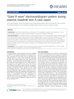
was 17 mmHg OD and 20 mmHg OS. Computed tomog-
raphy disclosed a distinct homogeneous lesion in the left
superior orbit, molding to the globe and other orbital
structures (Fig. 1). There was no bone erosion. The find-
ings favored the diagnosis of a lymphoid lesion and a
transpalpebral biopsy was indicated and performed.
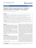
Histopathological evaluation revealed the presence of
structures resembling lymphoid follicles surrounded by
loose connective tissue (Fig. 2A). At higher magnification,
those structures were composed of numerous blood ves-
sels lined by plump endothelial cells with oval nuclei pro-
truding into the lumen (Fig. 2B). Surrounding the vessels,
there was a chronic inflammatory infiltrate composed of
lymphocytes, plasma cells and a large proportion of eosi-
nophils. Immunohistochemical studies, performed on
paraffin-embedded tissue, showed the following: Factor
VIII underscored the marked vascularity of the lesion (Fig.
2C), highlighting atypical vascular lining with "epithe-
lioid" or "histiocytoid" cells (Fig. 2D). Whole body gal-
lium scan failed to reveal lymph node involvement
elsewhere. Blood counts and urinalysis were normal.
Based on clinical and histopathological findings, the diag-
nosis of EH was made.
Conclusion
Epithelioid hemangioma (EH) and Kimura's Disease
(KD) share many clinical and histopathological features
[1] Although they were once considered different stages of
the same disease, they are now known to represent sepa-
rate entities [2] The former is a localized hyperplasia of
atypical endothelial cells with no systemic involvement.
On the other hand, the latter can course with lymphaden-
opathy, blood eosinophilia, and nephrotic syndrome due
to IgE deposition in the renal glomeruli [1]
EH was first described in 1969 [5] It presents as nodules
or erythematous subcutaneous papules, usually in the
head and neck region of young women [6] It can occur in
all races. Whenever the orbit is involved, common symp-
toms are proptosis, tearing, pruritus around the eye, and
blurred peripheral vision. [4] The case presented herein
had no associated symptoms besides the swelling of the
eyelid, which makes the presentation even more atypical.
Histologically, most lesions are well-circumscribed and
composed of vessels lined by plump endothelial cells that
protrude into the lumen in a "tombstone fashion" [7] Sur-
rounding the vessels, there is usually a prominent inflam-
matory infiltrate. A large proportion of eosinophils can
often be seen.
KD probably represents an allergic or autoimmune
response that typically presents as subcutaneous nodules
in the head and neck region of young Asian males [6] Sys-
temic associations include blood eosinophilia, nephrotic
syndrome due to IgE depostion in the renal glomeruli,
lymphadenopathy and, less common, asthma, tuberculo-
sis and Loffler syndrome.[1]
Although exams like blood count, urinalysis and whole
body scans can assist in the differential diagnosis, EH can
be diagnosed and differentiated from KD on histopatho-
logical grounds. The presence of vascular hyperplasia with
plump endothelial cells protruding into the lumen is the
most important discriminator in establishing the diagno-
sis of EH. Such distinction is crucial for the patient
because EH is not associated with any of the systemic
manifestations present in KD.
Competing interests
The author(s) declare that they have no competing inter-
ests.
Computerized TomographyFigure 1
Computerized Tomography. A homogeneous lesion in the
left superior orbit, molding to the globe and other orbital
structures.
Journal of Medical Case Reports 2007, 1:30 />Page 3 of 4
(page number not for citation purposes)
Authors' contributions
BF, TH, AAlk and MNBJr were the pathologists that per-
formed the histopathological evaluation. AAlm and BA
are ophthalmogists from the Oculoplastics departments
and were the attending physician responsible of providing
all the clinical information All authors participated in the
design of the manuscript. BF, AAlk and AAlm helped to
draft the manuscript while TH, BA and MNBJr done the
final revisions of the paper. All authors read and approved
the final manuscript.
Acknowledgements
Written patient consent was obtained.
References
1. Buggage RR, Spraul CW, Wojno TH, Grossniklaus HE: Kimura dis-
ease of the orbit and ocular adnexa. Surv Ophthalmol 1999,
44(1):79-91.
2. Seregard S: Angiolymphoid hyperplasia with eosinophilia
should not be confused with Kimura's disease. Acta Ophthalmol
Scand 2001, 79(1):91-93.
3. Weiss SW, Enzinger FM: Epithelioid hemangioendothelioma: a
vascular tumor often mistaken for a carcinoma. Cancer 1982,
50(5):970-981.
4. McEachren TM, Brownstein S, Jordan DR, Montpetit VA, Font RL:
Epithelioid hemangioma of the orbit. Ophthalmology 2000,
107(4):806-810.
5. Wells GC, Whimster IW: Subcutaneous angiolymphoid hyper-
plasia with eosinophilia. Br J Dermatol 1969, 81(1):1-14.
6. Acocella A, Catelani C, Nardi P: Angiolymphoid hyperplasia with
eosinophilia: a case report of orbital involvement. J Oral Max-
illofac Surg 2005, 63(1):140-144.
A) Low-power photomicrography showing structures resembling lymphoid follicles surrounded by loose connective tissue (H&E; original magnification × 25)Figure 2
A) Low-power photomicrography showing structures resembling lymphoid follicles surrounded by loose connective tissue
(H&E; original magnification × 25). B) Plump endothelial cells, surrounded by an inflammatory infiltrate containing a large
number of eosinophils (H&E; original magnification × 400). C) Factor VIII immunostaining, highlighting the florid vascular prolif-
eration (Original magnification × 25). D) The atypical endothelial cells all stained positive (Factor VIII, original magnification ×
400).
Publish with BioMed Central and every
scientist can read your work free of charge
"BioMed Central will be the most significant development for
disseminating the results of biomedical research in our lifetime."
Sir Paul Nurse, Cancer Research UK
Your research papers will be:
available free of charge to the entire biomedical community
peer reviewed and published immediately upon acceptance
cited in PubMed and archived on PubMed Central
yours — you keep the copyright
Submit your manuscript here:
/>BioMedcentral
Journal of Medical Case Reports 2007, 1:30 />Page 4 of 4
(page number not for citation purposes)
7. Calonje E, Fletcher CD: Tumors of Blood Vessels and Lymphat-
ics. In Diagnostic Histopathology of Tumors Volume 1. 2nd edition.
Edited by: Fletcher CD. London , Hartcourt Publishers Limited;
2000:55-56.