Báo cáo khoa hoc:" Pituitary macroadenomas: are combination antiplatelet and anticoagulant therapy contraindicated? A case report" pot
Bạn đang xem bản rút gọn của tài liệu. Xem và tải ngay bản đầy đủ của tài liệu tại đây (470.01 KB, 4 trang )
BioMed Central
Page 1 of 4
(page number not for citation purposes)
Journal of Medical Case Reports
Open Access
Case report
Pituitary macroadenomas: are combination antiplatelet and
anticoagulant therapy contraindicated? A case report
Tricia MM Tan*
1
, Carmela Caputo
1
, Amrish Mehta
2
, Emma CI Hatfield
1
,
Niamh M Martin
1
and Karim Meeran
1
Address:
1
Endocrine Unit, Hammersmith Hospitals NHS Trust, Imperial College Faculty of Medicine, London, UK and
2
Department of Radiology,
Hammersmith Hospitals NHS Trust, Imperial College Faculty of Medicine, London, UK
Email: Tricia MM Tan* - ; Carmela Caputo - ; Amrish Mehta - ;
Emma CI Hatfield - ; Niamh M Martin - ; Karim Meeran -
* Corresponding author
Abstract
Background: Pituitary apoplexy is a life-threatening endocrine emergency that is caused by
haemorrhage or infarction of the pituitary gland, commonly within a pituitary adenoma. Patients
classically present with headache, ophthalmoplegia, visual field defects and altered mental state, but
may present with a typical symptoms such as fever and altered conscious level.
Case presentation: A 57-year-old female with a known pituitary macroadenoma was treated for
suspected acute coronary syndrome with aspirin, clopidogrel and full dose enoxaparin. She
developed a severe and sudden headache, nausea and vomiting and visual deterioration. A CT scan
showed haemorrhage into the pituitary macroadenoma. She underwent neurosurgical
decompression. Post-operatively her visual fields and acuity returned to baseline. She was
continued on hydrocortisone and thyroxine replacement on discharge.
Conclusion: This case illustrates the risks of anticoagulation in a patient with a known pituitary
macroadenoma, and raises the issue of whether these tumours present a relative contraindication
to the use of dual antiplatelet and anticoagulation in acute coronary syndrome.
Background
Pituitary apoplexy is defined as haemorrhage or infarction
of the pituitary gland. This occurs often in the context of a
pituitary adenoma, although it can occur in normal pitui-
taries in patients with post partum haemorrhage (Shee-
han's syndrome) [1]. This is an emergency because of the
combination of secondary adrenal insufficiency, with
compression of the optic chiasm and the III, IV, V and VI
cranial nerves [2].
The prevalence of classical pituitary apoplexy in retrospec-
tive case series of patients undergoing pituitary surgery
varies from 5% [3] to 9.1% [4]. In patients with non-func-
tioning macroadenomas, who were not operated on and
followed up for 85 +/- 13 months, 14% developed pitui-
tary apoplexy [5]. The majority present with no previous
history of pituitary adenoma, and their tumour is discov-
ered when the apoplexy occurs [6]. Many precipitating
factors have been described, from dopamine agonists [7],
anticoagulation [8], head trauma [9], pituitary irradiation
Published: 30 August 2007
Journal of Medical Case Reports 2007, 1:74 doi:10.1186/1752-1947-1-74
Received: 15 May 2007
Accepted: 30 August 2007
This article is available from: />© 2007 Tan et al; licensee BioMed Central Ltd.
This is an Open Access article distributed under the terms of the Creative Commons Attribution License ( />),
which permits unrestricted use, distribution, and reproduction in any medium, provided the original work is properly cited.
Journal of Medical Case Reports 2007, 1:74 />Page 2 of 4
(page number not for citation purposes)
[10], to dynamic endocrine testing, most likely triggered
by TRH administration [11].
Case presentation
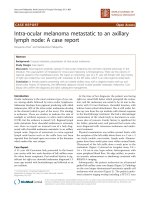
A 57-year-old menopausal female, during evaluation for
headache, was incidentally found to have a 20 × 16 mm
mixed solid and cystic pituitary mass abutting the optic
chiasm on MRI scanning (Figure 1). Endocrine testing
showed that she had normal pituitary hormone levels
with the exception of gonadotrophin deficiency. Formal
perimetry initially showed full visual fields. Within 12
months, however, she developed a mild superior bi-tem-
poral hemianopia. Despite this, she elected not to have
surgery.
Several months later, she was admitted to hospital with
chest pain and treated for acute coronary syndrome with
aspirin, clopidogrel and 1 mg/kg enoxaparin bd. She had
no ECG changes suggestive of myocardial ischaemia or
infarction. Her troponin level at 12 hours was undetecta-
ble. On the 2nd day of admission she developed a severe
and sudden headache associated with nausea and vomit-
ing. At this stage, her blood pressure was 144/85 mmHg
and there were no neuro-ophthalmological symptoms or
signs. The next day her symptoms continued and she
noticed a constriction in her visual fields. She was febrile
with an elevated C-reactive protein of 155 mg/l (normal <
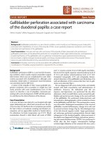
5 mg/l). An ECG showed a new finding of global ST
depression, but without chest pain (Figure 2).
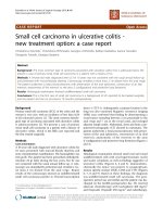
Perimetry confirmed deterioration in the patient's visual
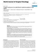
fields and acuities (Figure 3), and a CT scan showed
haemorrhage into the pituitary macroadenoma (Figure
4). She was given intramuscular hydrocortisone, and
underwent urgent trans-sphenoidal surgery and decom-
pression of the apoplectic pituitary.
Post-operatively her visual fields and acuity returned to
baseline (Figure 3). Her pyrexia ceased and CRP
ECG taken during apoplectic crisisFigure 2
ECG taken during apoplectic crisis. Global ST depression is demonstrated, particularly in leads V2-V6. These changes
were not present on her admission ECG and resolved after surgery.
MRI scan demonstrating pituitary macroadenomaFigure 1
MRI scan demonstrating pituitary macroadenoma. A
mixed cystic/solid mass is demonstrated lying within the pitu-
itary fossa, bowing and indenting the optic chiasm.
Journal of Medical Case Reports 2007, 1:74 />Page 3 of 4
(page number not for citation purposes)
decreased. The ECG returned to normal. Cardiological
investigations including exercise stress testing and a myo-
cardial perfusion scan did not show evidence of ischaemic
heart disease, implying that the global ST depression
noted pre-operatively was the result of her pituitary apo-
plexy. She was discharged home on hydrocortisone and
thyroxine replacement.
Discussion
We describe a case of pituitary apoplexy in a patient who
was already known to have a pituitary adenoma, and who
was treated for acute coronary syndrome. With the mod-
ern treatment of acute coronary syndrome, an anticoagu-
lation cocktail that includes aspirin, heparin and
clopidogrel is employed. The addition of clopidogrel to
aspirin and heparin has been demonstrated to reduce the
incidence of further vascular events over aspirin and
heparin alone. However, this is at the expense of a signif-
icant increase in rates of major bleeding (mainly gastroin-
testinal) from 2.7% to 3.7% [12]. The apoplectic crisis
initially presented with fever, headache, nausea, and vom-
iting, and ST segment depression on ECG, a finding that
has been reported with subarachnoid haemorrhage [13],
but has not previously been reported in association with
pituitary apoplexy. The risks of death or serious visual loss
in the event of apoplexy are considerable, especially if
there is diagnostic delay occasioned by a non-classical
presentation, e.g. with fever of unknown origin, hyponat-
raemia and altered consciousness [6]. This situation can
be compounded by non-diagnostic investigations such as
non-specific changes in the CSF [14], or an apparently
normal CT scan of the brain, which has been shown to be
of lower sensitivity in detecting pituitary apoplexy com-
pared to MRI scanning [15].
Conclusion
Anticoagulation is well known as a precipitating factor for
pituitary apoplexy. Like our patient, one case study has
reported a patient with pituitary apoplexy precipitated by
aspirin, clopidogrel and enoxaparin, although in that case
the patient did not have a previously known pituitary ade-
noma [16]. Our case therefore highlights some important
practice points: in patients who are already known to have
a pituitary adenoma, this condition should be considered
a relative contraindication for anticoagulation. These
patients should be warned about the potential risks of
anticoagulation with respect to their pituitary adenoma. If
these patients are anticoagulated, a high index of suspi-
cion of pituitary apoplexy should guide the clinician if
CT scan of the pituitary demonstrating haemorrhageFigure 4
CT scan of the pituitary demonstrating haemor-
rhage. Patchy enhancement of the pituitary mass is seen
indicating haemorrhage within the pituitary macroadenoma.
Visual field tests before and after pituitary surgeryFigure 3
Visual field tests before and after pituitary surgery.
The results of visual field testing are shown. The upper set,
taken before the operation, demonstrate a temporal hemian-
opia in the left eye field and closure of three quadrants in the
right eye field, sparing the inferior nasal quadrant. The lower
set demonstrates the improved visual fields after surgery.
Left Right
Pre-operation
Left Right
Post-operation
Publish with BioMed Central and every
scientist can read your work free of charge
"BioMed Central will be the most significant development for
disseminating the results of biomedical research in our lifetime."
Sir Paul Nurse, Cancer Research UK
Your research papers will be:
available free of charge to the entire biomedical community
peer reviewed and published immediately upon acceptance
cited in PubMed and archived on PubMed Central
yours — you keep the copyright
Submit your manuscript here:
/>BioMedcentral
Journal of Medical Case Reports 2007, 1:74 />Page 4 of 4
(page number not for citation purposes)
they fall acutely ill: early hydrocortisone replacement
should be instituted.
Competing interests
The author(s) declare that they have no competing inter-
ests.
Authors' contributions
All authors participated in the care of the patient
described. TMMT drafted the manuscript. CC, ECIH,
NMM, KM critically revised the content of the manuscript.
All authors have read and approved the final version of
the manuscript.
Acknowledgements
Written consent has been obtained from the patient described in this case
report.
References
1. Asa SL: Pituitary histopathology in man: normal and abnor-
mal. In Endotext.com: Pituitary Disease and Neuroendocrinology Edited
by: DeGroot LJ, Grossman AB. South Dartmouth, MA, USA:
MDText.com, Inc; 2002.
2. Cardoso ER, Peterson EW: Pituitary apoplexy: a review. Neuro-
surgery 1984, 14:363-373.
3. Bonicki W, Kasperlik-Zaluska A, Koszewski W, Zgliczynski W, Wis-
lawski J: Pituitary apoplexy: endocrine, surgical and oncologi-
cal emergency. Incidence, clinical course and treatment with
reference to 799 cases of pituitary adenomas. Acta Neurochir
(Wien) 1993, 120:118-122.
4. Wakai S, Fukushima T, Teramoto A, Sano K: Pituitary apoplexy: its
incidence and clinical significance. J Neurosurg 1981, 55:187-193.
5. Dekkers OM, Hammer S, de Keizer RJ, Roelfsema F, Schutte PJ, Smit
JW, Romijn JA, Pereira AM: The natural course of non-function-
ing pituitary macroadenomas. Eur J Endocrinol 2007,
156:217-224.
6. Sibal L, Ball SG, Connolly V, James RA, Kane P, Kelly WF, Kendall-Tay-
lor P, Mathias D, Perros P, Quinton R, Vaidya B: Pituitary apoplexy:
a review of clinical presentation, management and outcome
in 45 cases. Pituitary 2004, 7:157-163.
7. Yamaji T, Ishibashi M, Kosaka K, Fukushima T, Hori T, Manaka S, Sano
K: Pituitary apoplexy in acromegaly during bromocriptine
therapy. Acta Endocrinol (Copenh) 1981, 98:171-177.
8. Oo MM, Krishna AY, Bonavita GJ, Rutecki GW: Heparin therapy
for myocardial infarction: an unusual trigger for pituitary
apoplexy. Am J Med Sci 1997, 314:351-353.
9. Holness RO, Ogundimu FA, Langille RA: Pituitary apoplexy fol-
lowing closed head trauma. Case report. J Neurosurg 1983,
59:677-679.
10. Weisberg LA: Pituitary apoplexy. Association of degenerative
change in pituitary adenoma with radiotherapy and detec-
tion by cerebral computed tomography. Am J Med 1977,
63:109-115.
11. Masago A, Ueda Y, Kanai H, Nagai H, Umemura S: Pituitary apo-
plexy after pituitary function test: a report of two cases and
review of the literature. Surg Neurol 1995, 43:158-64. discussion
165
12. Peters RJ, Mehta SR, Fox KA, Zhao F, Lewis BS, Kopecky SL, Diaz R,
Commerford PJ, Valentin V, Yusuf S: Effects of aspirin dose when
used alone or in combination with clopidogrel in patients
with acute coronary syndromes: observations from the
Clopidogrel in Unstable angina to prevent Recurrent Events
(CURE) study. Circulation 2003, 108:1682-1687.
13. Kawasaki T, Azuma A, Sawada T, Sugihara H, Kuribayashi T, Satoh M,
Shimizu Y, Nakagawa M: Electrocardiographic score as a predic-
tor of mortality after subarachnoid hemorrhage. Circ J 2002,
66:567-570.
14. Embil JM, Kramer M, Kinnear S, Light RB: A blinding headache.
Lancet 1997, 350:182.
15. Randeva HS, Schoebel J, Byrne J, Esiri M, Adams CB, Wass JA: Clas-
sical pituitary apoplexy: clinical features, management and
outcome. Clin Endocrinol (Oxf) 1999, 51:181-188.
16. Nagarajan DV, Bird D, Papouchado M: Pituitary apoplexy follow-
ing anticoagulation for acute coronary syndrome. Heart 2003,
89:10.