Báo cáo khoa hoc:" Sublingual epidermoid cyst: a case report" pps
Bạn đang xem bản rút gọn của tài liệu. Xem và tải ngay bản đầy đủ của tài liệu tại đây (274.75 KB, 4 trang )
BioMed Central
Page 1 of 4
(page number not for citation purposes)
Journal of Medical Case Reports
Open Access
Case report
Sublingual epidermoid cyst: a case report
Tolga Kandogan*
1
, Murat Koç
1
, Enver Vardar
2
, Elif Selek
2
and Özlem Sezgin
3
Address:
1
Department of Otolaryngology, İzmir Teaching and Research Hospital, Bozyaka 35220 İzmir, Turkey,
2
Department of Pathology, İzmir
Teaching and Research Hospital, Bozyaka 35220 İzmir, Turkey and
3
Department of Radiology, İzmir Teaching and Research Hospital, Bozyaka
35220 İzmir, Turkey
Email: Tolga Kandogan* - ; Murat Koç - ; Enver Vardar - ;
Elif Selek - ; Özlem Sezgin -
* Corresponding author
Abstract
Epidermoid and dermoid cysts represent less than 0.01% of all oral cavity cysts. The cysts can be
defined as epidermoid when the lining presents only epithelium, dermoid cysts when skin adnexa
are found, and teratoid cysts when other tissue such as muscle, cartilage, and bone are present.
In this article, we present the case of an epidermoid cyst, with an oral as well as a submental
component, in an 11 year old boy who presented with complaints of a mass in the oral cavity,
difficulty chewing and swallowing of solid foods for about 3 years. He was admitted to the
otolaryngology department. On examination, a mass displacing the tongue superiorly and
posteriorly was noticed. An MRI scan was done and showed a 40 × 35 mm well-circumscribed non-
enhancing cystic mass extending from the sublingual area to the level of the thyroid notch. The
content of the cyst was homogenous. On examining the neck, a firm swelling was also noticed in
the submental area, extending down to the thyroid notch. Under general anesthesia and with
nasotracheal intubation, the patient underwent surgical removal of the mass. Extraorally, a midline
submental horizontal incision was performed through the mucosa overlying the swelling and the
cyst was dissected from the surrounding tissues and removed. On histological examination,
acidophilic stratum corneum and basophilic dot like staining of stratum granulosum, which is the
hallmark of an epidermoid cyst, were seen. The patient did well postoperatively, and no recurrence
was noticed at the 6-months follow-up.
Introduction
Epidermoid and dermoid cysts are benign lesions encoun-
tered throughout the body, with 7% occurring in the head
and neck area and 1.6% within the oral cavity [1]. They
represent less than 0.01% of all oral cavity cysts [2]. The
cysts can be defined as epidermoid when the lining
presents only epithelium, dermoid cysts when skin adn-
exa are found, and teratoid cysts when other tissue such as
muscle, cartilage, and bone are present [3].
The pathogenesis of midline cysts of the floor of the
mouth is not well established, and dysontogenetic, trau-
matic, and thyroglossal anomaly theories have been sug-
gested. Histologically, Meyer divided the cysts of the floor
of the mouth into three groups: epidermoid, dermoid,
and teratoid [4]. Although dermoid cysts represent a sep-
arate entity, the term "dermoid" is typically used to indi-
cate all three categories [4]. In fact, dermoid cysts occur
primarily in the testes and ovaries, and the most common
Published: 17 September 2007
Journal of Medical Case Reports 2007, 1:87 doi:10.1186/1752-1947-1-87
Received: 4 June 2007
Accepted: 17 September 2007
This article is available from: />© 2007 Kandogan et al; licensee BioMed Central Ltd.
This is an Open Access article distributed under the terms of the Creative Commons Attribution License ( />),
which permits unrestricted use, distribution, and reproduction in any medium, provided the original work is properly cited.
Journal of Medical Case Reports 2007, 1:87 />Page 2 of 4
(page number not for citation purposes)
location in the head and neck is the external third of the
eyebrow [4].
Dermoid cysts generally present with slow and progressive
growth, and even if they are congenital, the diagnosis is
usually possible in the second or third decade of life [5].
The treatment of dermoid cysts of the floor of the mouth
is surgical and can be by an intraoral or extraoral route
according to the localization and the size of the mass [6].
Dermoid cysts usually present early in life as an asympto-
matic mass and are treated by simple excision. However,
they may reach a large size, involve more than one ana-
tomical area and/or abut the hyoid bone when in the neck
[7].
Such a swelling on the floor of the mouth can occasionally
cause serious problems for swallowing and speaking [8,9].
In this article, we outline the case of an epidermoid cyst
with an oral as well as a submental component diagnosed
in an 11 year old boy.
Case presentation
An 11 year old male patient was admitted to the otolaryn-
gology department with complaints of a mass in the oral
cavity and difficulty chewing and swallowing of solid
foods for the past 3 years. The patient had no dyspnea or
pain. There was no history of previous surgery or trauma
to the oral cavity or neck. On examination, there was a 40
× 35 mm sublingual mass with normal covering mucosa
displacing the tongue superiorly and posteriorly.
On examining the neck, a firm swelling was also noticed
in the submental area, extending down to the thyroid
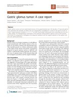
notch. An MRI scan was done and showed an 40 × 35 mm
well-circumscribed non-enhancing cystic mass extending
from the sublingual area to the thyroid notch level (Figure
1). The content of the cyst was homogenous. In order to
exclude any thyroid pathology, thyroid scintigraphy was
taken. This was also normal.
Under general anesthesia and with nasotracheal intuba-
tion, the patient underwent surgical removal of the mass.
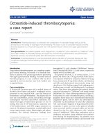
Extraorally, a midline submental horizontal incision was
performed through the mucosa overlying the swelling and
the cyst was dissected from the surrounding tissues and
removed (Figure 2). The wound was closed primarily. The
postoperative period was without any complication and
the tongue went back to its normal position. On histolog-
ical examination, acidophilic stratum corneum and
basophilic dot like staining of stratum granulosum were
seen (Figure 3). Stratum granulosum is the hallmark of
epidermoid cyst. (H-E ×200). It confirmed the diagnosis
of an epidermoid cyst. The patient did well postopera-
tively, and no recurrence was noticed at the 6-months fol-
low-up.
Discussion
Epidermoid cysts may be classified as congenital or
acquired, even if there is no difference between the two on
presentation or histologically. Many etiopathogenetical
theories have been proposed. Congenital cysts are dysem-
bryogenetic lesions that arise from ectodermic elements
entrapped during the midline fusion of the first and sec-
ond branchial arches between the third and fourth weeks
of intrauterine life. Alternatively, they may arise from the
tuberculum impar of His which, with each mandibular
arch, forms the floor of the mouth and the body of the
tongue. Acquired cysts derive from traumatic or iatrogenic
inclusion of epithelial cells or from the occlusion of a
sebaceous gland duct. Moreover, others authors proposed
that midline cysts may represent a variant form of thy-
roglossal duct cyst [4,6,8,10].
Congenital cysts of ectodermal origin are uncommon in
the oral cavity (1.6%), with epidermoid cysts rarely occur-
ring there [11]. Midline cysts of the floor of the mouth are
painless lesions that swell from the anterior portion of
this region. Because they can displace the tongue, patients
usually present with dysphagia, dysphonia, and dyspnea,
and in the case of lower localization, they present a char-
acteristic double chin [6].
Dermoid cysts are generally diagnosed in young adults in
the second and third decades of life [6]; although the case
presented here was an 11 year old boy.
There are no rules regarding the timing for operation;
because dermoid cysts are mainly congenital, they can
appear in every age of life, so the time when they appear
(generally with dysphagia, dysphonia, and dyspnea) is
generally the right time to operate on them. Also, in very
young patients, a problem can arise from the anesthesio-
logic risk, which is generally quite low in patients weigh-
ing more than 20 kg [6].
Histologically, midline dermoid cysts of the floor of the
mouth are classified according to Meyer's classification,
thus dividing them into three groups: epidermoid cysts,
which consist of an epithelial-lined wall that may be
partly keratinized; dermoid cysts, which are epidermoid-
like cysts but show evidence of skin appendages, such as
hair follicles, hair, sweat, and sebaceous glands; and ter-
atomas, which contain, in addition to skin appendages,
mesodermal elements such as bone, muscle, respiratory
and gastrointestinal tissues, and a fibrous capsule. The lat-
ter type is the only variety that may have a malignant
change [3,6,8,12].
Journal of Medical Case Reports 2007, 1:87 />Page 3 of 4
(page number not for citation purposes)
Anatomic classification divides the cysts of the floor of the
mouth into three groups according to their relation to the
muscles of the floor of the mouth : sublingual or median
genioglossal cysts, located above the geniohyoid muscles;
median geniohyoid cysts, located in the submental region
between the geniohyoid and mylohyoid muscles; and lat-
eral cysts, located in the submaxillary region [6].
The differential diagnosis of sublingual lesions includes:
infectious process, ranula, lymphatic malformation, der-
moid cyst, epidermoid cyst, heterotopic gastrointestinal
cyst and duplication foregut cyst. For this reason, biman-
ual palpation and conventional radiography are not
always sufficient in making differential diagnoses. In
these cases, it is necessary to use ultrasonography, com-
puted tomography, or magnetic resonance imaging
together with cytologic examination by fine-needle aspi-
ration biopsy [8]. Ultrasonography represents the first
choice of imaging technique because it is reliable, eco-
nomical, and without x-ray exposure, so it is easily suita-
ble for young patients also. Computed tomography and
magnetic resonance imaging allow more precise localiza-
tion of the lesion in relationship to geniohyoid and mylo-
hyoid muscles, and they also enable the surgeon to
choose the most appropriate surgical approach, especially
for very large lesions [6].
Surgical enucleation is the only effective treatment for
these kinds of lesions. Several techniques are reported in
the literature, which may be divided into intraoral and
extraoral techniques depending on which approach is
used [6]. The extraoral approach is generally preferred in
the case of median geniohyoid or very large sublingual
cysts, whereas the intraoral approach is typically used for
smaller sublingual cysts [13].
Prognosis is very good, with a very low incidence of
relapse, usually related to the genial tubercles or to the
Acidophilic stratum corneum and basophilic dot like staining of stratum granulosum were seenFigure 3
Acidophilic stratum corneum and basophilic dot like staining
of stratum granulosum were seen. Stratum granulosum is
hallmark of epidermoid cyst. (H-E –200).
A MRI scan showing an 40 × 35 mm well-circumscribed non-enhancing cystic mass extending from the sublingual area to the thyroid notch levelFigure 1
A MRI scan showing an 40 × 35 mm well-circumscribed non-
enhancing cystic mass extending from the sublingual area to
the thyroid notch level.
A per-operative view to the cystFigure 2
A per-operative view to the cyst.
Publish with BioMed Central and every
scientist can read your work free of charge
"BioMed Central will be the most significant development for
disseminating the results of biomedical research in our lifetime."
Sir Paul Nurse, Cancer Research UK
Your research papers will be:
available free of charge to the entire biomedical community
peer reviewed and published immediately upon acceptance
cited in PubMed and archived on PubMed Central
yours — you keep the copyright
Submit your manuscript here:
/>BioMedcentral
Journal of Medical Case Reports 2007, 1:87 />Page 4 of 4
(page number not for citation purposes)
hyoid bone. Malignant changes have been recorded in
dermoid cysts by New and Erich but not in the floor of the
mouth, although a 5% rate of malignant transformation
of oral dermoid cysts of the teratoid type has been
reported by other authors [5].
Conclusion
Appropriate imaging techniques and thyroid scintigraphy
are necessary in the preoperative diagnosis of cysts of the
floor of the mouth. Surgical enucleation is the only effec-
tive treatment for these kinds of lesions.
Competing interests
The author(s) declare that they have no competing inter-
ests.
Authors' contributions
TK, MK, EV, ES and ÖS drafted the manuscript and
designed the case report. All authors read and approved
the final manuscript.
Acknowledgements
Written informed patient consent was obtained for publication.
References
1. Turetschek K, Hospodka H, Steiner E: Case report: epidermoid
cyst of the floor of the mouth: diagnostic imaging by sonog-
raphy, computed tomography and magnetic resonance
imaging. Br J Radiol 1995, 68:205-207.
2. Rajayogeswaran V, Eveson JW: Epidermoid cyst of the buccal
mucosa. Oral Surg Oral Med Oral Pathol 1989, 67:181-184.
3. Calderon S, Kaplan I: Concomitant sublingual and submental
epidermoid cysts: a case report. J Oral Maxillofac Surg 1993,
51:790-792.
4. Howell CJT: The sublingual dermoid cyst: Report of five cases
and review of the literature. Oral Surg Oral Med Oral Pathol 1985,
59:578.
5. Zachariades N, Skoura-Kafoussia C: A life threatening epider-
moid cyst of the floor of the mouth: Report of a case. J Oral
Maxillofac Surg 1990, 48:400.
6. Longo F, Maremonti P, Mangone GM, De Maria G, Califano L: Mid-
line (dermoid) cysts of the floor of the mouth: report of 16
cases and review of surgical techniques. Plast Reconstr Surg
2003, 112:1560-1565.
7. Bitar MA, Kumar S: Plunging congenital epidermoid cyst of the
oral cavity. Eur Arch Otorhinolaryngol 2003, 260:223-225.
8. Walstad WR, Solomon JM, Schow SR, Ochs MW: Midline cystic
lesion of the floor of the mouth. J Oral Maxillofac Surg 1998,
56:70-74.
9. Koca H, Seckin T, Sipahi A, Kazanc A: Epidermoid cyst in the floor
of the mouth: Report of a case. Quintessence Int 2007,
38:473-477.
10. De Ponte FS, Brunelli A, Marchetti E, Bottini DJ: Sublingual epider-
moid cyst. J Craniofac Surg 2002, 13:308-310.
11. Al-Khayat M, Kenyon GS: Midline sublingual dermoid cyst. J
Laryngol Otol 1990, 104:578-580.
12. Yilmaz I, Yilmazer C, Yavuz H, Bal N, Ozluoglu LN: Giant sublingual
epidermoid cyst: a report of two cases. J Laryngol Otol 2006,
120:E19.
13. Lowry RE, Tempero RM, Davis LF: Epidermoid cyst of the floor
of the mouth. J Oral Surg 1979, 37:271.