Báo cáo khoa hoc:" Small cell carcinoma arising in Barrett''''s esophagus: a case report and review of the literature" pptx
Bạn đang xem bản rút gọn của tài liệu. Xem và tải ngay bản đầy đủ của tài liệu tại đây (413.33 KB, 4 trang )
BioMed Central
Page 1 of 4
(page number not for citation purposes)
Journal of Medical Case Reports
Open Access
Case report
Small cell carcinoma arising in Barrett's esophagus: a case report
and review of the literature
Haridimos Markogiannakis*
1
, Dimitrios Theodorou
1
,
Konstantinos G Toutouzas
1
, Andreas Larentzakis
1
, Michael Pattas
1
,
Angeliki Bousiotou
2
, Pavlos Papacostas
3
, Konstantinos Filis
1
and
Stilianos Katsaragakis
1
Address:
1
1st Department of Propaedeutic Surgery, Hippokrateion Hospital, Athens Medical School, University of Athens, Q. Sofias 114 av.,
11527, Athens, Greece,
2
Department of Histopathology, Hippokrateion Hospital, Athens, Greece and
3
Department of Oncology, Hippokrateion
Hospital, Athens, Greece
Email: Haridimos Markogiannakis* - ; Dimitrios Theodorou - ;
Konstantinos G Toutouzas - ; Andreas Larentzakis - ; Michael Pattas - ;
Angeliki Bousiotou - ; Pavlos Papacostas - ; Konstantinos Filis - ;
Stilianos Katsaragakis -
* Corresponding author
Abstract
Introduction: Gastrointestinal tract small cell carcinoma is an infrequent and aggressive neoplasm that represents 0.1–
1% of gastrointestinal malignancies. Very few cases of small cell esophageal carcinoma arising in Barrett's esophagus have
been reported in the literature. An extremely rare case of primary small cell carcinoma of the distal third of the
esophagus arising from dysplastic Barrett's esophagus is herein presented.
Case presentation: A 62-year-old man with gastroesophageal reflux history presented with epigastric pain, epigastric
fullness, dysphagia, anorexia, and weight loss. Esophagogastroscopy revealed an ulceroproliferative, intraluminar mass in
the distal esophagus obstructing the esophageal lumen. Biopsy showed small cell esophageal carcinoma. Contrast-
enhanced chest and abdominal computed tomography demonstrated a large tumor of the distal third of the esophagus
without any lymphadenopathy or distant metastasis. Preoperative chemotherapy with cisplatine and etoposide for 3
months resulted in a significant reduction of the tumor. After en block esophagectomy with two field lymph node
dissection, proximal gastrectomy, and cervical esophagogastric anastomosis, the patient was discharged on the 14
th
postoperative day. Histopathology revealed a primary small cell carcinoma of the distal third of the esophagus arising
from dysplastic Barrett's esophagus. The patient received another 3 month course of postoperative chemotherapy with
the same agents and remained free of disease at 12 month review.
Conclusion: Although small cell esophageal carcinoma is rare and its association with dysplastic Barrett's esophagus is
extremely infrequent, the high carcinogenic risk of Barrett's epithelium should be kept in mind. Prognosis is quite
unfavorable; a better prognosis might be possible with early diagnosis and treatment strategies incorporating
chemotherapy along with oncological radical surgery and/or radiotherapy as part of a multimodality approach. Since
treatment protocols are not well established due to the rarity of the neoplasm, multi-institutional studies are needed to
obtain sufficiently large populations for investigation and optimization of therapy of the disease.
Published: 22 January 2008
Journal of Medical Case Reports 2008, 2:15 doi:10.1186/1752-1947-2-15
Received: 12 May 2007
Accepted: 22 January 2008
This article is available from: />© 2008 Markogiannakis et al; licensee BioMed Central Ltd.
This is an Open Access article distributed under the terms of the Creative Commons Attribution License ( />),
which permits unrestricted use, distribution, and reproduction in any medium, provided the original work is properly cited.
Journal of Medical Case Reports 2008, 2:15 />Page 2 of 4
(page number not for citation purposes)
Introduction
Gastrointestinal tract small cell carcinoma is an infre-
quent and aggressive malignancy. It represents 0.1–1% of
gastrointestinal malignancies, with the esophagus being
the most common primary site [1,2]. Small cell esopha-
geal carcinoma is a rare tumor constituting 0.8–2.4% of
all esophageal carcinomas [1,2]. It is characterized by
aggressive progression, high incidence of metastatic dis-
ease at presentation, and a poor overall prognosis [1,2].
Since McKeown's first description in 1952 [3], few cases of
small cell esophageal carcinoma have been reported in the
literature. Furthermore, only 4 cases of the neoplasm aris-
ing in Barrett's esophagus have been reported [4-7]. An
extremely rare case of primary small cell carcinoma of the
distal third of the esophagus arising from dysplastic Bar-
rett's mucosa is presented and the relevant literature is
reviewed.
Case presentation
A 62-year-old man with a history of gastroesophageal
reflux disease presented with a one month history of epi-
gastric pain, epigastric fullness, dysphagia, anorexia, and
weight loss of 9 kgs. Clinical examination and blood tests
were normal apart from elevated carcinoembryonic anti-
gen (42.8 ng/ml; normal value < 5.0 ng/ml). Chest x-ray,
electrocardiogram, echocardiography, and abdominal
ultrasound (US) were normal. Esophagogastroscopy
revealed an ulceroproliferative, intraluminar mass in the
distal third of the esophagus obstructing the esophageal
lumen. Biopsy showed small cell esophageal carcinoma.
Due to esophageal lumen obstruction, endoscopic US was
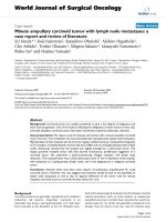
not feasible. Contrast-enhanced chest and abdominal
computed tomography (CT) demonstrated a large tumor
of the distal third of the esophagus without any lymphad-
enopathy or distant metastasis (Figure 1).
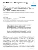
The patient received preoperative chemotherapy with cis-
platine and etoposide for 3 months with a significant
reduction of the tumor size (Figure 2). En block
esophagectomy with two field lymph node dissection,
proximal gastrectomy, and cervical esophagogastric anas-
tomosis were performed. After an uneventful postopera-
tive period, he was discharged on the 14
th
postoperative
day.
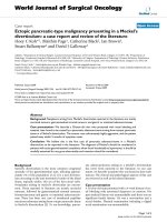
Histopathology revealed a tumor (2.5 × 1.5 cm), 2 cm
above the gastroesophageal junction, that was a small cell
esophageal carcinoma infiltrating the submucosal and,
focally, the muscular layer (Figure 3). The margins of the
specimen were free of tumor. Metastasis in two
paraesophageal lymph nodes was found. Moreover, Bar-
rett's esophagus, of intestinal type, with predominantly
low grade and, focally, high grade dysplasia was identified
in the lower esophagus. Immunohistochemical staining
of the tumor cells was positive for chromogranin and neu-
ron-specific enolase (NSE).
A diagnosis of primary small cell carcinoma of the distal
third of the esophagus arising from dysplastic Barrett's
esophagus was made. The patient received another 3
month course of postoperative chemotherapy with the
same agents (cisplatine and etoposide) and was free of
disease at review after 12 months.
Discussion
Small cell esophageal carcinoma is a rare neoplasm [1,2].
Although the rarity of this tumor has impeded statistical
Post-chemotherapy CT scan showed a significant reduction of the tumor sizeFigure 2
Post-chemotherapy CT scan showed a significant reduction
of the tumor size.
Initial CT scan revealed a large mass in the distal third of the esophagusFigure 1
Initial CT scan revealed a large mass in the distal third of the
esophagus.
Journal of Medical Case Reports 2008, 2:15 />Page 3 of 4
(page number not for citation purposes)
evaluation, it is generally associated with a poor prognosis
because of aggressive biologic behavior and early wide-
spread dissemination [1,2].
Small cell esophageal carcinoma is an exceptional finding
in patients with Barrett's esophagus [4-7]. The presented
case is indicative of the high carcinogenic risk of Barrett's
epithelium. The spectrum of differentiation in Barrett's
esophagus-associated carcinomas is attributed to the
totipotential cell population at the squamocolumnar
junction and to the considerable histologic heterogeneity
of Barrett's mucosa.
The most common symptoms are dysphagia, anorexia,
weight loss, chest pain, and regurgitation [1,2,6]. The neo-
plasm often appears as an exophytic mass usually located
in the middle or lower esophagus while ulceration of the
overlying mucosa is common [1,2,6]. Diagnosis is estab-
lished by the characteristic silver affinity of the tumor
cells, the ultrastructural occurrence of neurosecretory
granules, and immunohistochemically detectable mark-
ers, including neuron-specific enolase, chromogranin,
and synaptophysin [1,2,4]. Ectopic hormonal secretion,
including adrenocorticotropic hormone, calcitonin, gas-
trin, somatostatin, and antidiuretic hormone may occur
[1,2].
Although treatment protocols are not well established
because of the paucity of cases and the lack of large stud-
ies, chemotherapy remains the treatment of choice given
the systemic nature of the disease [1,2]. Since small cell
carcinoma of the esophagus is histologically identical to
small cell carcinoma of the lung and, furthermore, their
aggressive behavior but also chemosensitivity are similar,
the chemotherapeutic agents used for small cell esopha-
geal carcinoma are similar to those for its lung counterpart
[1,2]. Radiotherapy has been used concurrently with
chemotherapy to enhance local control [1,2]. In locore-
gional disease, the literature suggests that treatment be
initiated using chemotherapy and then, if metastatic dis-
ease is still excluded, radical surgical resection be consid-
ered as a second therapy that may have a potential impact
on long-term remission and long-term survival [1,2]. Sur-
vival ranges from several weeks for untreated patients to
6–24 months for those receiving therapy [1,2].
The reported patient presented with a large tumor of the
distal esophagus. Our initial treatment consisted of a 3
month course of preoperative chemotherapy with cispla-
tine and etoposide that resulted in a significant reduction
of the neoplasm. Given the response to chemotherapy,
and since no metastatic disease was identified in the post-
chemotherapy investigation, radical surgical resection,
including en block esophagectomy with two field lymph
node dissection and proximal gastrectomy, was then per-
formed. Based on the promising results of preoperative
chemotherapy, another 3 month course of postoperative
chemotherapy with the same agents was administered.
Although conclusions regarding treatment of such a rare
clinical entity cannot be drawn from a case report, the
effects of our treatment strategy seem encouraging since
our patient remained free of disease at review at 12
months.
Conclusion
Small cell esophageal carcinoma is rare and its association
with dysplastic Barrett's esophagus is extremely infre-
quent. The high carcinogenic risk of Barrett's epithelium,
though, should be kept in mind. Prognosis is quite unfa-
vorable and treatment protocols are not well established.
A better prognosis might be possible with early diagnosis
and treatment strategies incorporating chemotherapy
along with oncological radical surgery and/or radiother-
apy as part of a multimodality approach. Our treatment
strategy of preoperative chemotherapy followed by radical
surgical resection and postoperative chemotherapy in the
reported patient may have yielded promising results.
Multi-institutional studies are needed to obtain suffi-
ciently large populations for investigation and optimiza-
tion of therapy of the disease.
Competing interests
The author(s) declare that they have no competing inter-
ests.
Authors' contributions
HM contributed to manuscript conception, research,
acquisition of data, drafting and writing of the manu-
script. DT carried out the operation and contributed to
acquisition of consent and critical review of the manu-
Infiltration of the esophageal wall from small cell carcinoma (H-E ×40)Figure 3
Infiltration of the esophageal wall from small cell carcinoma
(H-E ×40).
Publish with BioMed Central and every
scientist can read your work free of charge
"BioMed Central will be the most significant development for
disseminating the results of biomedical research in our lifetime."
Sir Paul Nurse, Cancer Research UK
Your research papers will be:
available free of charge to the entire biomedical community
peer reviewed and published immediately upon acceptance
cited in PubMed and archived on PubMed Central
yours — you keep the copyright
Submit your manuscript here:
/>BioMedcentral
Journal of Medical Case Reports 2008, 2:15 />Page 4 of 4
(page number not for citation purposes)
script. KGT assisted in the operation, contributed to
organising and drafting of the manuscript, and critically
revised the manuscript. AL contributed to manuscript
conception, research, acquisition of data, drafting and
writing of the manuscript. MP contributed to manuscript
conception, research, acquisition of data, drafting and
writing of the manuscript. AB carried out the histopatho-
logic evaluation and contributed to writing of the manu-
script. PP contributed to the preoperative and
postoperative management of the patient and to critical
review of the manuscript. KF contributed to organising
and drafting of the manuscript, and critically revised the
manuscript. SK assisted in the operation and contributed
to critical review of the manuscript.
All authors read and approved the final manuscript.
Consent
Written informed consent was obtained from the patient
for publication of this Case report and any accompanying
images. A copy of the written consent is available for
review by the Editor-in-Chief of this journal.
References
1. Huncharek M, Muscat J: Small cell carcinoma of the esophagus.
The Massachusetts General Hospital experience, 1978 to
1993. Chest 1995, 107(1):179-181.
2. Pantvaidya GH, Pramesh CS, Deshpande MS, Jambhekar NA, Sharma
S, Deshpande RK: Small cell carcinoma of the esophagus: the
Tata Memorial Hospital experience. Ann Thorac Surg 2002,
74(6):1924-1927.
3. McKeown F: Oat-cell carcinoma of the oesophagus. J Pathol Bac-
teriol 1952, 64(4):889-891.
4. Noguchi T, Takeno S, Sato T, Uchida Y, Daa T, Yokoyama S: Coex-
istent multiple adenocarcinomas arising in Barrett's esopha-
gus 23 years after total gastrectomy and esophageal small
cell carcinoma. Jpn J Thorac Cardiovasc Surg 2003, 51(6):259-262.
5. Gonzalez LM, Sanz-Esponera J, Saez C, Alvarez T, Sierra E, Sanz-
Ortega J: Case report: esophageal collision tumor (oat cell
carcinoma and adenocarcinoma) in Barrett's esophagus:
immunohistochemical, electron microscopy and LOH analy-
sis. Histol Histopathol 2003, 18(1):1-5.
6. Chen KT: Cytology of small-cell carcinoma arising in Barrett's
esophagus. Diagn Cytopathol 2000, 23(3):180-182.
7. Saint Martin MC, Chejfec G: Barrett esophagus-associated small
cell carcinoma. Arch Pathol Lab Med 1999, 123(11):1123.