Báo cáo y học: "Effects of monoclonal anti-PcrV antibody on Pseudomonas aeruginosa-induced acute lung injury in a rat model" ppt
Bạn đang xem bản rút gọn của tài liệu. Xem và tải ngay bản đầy đủ của tài liệu tại đây (351.35 KB, 9 trang )
BioMed Central
Page 1 of 9
(page number not for citation purposes)
Journal of Immune Based Therapies
and Vaccines
Open Access
Original research
Effects of monoclonal anti-PcrV antibody on Pseudomonas
aeruginosa-induced acute lung injury in a rat model
Karine Faure
1,4
, Junichi Fujimoto
1,5
, David W Shimabukuro
1
,
Temitayo Ajayi
1,3
, Nobuaki Shime
1,6
, Kiyoshi Moriyama
1
, Edward G Spack
7
,
Jeanine P Wiener-Kronish
1,2,3
and Teiji Sawa*
1
Address:
1
Department of Anesthesia and Perioperative Care, University of California, San Francisco, CA94143-0542, U.S.A,
2
Department of
Medicine, University of California, San Francisco, CA94143-0542, U.S.A,
3
Cardiovascular Research Institute, University of California, San
Francisco, CA94143-0542, U.S.A,
4
Laboratoire de Recherche en Pathologie Infectieuse, EA2689, Lille, France,
5
Department of Anesthesiology,
School of Medicine, Yokohama City University, Yokohama 236-0004, Japan,
6
Department of Anesthesiology, Kyoto Prefectural University of
Medicine, Kyoto 602-8566, Japan and
7
InterMune, Inc., Brisbane, CA94010-1317, U.S.A
Email: Karine Faure - ; Junichi Fujimoto - ;
David W Shimabukuro - ; Temitayo Ajayi - ; Nobuaki Shime - ;
Kiyoshi Moriyama - ; Edward G Spack - ; Jeanine P Wiener-
Kronish - ; Teiji Sawa* -
* Corresponding author
Abstract
Background: The effects of the murine monoclonal anti-PcrV antibody Mab166 on acute lung
injury induced by Pseudomonas aeruginosa were analyzed in a rat model.
Methods: Lung injury was induced by the instillation of P. aeruginosa strain PA103 directly into the
left lungs of anesthetized rats. One hour after the bacterial instillation, rabbit polyclonal anti-PcrV
IgG, murine monoclonal anti-PcrV IgG Mab166 or Mab166 Fab-fragments were administered
intratracheally directly into the lungs. The degree of alveolar epithelial injury, amount of lung
edema, decrease in oxygenation and extent of lung inflammation by histology were evaluated as
independent parameters of acute lung injury.
Results: These parameters improved in rats that had received intratracheal instillation of either
rabbit polyclonal anti-PcrV IgG, murine monoclonal anti-PcrV IgG Mab166 or Mab166 Fab-
fragments in comparison with the control group.
Conclusion: Mab166 and its Fab fragments have potential as adjuvant therapy for acute lung injury
due to P. aeruginosa pneumonia.
Background
Pseudomonas aeruginosa (P. aeruginosa) pneumonia fre-
quently causes bacteremia and sepsis in immunocompro-
mised and mechanically ventilated patients [1–7]. This
leads to an increased morbidity and mortality compared
with pneumonia caused by other pathogens [1–5]. The
rapid systemic dissemination of P. aeruginosa is associated
with the fact that some strains of P. aeruginosa cause acute
lung epithelial injury by inducing the necrosis of the lung
epithelium [8,9]. To protect patients who are at risk for
the development of P. aeruginosa pneumonia and sepsis,
therapy would have to be given prior to the development
Published: 13 August 2003
Journal of Immune Based Therapies and Vaccines 2003, 1:2
Received: 02 July 2003
Accepted: 13 August 2003
This article is available from: />© 2003 Faure et al; licensee BioMed Central Ltd. This is an Open Access article: verbatim copying and redistribution of this article are permitted in all
media for any purpose, provided this notice is preserved along with the article's original URL.
Journal of Immune Based Therapies and Vaccines 2003, 1 />Page 2 of 9
(page number not for citation purposes)
of extensive lung injury, as dissemination and multi-sys-
tem organ failure occur once significant lung epithelial
injury is produced [10]. In addition, resistance to antibi-
otics is a major problem in the therapy of P. aeruginosa
infections in critically ill patients. Therefore, a need for
non-antibiotic based adjuvant therapies for virulent P.
aeruginosa has created more interest in generating anti-
body reagents against the Pseudomonal virulence factors
causing acute lung injury.
The pathogenicity of P. aeruginosa appears to be related to
its repertoire of toxins. Type III secretion is a recently iden-
tified toxin secretion system found in most pathogenic
gram-negative bacteria [11,12]. Requiring intimate con-
tact with eukaryotic cell surfaces, this bacterial secretion
system delivers its toxins directly into the cytosol of the
eukaryotic cells, thereby modulating the host immune
response [13]. The virulence of type III secretory cytotox-
ins in P. aeruginosa is associated with acute lung epithelial
damage and dissemination of inflammatory cytokines
and bacteria from the lungs to the circulation [10]. To
date, four type III secretory toxins (ExoS, ExoT, ExoU and
ExoY) have been identified in P. aeruginosa. Cytotoxic P.
aeruginosa possesses the type III secreted cytotoxin ExoU,
which is necessary for causing acute necrotic cell death
[14–16].
We have documented that clinical isolates of P. aeruginosa
expressing the type III secretory proteins was associated
with higher morbidity and poorer outcome than that for
patients infected with P. aeruginosa strains that did not
secrete these proteins [17]. In addition, a correlation
between poor prognosis of patients with ventilator-associ-
ated pneumonia caused by P. aeruginosa and the bacterial
expression of type III secretion was also reported [18].
PcrV is one component of the P. aeruginosa type III secre-
tion system and is homologous to the Yersinia V-antigen
(LcrV) [16]. PcrV appears to be an integral component of
the translocation apparatus of the type III secretion system
mediating the delivery of the type III secretory toxins into
target eukaryotic cells [19]. Active and passive immuniza-
tion against PcrV improved acute lung injury and mortal-
ity of mice infected with cytotoxic P. aeruginosa [19]. The
major effect of immunization against PcrV was due to the
blockade of translocation of the type III secretory toxins
into eukaryotic cells [19]. Furthermore, we demonstrated
that the therapeutic administration of a polyclonal anti-
PcrV IgG prevented septic shock and acute lung injury in
a rabbit model of P. aeruginosa pneumonia, and that the
effects of the anti-PcrV antibody were independent of the
Fc-fragments of IgG [20].
We recently generated a murine monoclonal anti-PcrV
antibody, Mab166, that was found to be protective against
P. aeruginosa-induced mortality when coinstilled with the
bacteria in lungs or intraperitoneally administered to
mice before infection [21]. More recently, major advances
have been made in the development of antibodies safe for
human patients; this has been accomplished by engineer-
ing recombinant antibodies to decrease the immuno-
genicity of murine antibodies (chimeric and humanized
antibodies) and by developing transgenic animals that
produce human monoclonal antibodies. Mab166 could
be humanized if it proves to be effective in protecting ani-
mals from virulent P. aeruginosa. Our objective in this
study was to test the protective effects of a murine mono-
clonal antibody, an antibody that could be humanized, in
a model of early P. aeruginosa lung infection. If effective in
early infection, the monoclonal antibody would be as
effective or even more protective when given prior to the
development of infection. Therefore, we investigated the
protective properties of intratracheally administered
Mab166 and its Fab fragments on acute lung injury in a rat
model of P. aeruginosa pneumonia.
Methods
Animals
Certified pathogen-free, Sprague Dawley male rats (body
weight, 280–380 g) were purchased from Charles River
Laboratories (Wilmington, MA). The rats were housed in
cages with filter tops in specific pathogen-free conditions.
Sterile food and water were provided ad lib. All experi-
ments were done in compliance with Animal Care Com-
mittee rules of the University of California at San
Francisco, U.S.A., and all protocols were approved prior to
the start of the experiments.
P. aeruginosa strain and culture conditions
P. aeruginosa PA103 was used in this study. Bacteria from
frozen stocks, stored at -70°C in 10% sterile skim milk
solutions, were streaked onto trypticase soy agar plates.
Five milliliters of a deferrated dialysate of trypticase soy
broth supplemented with 10 mM nitrilotriacetic acid
(Sigma Chemical, St. Louis, MO), 1% glycerol, and 100
mM monosodium glutamate was inoculated with a loop
of bacteria and grown at 33°C for 13 h under shaking con-
ditions. Cultures were centrifuged at 8,500 × g for 5 min
and the media discarded. The bacterial pellet was washed
twice in lactated Ringer's (L/R) solution and diluted to the
appropriate concentration of CFU/ml in L/R solution, as
determined by spectrophotometry. Plating out the known
dilutions on sheep blood agar plates confirmed the bacte-
rial concentrations.
Surgical preparation and ventilation
The rat model for P. aeruginosa pneumonia was reported
previously [22,23]. Briefly, rats were anesthetized with
100 mg/kg of pentobarbital sodium administered intra-
peritoneally. An endotracheal tube (PE-240, Clay Adams,
Parsippany, NJ) was inserted into the trachea via an open
Journal of Immune Based Therapies and Vaccines 2003, 1 />Page 3 of 9
(page number not for citation purposes)
tracheostomy. The rats were ventilated with a constant-
volume respirator (Harvard Apparatus, South Natick, MA)
with an inspired O
2
fraction of 1.0, peak airway pressures
of 8–12 cmH
2
O and a 2 cm positive end expiratory pres-
sure (PEEP). The respiratory rate was adjusted to maintain
PaCO
2
between 35 and 45 mmHg. The rats remained
anesthetized, intubated and ventilated throughout the
entire experiment. The right carotid artery was canulated
with a polyethylene tube (PE-50, Clay Adams) to monitor
systemic arterial pressure, administrate drugs and obtain
blood samples.
Bacterial instillate preparation and administration
The instillate consisted of 5% bovine serum albumin
(BSA), 2 mg of Evans blue dye, and 1 µCi of
131
I-labeled
albumin, and P. aeruginosa, at a final concentration of 5
× 10
7
CFU/ml in L/R solution to a total volume of 1 mil-
liliter; Colloid osmotic pressure of the instillate was
adjusted by adding 5% BSA as an established method to
quantify liquid clearance of lung epithelial barriers as an
index of lung edema [24]. The bacteria were added just
before airspace instillation if the experiment was to
include bacteria. A sample of the instillate was saved for
radioactivity measurement (counts/min/g) in a γ-ray
counter (Auto-Gamma, model 5550, Packard, Downers
Grove, IL) and quantitative bacterial cultures on sheep
blood agar plates to assure accurate inoculations. The
instillates were delivered slowly, over a 30 min period
using a polyethylene tube (PE-10, Clay Adams) into the
left lungs.
Interventions
Mab166 IgG (IgG2bκ) or its Fab fragments were previ-
ously prepared in PBS and stored at -70°C [21]. Experi-
mental groups are listed in Table 1. One additional group
of rats was used as the sham control group; rats received
L/R solution not containing IgG. In three groups of rats,
we co-instilled P. aeruginosa PA103 (5 × 10
7
CFU) with 4
mg/kg of either mouse monoclonal isotype-matched con-
trol IgG (IgG2b, clone #20116.11, R&D System, Minneap-
olis, MN), rabbit anti-PcrV polyclonal IgG, or murine
monoclonal Mab166 IgG intratracheally. In another three
groups, rats received either PBS or 4 mg/kg of either anti-
PcrV polyclonal IgG, Mab166 IgG, or Mab166 Fab frag-
ments one hour after the airspace instillation of P. aerugi-
nosa PA103 (5 × 10
7
CFU).
General experimental protocol
After surgical preparation, blood pressure and gas
exchanges were allowed to stabilize. Systemic arterial
pressure and airway pressure were continuously moni-
tored using an on-line data logging system (Powerlab,
ADInstruments, Mountain View, CA). Blood samples
were collected every hour for gas exchange measurement,
131
I-albumin radioactivity count and bacterial culture. The
rats were kept anesthetized and paralyzed throughout the
experiment. Four hours after bacterial instillation, rats
were deeply anesthetized and exsanguinated. Pleural flu-
ids were obtained for radioactivity counts. The lungs were
removed through a sternotomy; the left and right lobes
were weighed and homogenized separately for water to
dry weight ratio measurement and radioactivity counts.
Measurement of lung injury
Lung injury was quantified in two different ways, as previ-
ously described [22,23]. The first method evaluates the
integrity of the lung epithelial barrier by quantifying the
efflux of
131
I-albumin from the alveolar to the blood-
stream. Total
131
I-albumin instilled into the lung was
determined by measuring duplicate samples of the
Table 1: Experimental groups.*
Groups Infection (P. aeruginosa) Intervention n
Control None 3
Co-instillation Antibodies were premixed with PA103
Control IgG PA103 (5 × 10
7
CFU), IT Control IgG (IgG2b), 4 mg/ml IT 3
Rab anti-PcrV PA103 (5 × 10
7
CFU), IT Polyclonal anti-PcrV IgG, 4 mg/ml IT 3
Mab166 PA103 (5 × 10
7
CFU), IT Monoclonal Mab166, 4 mg/ml IT 3
Therapeutic Antibodies were intratracehally instilled 1 h after the instillation of PA103
w/o IgG PA103 (5 × 10
7
CFU), IT Phosphate-buffered saline 5
Rab anti-PcrV PA103 (5 × 10
7
CFU), IT Polyclonal anti-PcrV IgG, 4 mg/ml IT 3
Mab166 PA103 (5 × 10
7
CFU), IT Monoclonal Mab166, 4 mg/ml IT 5
Mab166 Fab PA103 (5 × 10
7
CFU), IT Fab fragments of Mab166, 4 mg/ml IT 3
*IT: Intratracheal administration
Journal of Immune Based Therapies and Vaccines 2003, 1 />Page 4 of 9
(page number not for citation purposes)
instillate for total radioactivity (cpm/g) and multiplying
this amount by the total volume instilled into the lung.
Circulating plasma
131
I-albumin was measured from
blood samples obtained every hour and at the end of the
experiment. The plasma fraction was calculated by multi-
plying the counts per gram times the plasma volume
[body weight × 0.07 (1-hematocrit)]. The second method,
the water to dry weight ratio, is a well-accepted index of
lung edema. Lung homogenates were placed in pre-
weighed aluminum pans and dried to a constant weight in
an oven at 80°C for 3 days. The excess water in the exper-
imental lung was calculated with an equation described
previously [22,23].
Histology analysis
Lungs were perfused with 10% buffered formalin phos-
phate for fixation and were embedded in paraffin.
Mounted sections were stained with hematoxylin-eosin
and observed under light microscopy.
Statistical analysis
Results are presented as mean ± standard errors. The dif-
ference between the control IgG-treated group and the
Mab166 IgG or Mab166 Fab fragments-treated group was
analyzed. Two-way analysis of variance (ANOVA),
repeated measure, followed by the Newman-Keuls t-test
or unpaired Student's t-test was used for comparisons of
data. Significance was accepted at P value of < 0.05.
Results
Coinstillation of Mab166 with P. aeruginosa decreased
induced acute lung injury
First, to evaluate the maximal blocking effects of anti-PcrV
IgGs on P. aeruginosa-induced acute lung injury, we coin-
stilled either rabbit-derived polyclonal anti-PcrV IgG,
murine monoclonal anti-PcrV IgG Mab166 (IgG2b), or
irrelevant monoclonal control IgG (IgG2b) (4 mg/kg,
respectively) with P. aeruginosa PA103 (5 × 10
7
CFU) into
the lungs of the anesthetized ventilated rats under artifi-
cially controlled ventilation. The antibodies were
premixed with P. aeruginosa three min before the instilla-
tion. The acute alveolar lung injury was quantified as the
efflux of the coinstilled radioactive alveolar protein tracers
(
131
I-albumin) into the circulation every one-hour during
the 4-h experimental period.
The control rats that received the lactated Ringer's solu-
tion supplemented with 5% bovine serum albumin but
without bacteria did not show any lung epithelial injury
(Fig. 1). Wet to dry weight ratios of the lungs increased to
approximately 6 in the control rats (Fig. 2). Note a wet to
dry weight ratio of the lung of a normal rat is between
3.5–4.0 (data not shown). The rats that received P. aerugi-
nosa mixed with control irrelevant monoclonal IgG intrat-
racheally developed significant acute epithelial injury in 4
h (Fig. 1). Severe lung edema was observed in this group
of rats 4 h after bacterial instillation (Fig. 2). The arterial
blood pressure decreased below 80 mmHg after 4 h time
point (Fig. 3). Arterial blood oxygenation severely
decreased to approx. 100 mmHg immediately after bacte-
rial instillation, never normalized (Fig. 4). Metabolic aci-
dosis gradually developed over the 4 h in this group of rats
(Fig. 5).
The rats that received P. aeruginosa premixed with rabbit
polyclonal anti-PcrV IgG intratracheally developed signif-
icantly lower levels of lung injury. Alveolar epithelial
injury was significantly lower than that of rats that had
received control IgG (Fig. 1), and lung edema was less,
although not significantly (Fig. 2). Blood pressure was
normal for the 4 h (Fig. 3), arterial blood oxygenation
recovered by the 4 h time point (Fig. 4), and acidosis did
not developed (Fig. 5). Finally, in the rats which received
P. aeruginosa premixed with murine monoclonal anti-PcrV
IgG Mab166, the lung epithelial injury and lung edema
were significantly less than in the other groups (Fig. 1 and
2). Arterial blood pressures and acid-base status of these
rats were normal for 4 h (Fig. 3 and 5), and the arterial
blood oxygenation was the best among the three groups
(Fig. 4). Thus, co-instillation of Mab166 with P. aeruginosa
was the most protective.
Therapeutic administration of Mab166 intratracheally
protects against P. aeruginosa-induced acute lung injury
Next, we evaluated the therapeutic administration of anti-
PcrV IgG in our rat model. In this series of the experi-
ments, we administered either rabbit polyclonal anti-PcrV
IgG, murine monoclonal anti-PcrV IgG Mab166, Fab frag-
ments of Mab166 (4 mg/kg, respectively), or PBS alone
without IgG 1 h after the instillation of P. aeruginosa (5 ×
10
7
CFU) into the lungs of the anesthetized ventilated
rats. The rats that received PBS alone 1 h after bacterial
instillation showed a significant increase in lung epithe-
lial injury and lung edema after 4 h. The arterial blood
pressure gradually decreased to 80 mmHg over the exper-
imental periods. The arterial blood oxygenation remained
significantly decreased (Fig. 4). Severe metabolic acidosis
developed over the 4 h (Fig. 5).
The rats that had received either rabbit polyclonal anti-
PcrV IgG, murine monoclonal anti-PcrV IgG Mab166, or
Fab fragments of Mab166 (4 mg/kg) intratracheally
showed significant improvement of alveolar epithelial
injury and lung edema 4 h after bacterial instillation (Fig.
1). The protective effect of Mab166 Fab fragments on lung
epithelial injury was the most significant among the three
antibodies, while rabbit polyclonal and murine mono-
clonal anti-PcrV IgGs were better in improving lung
edema than Mab166 Fab fragments (Fig. 2). Hypotension
did not develop in the three groups of rats that received
Journal of Immune Based Therapies and Vaccines 2003, 1 />Page 5 of 9
(page number not for citation purposes)
any anti-PcrV antibodies (Fig. 3). The arterial oxygenation
in the three treated groups of rats was significantly
improved compared to the untreated rats (Fig. 4).
Although mild metabolic acidosis did develop in the rats
that had received either rabbit polyclonal anti-PcrV IgG or
Mab166 Fab, the rats that had received Mab166 did not
become acidotic (Fig. 5).
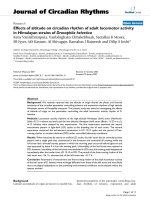
We compared the lung histology between the rats treated
with Mab166 and the rats treated with control IgG (Fig.
6). While the rats that received control IgG one hour after
bacterial instillation showed severe neutrophil recruit-
ment and destruction of alveolar structures (Fig. 6A), the
rats that received Mab166 had almost no neutrophils in
their airspaces and had preservation of normal alveolar
structures (Fig. 6B). As a result, therapeutic administration
of Mab166 showed comparable effects to rabbit
polyclonal anti-PcrV IgG in preventing acute lung injury
and subsequently occurring systemic distress. The thera-
peutic administration of Mab166 Fab fragments also had
the same or better effects than the administration of
Mab166 IgG.
Quantification of acute lung epithelial injuryFigure 1
Quantification of acute lung epithelial injury. The efflux of alveolar protein tracer (
131
I-albumin) from lungs to the circu-
lation was calculated in 4-h experiments of rats as an index of acute lung epithelial injury. In the control group (Control, no
bacteria), only lactated Ringer's solution was instilled into the airspace of the rats and no therapeutic intervention was taken.
Three sets of rats were co-instilled P. aeruginosa PA103 with 4 mg/kg of either irrelevant monoclonal IgG (Control IgG), rabbit
polyclonal anti-PcrV IgG (Rab anti-PcrV), or murine monoclonal anti-PcrV IgG (Mab166). Four sets of rats were intratracheally
administered either PBS, rabbit polyclonal anti-PcrV IgG (Rab anti-PcrV)(4 mg/kg), murine monoclonal anti-PcrV IgG
(Mab166)(4 mg/kg), or Mab166 Fab (4 mg/kg). Data are shown as means+standard errors. The numbers of rats are listed in
Table 1. +P < 0.05 to the control group (no bacteria) and *P < 0.05 to the control IgG group in co-instillation and to the group
without IgG (PBS) in therapeutic administration by two way-ANOVA, repeated measure, followed by the Newman-Keuls t-
test.
0
25
20
15
10
5
Control
(no bacteria)
2h
3h
4h
2h
3h
4h
2h
3h
4h
2h
3h
4h
2h
3h
4h
2h
3h
4h
2h
3h
4h
2h
3h
4h
Co-instillation
Therapeutic
(1h after infection)
Control IgG
R
ab anti-PcrV
Mab166
R
ab anti-PcrV
Mab166
Mab166 Fab
w/o IgG
Alveolar protein tracer efflux
(%)
*
*
*
*
*
+
+
Journal of Immune Based Therapies and Vaccines 2003, 1 />Page 6 of 9
(page number not for citation purposes)
Discussion
The widespread use of antibiotics has generated multiple
antibiotic-resistant microorganisms, and there is a new
need for non-antibiotic based adjuvant therapies for
microbial infections. Antibody-based immunotherapy is
one of the adjuvant therapies that can help treat antibi-
otic-resistant bacterial infections. In this investigation, we
showed that intratracheal administration of murine mon-
oclonal anti-PcrV IgG Mab166 improved acute lung injury
in infected animals. An important consideration in the
comparisons of effectiveness of various treatments of lung
infections in experimental animal models is the ability to
produce a consistent quantity of bacterial induced lung
injury. In our rat model, the administration of P. aerugi-
nosa (5 × 10
7
CFU) for an interval (4 h) consistently leads
to modest quantities of lung injury. Using independent
measurement of lung epithelial injury and of lung edema,
we have been able to evaluate the therapeutic effects of
various antibodies on acute lung injury [22]. The effects of
Mab166 were comparable to the administration of rabbit
polyclonal anti-PcrV IgG. The intratracheal
Quantification of lung edemaFigure 2
Quantification of lung edema. Water-to-wet weight
ratios of the lungs were measured at 4-h time points in the
rats infected with P. aeruginosa. as an index of acute lung
edema. In the control group (Control, no bacteria), only lac-
tated Ringer's solution was instilled into the airspace of the
rats and no therapeutic intervention was taken. Three sets of
rats were co-instilled P. aeruginosa PA103 with 4 mg/kg of
either irrelevant monoclonal IgG (Control IgG), rabbit poly-
clonal anti-PcrV IgG (Rab anti-PcrV), or murine monoclonal
anti-PcrV IgG (Mab166). Four sets of rats were intratrache-
ally administered either phosphate-buffered saline (PBS), rab-
bit polyclonal anti-PcrV IgG (Rab anti-PcrV)(4 mg/kg), murine
monoclonal anti-PcrV IgG (Mab166)(4 mg/kg), or Mab166
Fab (4 mg/kg). Data are shown as means+standard errors.
The numbers of animals are listed in Table 1. +P < 0.05 to
the control group (no bacteria) and *P < 0.05 to the control
IgG group in co-instillation and to the group without IgG
(PBS) in therapeutic administration by two way-ANOVA, fol-
lowed by the Newman-Keuls t-test.
3
4
5
6
7
8
9
Wet to dry weight ratio
Control
(no bacteria)
Control IgG
Rab anti-PcrV
Mab166
w/o IgG
Rab anti-PcrV
Mab166
Mab166 Fab
Co-instillation
Therapeutic
*
*
*
*
+
+
Mean arterial blood pressureFigure 3
Mean arterial blood pressure. The mean arterial blood
pressure was measured for 4 h in the rats. A. Either irrele-
vant monoclonal IgG (Control IgG, filled squares), rabbit pol-
yclonal anti-PcrV IgG (Rab anti-PcrV, open diamonds), or
murine monoclonal anti-PcrV IgG (Mab166, open circles) (4
mg/kg, respectively) was co-instilled with P. aeruginosa PA103
(5 × 10
7
CUF) in the airspaces of the rats. B. Either PBS with-
out IgG (w/o IgG, filled squares), rabbit polyclonal anti-PcrV
IgG (Rab anti-PcrV, open diamonds), murine monoclonal
anti-PcrV IgG (Mab166, open circles), or Fab fragments
Mab166 (Mab166 Fab, open triangles) (4 mg/kg, respectively)
was intratracheally instilled one hour after the airspace instil-
lation of P. aeruginosa PA103 (5 × 10
7
CUF). Data are shown
as means ± standard errors. The numbers of animals are
listed in Table 1. *P < 0.05 in the Mab166 group to the con-
trol group (w/o IgG) at the 4 h time point by unpaired t-test.
160
140
120
100
80
60
0
1234
Time after infection (h)
Mean arterial pressure
(mmHg)
Mab166
Rab anti-Pc
rV
Control IgG
0
1234
Time after infection (h)
160
140
120
100
80
60
Mean arterial pressure
(mmHg)
Rab anti-Pcr
V
Mab166
w/o IgG
IgG it
A.
Co-instillation
B
.
Therapeutic
180
Mab166 Fab
*
Journal of Immune Based Therapies and Vaccines 2003, 1 />Page 7 of 9
(page number not for citation purposes)
administration of Mab166 (4 mg/kg) significantly
improved the lung epithelial injury caused by cytotoxic P.
aeruginosa. Lung edema, measured as wet/dry ratios of the
lungs, decreased significantly in the rats treated with
intratracheal Mab166. The lung wet/dry ratios of the rats
instilled with bacteria and treated with any of anti-PcrV
IgGs were lower that those of the control rats (no bacteria)
probably due to the ability of Pseudomonal exotoxin A to
increase lung liquid clearance. Note P. aeruginosa strain
PA103 used in this study is a high producer of type II
secretory exotoxin A and P. aeruginosa treated with anti-
PcrV IgG would still secrete exotoxin A which has been
shown to increase the lung liquid clearance (and decrease
lung edema) [25] although exotoxin A itself does not
cause neither lung epithelial injury nor lung edema [26].
Hemodynamics, oxygenation, and metabolic acidosis
were improved by the treatment with intratracheal
Mab166. Lung histology in the rat treated with Mab166
showed significant improvement and preservation of nor-
mal structures. We previously showed that F(ab')
2
frag-
ments of rabbit polyclonal anti-PcrV IgG prevented sepsis
and allowed survival in a rabbit model of P. aeruginosa
infection [20]. Similarly, the Fab fragments of the murine
monoclonal anti-PcrV IgG (Mab166 Fab) had comparable
The oxygenation of arterial bloodFigure 4
The oxygenation of arterial blood. The oxygen pressure
of the arterial blood was measured for 4 h in the rats. A.
Either irrelevant monoclonal IgG (Control IgG, filled
squares), rabbit polyclonal anti-PcrV IgG (Rab anti-PcrV,
open diamonds), or murine monoclonal anti-PcrV IgG
(Mab166, open circles) (4 mg/kg, respectively) was co-
instilled with P. aeruginosa PA103 (5 × 10
7
CUF) in the air-
spaces of the rats. B. Either PBS without IgG (w/o IgG, filled
squares), rabbit polyclonal anti-PcrV IgG (Rab anti-PcrV,
open diamonds), murine monoclonal anti-PcrV IgG (Mab166,
open circles), or Fab fragments Mab166 (Mab166 Fab, open
triangles) (4 mg/kg, respectively) was intratracheally instilled
one hour after the airspace instillation of P. aeruginosa PA103
(5 × 10
7
CUF). Data are shown as means ± standard errors.
The numbers of animals are listed in Table 1.
(mmHg)
Mab166
Rab anti-PcrV
Control IgG
PaO2
(mmHg)
IgG it
A.
Co-instillation
B.
Therapeutic
0
1
234
Time after infection (h)
0
1
234
Time after infection (h)
600
500
400
300
200
100
0
600
500
400
300
200
100
0
PaO2
Rab anti-Pc
rV
Mab166
w/o IgG
Mab166 Fab
Metabolic acidosisFigure 5
Metabolic acidosis. Base excess was measured for 4 h in
the rats as an index of metabolic acidosis. A. Either irrele-
vant monoclonal IgG (Control IgG, filled squares), rabbit pol-
yclonal anti-PcrV IgG (Rab anti-PcrV, open diamonds), or
murine monoclonal anti-PcrV IgG (Mab166, open circles) (4
mg/kg, respectively) was co-instilled with P. aeruginosa PA103
(5 × 10
7
CUF) in the airspaces of the rats. B. Either PBS with
out IgG (w/o IgG, filled squares), rabbit polyclonal anti-PcrV
IgG (Rab anti-PcrV, open diamonds), murine monoclonal
anti-PcrV IgG (Mab166, open circles), or Fab fragments
Mab166 (Mab166 Fab, open triangles) (4 mg/kg, respectively)
was intratracheally instilled one hour after the airspace instil-
lation of P. aeruginosa PA103 (5 × 10
7
CFU). Data are shown
as means ± standard errors. The numbers of animals are
listed in Table 1. *P < 0.05 in the Mab166 group to the con-
trol group (w/o IgG) at the 4 h time point by unpaired t-test.
IgG it
A
. Co-instillation
B
. Therapeutic
0
1234
0
1234
Time after infection (h)
5.0
2.5
0
-2.5
-5.0
-7.5
Base excess
-10.0
Time after infection (h)
5.0
2.5
-2.5
-5.0
-7.5
Base excess
-10.0
0
Rab anti-Pc
rV
Mab166
Control IgG
*
Rab anti-Pc
rV
Mab166
w/o IgG
Mab166 Fab
Journal of Immune Based Therapies and Vaccines 2003, 1 />Page 8 of 9
(page number not for citation purposes)
therapeutic effects to the whole IgG molecules of Mab166
in preventing P. aeruginosa-induced acute lung injury.
Because, Fab portions had the same therapeutic effects as
whole IgG in P. aeruginosa-induced lung injury, the Fc-
dependent opsonization of the bacteria does not seem
critical for the efficacy of the anti-PcrV antibodies.
Intratracheal administration of Fab is attractive for the fol-
lowing reasons: 1) Direct delivery of therapeutic agents in
the site of infection is advantageous pharmacokinetically.
Only limited amounts of systemically administered IgGs
(intravenously, or intramuscularly) reach the airspaces of
the lung. 2) The administration of the whole IgG may
cause some inflammatory side effects, because the Fc-por-
tion of IgG may induce unfavourable inflammatory
responses such as complement fixation, activation of mac-
rophages. In our study, Fab fragment had the same
therapeutic potency as the whole IgG and the therapeutic
administration of Fab fragments may overcome the disad-
vantages of the intratracheal administration of whole IgG.
Since the discovery of the production of monoclonal anti-
bodies by Kohler and Milstein in 1975, only a handful of
antibodies had been used in human therapy [27]. The
main difficulty with monoclonal antibodies is that mouse
antibodies are seen by the human immune system as for-
eign, and the patient mounts an immune response against
them, producing "human anti-mouse antibodies
(HAMA)". These not only cause the therapeutic antibod-
ies to be eliminated from the host, but also cause the for-
mation of immune complexes that damage the kidneys.
Therefore, technology has focused on methodology that
produces less immunogenic monoclonal antibodies.
More recently, the techniques to engineer recombinant
chimera and humanized antibodies have been developed
to decrease the immunogenicity of murine antibodies
[28]. Due to the multiple antibiotic resistance mecha-
nisms that P. aeruginosa possesses, the need for adjunctive
therapies is becoming more important. Therefore, anti-
PcrV antibody-based immunotherapies are potential ther-
apeutic options for immunocompromised patients
infected with P. aeruginosa.
Conclusions
Intratracheal administration of the murine monoclonal
anti-PcrV antibody Mab166 and its Fab fragments pro-
tected rats infected with Pseudomonas aeruginosa from
acute lung injury. Mab166 and its Fab fragments are
potential useful adjuvant therapies for acute lung injury
secondary to P. aeruginosa pneumonia.
Authors' contributions
K. Fuare carried out animal studies, and drafted the man-
uscript. J. Fujimoto, D. W. Shimabukuro, N. Shime and K.
Moriyama participated in the animal studies. T. Ajayi
edited the manuscript. E. G. Spack contributed to the
production and purification of antibodies. J. P. Wiener-
Kronish and T. Sawa conceived of the study, and partici-
pated in its design and coordination. All authors read and
approved the final manuscript.
Abbreviations
P. aeruginosa:Pseudomonas aeruginosa, IT: Intratracheal
administration
Acknowledgements
This research was supported by NIH grant HL067600 and American Lung
Association Research Grant RG-004-N to T. Sawa, NIH grants RO1
HL59239 & AI44101, and a grant sponsored by InterMune, Inc. (Brisbane,
California, U.S.A.) to J. P. Wiener-Kronish.
References
1. Almirall J, Mesalles E, Klamburg J, Parra O and Agudo A: Prognostic
factors of pneumonia requiring admission to the intensive
care unit. Chest 1995, 107:511-516.
2. Brun-Buisson C, Doyon F, Carlet J, Dellamonica P, Gouin F, Lepoutre
A, Mercier JC, Offenstadt G and Regnier B: Incidence, risk factors,
and outcome of severe sepsis and septic shock in adults. A
multicenter prospective study in intensive care units. French
ICU Group for Severe Sepsis. JAMA 1995, 274:968-974.
3. Crouch Brewer S, Wunderink RG, Jones CB and Leeper KV: Venti-
lator-associated pneumonia due to Pseudomonas aeruginosa.
Chest 1996, 109:1019-1029.
Lung histologyFigure 6
Lung histology. Four hours after the intratracheal instilla-
tion of P. aeruginosa PA103 (5 × 10
7
CFU), the rats were
euthanized and their lungs were perfused with 10% buffered
formalin phosphate for fixation and were embedded in paraf-
fin. Mounted sections were stained with hematoxylin-eosin
and observed in light microscopy. A. The rat received irrele-
vant control IgG (4 mg/kg) intratracheally one hour after bac-
terial instillation. B. The rat received Mab166 (4 mg/kg)
intratracheally one hour after bacterial instillation. Magnifica-
tion of objective lens 20× (left figures) and 40× (right figures).
A
. Control IgG
B
. Mab166
Publish with BioMed Central and every
scientist can read your work free of charge
"BioMed Central will be the most significant development for
disseminating the results of biomedical research in our lifetime."
Sir Paul Nurse, Cancer Research UK
Your research papers will be:
available free of charge to the entire biomedical community
peer reviewed and published immediately upon acceptance
cited in PubMed and archived on PubMed Central
yours — you keep the copyright
Submit your manuscript here:
/>BioMedcentral
Journal of Immune Based Therapies and Vaccines 2003, 1 />Page 9 of 9
(page number not for citation purposes)
4. Vidal F, Mensa J, Almela M, Martinez JA, Marco F, Casals C, Gatell JM,
Soriano E and Jimenez de Anta MT: Epidemiology and outcome
of Pseudomonas aeruginosa bacteremia, with special empha-
sis on the influence of antibiotic treatment. Analysis of 189
episodes. Arch Intern Med 1996, 156:2121-2126.
5. Fagon JY, Chastre J, Hance AJ, Montravers P, Novara A and Gilbert
C: Nosocomial pneumonia in ventilated patients: a cohort
study evaluating attributable mortality and hospital stay. Am
J Med 1993, 94:281-288.
6. Parrillo JE, Parker MM, Nathanson C, Suffredini AF, Danner RL, Cun-
nion RE and Ognibene FP: Septic shock in humans Advances in
the understanding of pathogenesis, cardiovascular dysfunc-
tion and therapy. Ann Intern Med 1990, 113:227-237.
7. Taylor GD, Buchanan-Chell M, Kirkland T, McKenzie M and Wiens R:
Bacteremic nosocomial pneumonia. A 7-year experience in
one institution. Chest 1995, 108:786-788.
8. Wiener-Kronish JP, Albertine KH and Matthay MA: Differential
responses of the endothelial and epithelial barriers of the
lung in sheep to Escherichia coli endotoxin. J Clin Invest 1991,
88:864-875.
9. Wiener-Kronish JP, Sakuma T, Kudoh I, Pittet JF, Frank D, Dobbs L,
Vasil ML and Matthay M: Alveolar epithelial injury and pleural
empyema in acute P. aeruginosa pneumonia in anesthetized
rabbits. J Appl Physiol 1993, 75:1661-1669.
10. Kurahashi K, Kajikawa O, Sawa T, Ohara M, Gropper MA, Frank DW,
Martin TR and Wiener-Kronish JP: Pathogenesis of septic shock
in Pseudomonas aeruginosa pneumonia. J Clin Invest 1999,
104:743-750.
11. Hueck CJ: Type III protein secretion systems in bacterial
pathogens of animals and plants. Microbiol Mol Biol Rev 1998,
62:379-433.
12. Wiener-Kronish JP, Frank DW and Sawa T: Mechanisms of lung
epithelial cell Injury by acute by Pseudomonas aeruginosa. In
Molecular biology of acute lung injury Edited by: Clark RSG, Carcillo JA. Bos-
ton: Kluwer Academic Publishers; 2001:149-161.
13. Galan JE and Collmer A: Type III secretion machines: bacterial
devices for protein delivery into host cells. Science 1999,
284:1322-1328.
14. Frank DW: The exoenzyme S regulon of Pseudomonas
aeruginosa. Mol Microbiol 1997, 26:621-629.
15. Yahr TL, Vallis AJ, Hancock MK, Barbieri JT and Frank DW: ExoY,
an adenylate cyclase secreted by the Pseudomonas aeruginosa
type III system. Proc Natl Acad Sci U S A 1998, 95:13899-13904.
16. Yahr TL, Mende-Mueller LM, Friese MB and Frank DW: Identifica-
tion of type III secreted products of the Pseudomonas aerugi-
nosa exoenzyme S regulon. J Bacteriol 1997, 179:7165-7168.
17. Roy-Burman A, Savel RH, Racine S, Swanson BL, Revadigar NS, Fuji-
moto J, Sawa T, Frank DW and Wiener-Kronish JP: Type III protein
secretion is associated with death in lower respiratory and
systemic Pseudomonas aeruginosa infection. J Infect Dis 2001,
183:1767-1774.
18. Hauser AR, Cobb E, Bodi M, Mariscal D, Valles J, Engel JN and Rello
J: Type III protein secretion is associated with poor clinical
outcomes in patients with ventilator-associated pneumonia
caused by Pseudomonas aeruginosa. Crit Care Med 2002,
30:521-528.
19. Sawa T, Yahr TL, Ohara M, Kurahashi K, Gropper MA, Wiener-Kro-
nish JP and Frank DW: Active and passive immunization with
the Pseudomonas V antigen protects against type III intoxica-
tion and lung injury. Nat Med 1999, 5:392-398.
20. Shime N, Sawa T, Fujimoto J, Faure K, Allmond LR, Karaca T, Swan-
son BL, Spack EG and Wiener-Kronish JP: Therapeutic adminis-
tration of anti-PcrV F(ab')
2
in sepsis associated with
Pseudomonas aeruginosa. J Immunol 2001, 167:5880-5886.
21. Frank DW, Vallis A, Wiener-Kronish JP, Roy-Burman A, Spack EG,
Mullaney BP, Megdoud M, Marks JD, Fritz R and Sawa T: Generation
and characterization of a protective monoclonal antibody to
Pseudomonas aeruginosa PcrV. J Infect Dis 2002, 186:64-73.
22. Ernst EJ, Hashimoto S, Guglielmo J, Sawa T, Pittet JF, Kropp H, Jack-
son JJ and Wiener-Kronish JP: Effects of antibiotic therapy on
Pseudomonas aeruginosa-induced lung injury in a rat model.
Antimicrob Agents Chemother 1999, 43:2389-2394.
23. Sawa T, Kurahashi K, Ohara M, Gropper MA, Doshi V, Larrick JW and
Wiener-Kronish JP: Evaluation of antimicrobial and lipopoly-
saccharide-neutralizing effects of a aynthetic CAP18 frag-
ment against Pseudomonas aeruginosa in a mouse model.
Antimicrob Agents Chemother 1998, 42:3269-3275.
24. Jayr C, Garat C, Meignan M, Pittet JF, Zelter M and Matthay MA:
Alveolar liquid and protein clearance in anesthetized venti-
lated rats. J Appl Physiol 1994, 76:2636-2642.
25. Pittet JF, Hashimoto S, Pian M, McElroy MC, Nitenberg G and
Wiener-Kronish JP: Exotoxin A stimulates fluid reabsorption
from distal airspaces of lung in anesthetized rats. Am J Physiol
1996, 270:L232-L241.
26. Kudoh I, Wiener-Kronish JP, Hashimoto S, Pittet JF and Frank D:
Exoproduct secretions of Pseudomonas aeruginosa strains
influence severity of alveolar epithelial injury. Am J Physiol 1994,
267:L551-L556.
27. Kohler G and Milstein C: Continuous cultures of fused cells
secreting antibody of predefined specificity. Nature 1975,
256:495-497.
28. Gavilondo JV and Larrick JW: Antibody engineering at the
millennium. Biotechniques 2000, 29:128-138.