Báo cáo y học: "Acute small bowel obstruction as a result of a Meckel''''s diverticulum encircling the terminal ileum: A case report" pdf
Bạn đang xem bản rút gọn của tài liệu. Xem và tải ngay bản đầy đủ của tài liệu tại đây (518.68 KB, 5 trang )
BioMed Central
Page 1 of 5
(page number not for citation purposes)
Journal of Medical Case Reports
Open Access
Case report
Acute small bowel obstruction as a result of a Meckel's diverticulum
encircling the terminal ileum: A case report
Avnesh S Thakor, Siong S Liau and Dermot C o'Riordan*
Address: Department of Surgery, West Suffolk Hospital, Bury St. Edmunds, IP33 2QZ, UK
Email: Avnesh S Thakor - ; Siong S Liau - ; Dermot C o'Riordan* - dermot.o'
* Corresponding author
Abstract
Background: In the developed world, small bowel obstruction accounts for 20% of all acute
surgical admissions. The aetiology for majority of these cases includes postoperative adhesions and
herniae. However, a relatively uncommon cause is a Meckel's diverticulum. Although this diagnosis
is primarily reported in the adolescent population, it should also be considered in adults.
Case Presentation: In the present report, we present a rare case where a fit and healthy 74-
year-old gentleman, with no previous history of abdominal surgery, presented with the cardinal
symptoms and signs of small bowel obstruction as the result of a Meckel's diverticulum encircling
his terminal ileum. Initial investigations included a supine abdominal x-ray showing dilated loops of
small bowel and computerised tomographic imaging of the abdomen, which revealed a stricture in
the terminal ileum of unknown aetiology. At laparotomy, multiple loops of distended small bowel
were seen from the duodeno-jeujenal junction to the terminal ileum, which was encircled by a
Meckel's diverticulum. The Meckel's diverticulum was then divided to release the obstruction,
mobilised and subsequently removed. Finally, the small bowel contents were decompressed into
the stomach and the nasogastric tube aspirated, before returning the loops of bowel into the
abdomen in sequence. The patient made a good postoperative recovery and was discharged home
5 days later.
Conclusion: This report highlights the importance of considering a Meckel's diverticulum as a
cause of small bowel obstruction in individuals from all age groups and especially in a person with
no previous abdominal pathology or surgery.
Case Presentation
Background
In the developed world, small bowl obstruction accounts
for 20% of all acute surgical admissions. The aetiology of
small bowel obstruction includes several pathological fac-
tors, with the most common cause being postoperative
adhesions followed by herniae [1]. However, in patients
who present with the symptoms and signs of bowel
obstruction and who have had no previous abdominal
surgery, or any detectable herniae on physical examina-
tion, other causes such as a Meckel's diverticulum should
be considered.
A Meckel's diverticulum is a congenital pouch on the wall
of the distal ileum, usually about 2 inches from the ileoce-
cal valve. It represents a vestigial remnant of the ompha-
lomesenteric duct and occurs in approximately 2% of the
population, found twice as frequently in males as females.
Published: 23 March 2007
Journal of Medical Case Reports 2007, 1:8 doi:10.1186/1752-1947-1-8
Received: 22 December 2006
Accepted: 23 March 2007
This article is available from: />© 2007 Thakor et al; licensee BioMed Central Ltd.
This is an Open Access article distributed under the terms of the Creative Commons Attribution License ( />),
which permits unrestricted use, distribution, and reproduction in any medium, provided the original work is properly cited.
Journal of Medical Case Reports 2007, 1:8 />Page 2 of 5
(page number not for citation purposes)
Of those individuals who have a Meckel's diverticulum,
only 2% are symptomatic and they tend to be typically
below the age of two, thereby accounting for why this con-
genital gastrointestinal anomaly is comparatively better
studied in adolescents compared to in adults.
The main complications caused by a Meckel's diverticu-
lum, include intersusseption and volvulus in adolescents
and acute bleeding in adults [2]. However, there are cases
reported in the literature of a Meckel's diverticulum caus-
ing small bowel obstruction [3-6], but this predominantly
occurs in adolescents where the bowel lumen is narrower
and the intra-abdominal contents are more closely packed
together.
Here, we present a case of a Meckel's diverticulum causing
acute small bowel obstruction in a 74-year-old gentleman
as a result of it encircling, and thus constricting, the termi-
nal ileum. To the authors' knowledge, and from an exten-
sive review of the literature, such an unusual presentation
of a Meckel's diverticulum has not been previously
reported.
Case Report
A fit and healthy 74-year-old gentleman presented to the
accident and emergency department at the West Suffolk
Hospital with a 3-day history of abdominal pain, vomit-
ing, absolute constipation and abdominal distension. The
abdominal pain initially started as a dull generalised dis-
comfort, but later became colicky in nature with a subjec-
tive severity of 7/10. There were no other abdominal or
genitourinary symptoms. The patient had an unremarka-
ble past surgical history, with no prior abdominal surgery,
and a past medical history of only hypercholesterolaemia.
On examination, positive findings included marked
abdominal distension, generalised abdominal tenderness,
tinkling bowel sounds and soft stools high in the rectum.
Important negative findings included no herniae and no
signs of peritonism.
Initial management of the patient involved intravenous
fluid resuscitation, nasogastric tube insertion, catheterisa-
tion, routine bloods and erect chest and supine abdomi-
nal x-rays. Significant elevations in blood concentrations
of urea, creatinine and C-reactive protein were noted, with
dilated loops of small bowel (Fig. 1) and no free air under
either diaphragm on x-ray. Over the next 12 hours, the
patient's vital signs remained stable and his condition did
not deteriorate further. To identify the cause of the small
bowel obstruction, computerised tomographic imaging of
the abdomen with oral contrast was performed which
revealed dilated loops of small bowel with a stricture in
the ileum and collapse of the distal ileum and large bowel
(Fig. 2). As the aetiology of the stricture remained uniden-
tified, the decision was made to perform a diagnostic
laparotomy and manage the patient accordingly.
Following general anaesthesia, a midline laparotomy was
performed on the patient. On entering the peritoneal cav-
ity, gross distension of the small bowel and collapse of the
large bowel was identified. The small bowel was subse-
quently delivered carefully and examined. Loops of dis-
tended small bowel were identified extending proximally
from the duodeno-jejunal junction to the distal ileum. At
approximately 10 cm from the ileo-caecal valve, there was
a long tubular structure encircling and obstructing the ter-
minal ileum, which proved to be a Meckel's diverticulum.
The base of the Meckel's diverticulum arose approxi-
mately 40 cm proximal from the ileo-caecal valve. The
encircling Meckel's diverticulum did not appear inflamed
or thickened and was divided at the base using a linear sta-
pler (TLC55, Ethicon) to release the obstruction. Care was
taken not to compromise the lumen of the ileum. The tip
of the diverticulum was then dissected off the terminal
ileum and the anastomosis over sewn with continuous 3/
0 sutures. The small bowel was then decompressed and
the content milked gently into the stomach before being
aspirated via the nasogastric tube. The loops of bowel
were then returned into the abdomen in sequence. Clo-
sure of the abdomen was performed using loop sutures.
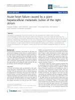
This x-ray shows multiple loops of dilated small bowelFigure 1
This x-ray shows multiple loops of dilated small bowel.
Journal of Medical Case Reports 2007, 1:8 />Page 3 of 5
(page number not for citation purposes)
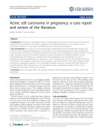
Preoperative helical computed tomography transverse scan of the abdomenFigure 2
Preoperative helical computed tomography transverse scan of the abdomen. This image shows small bowel
obstruction as a result of a stricture in the terminal ileum.
Meckel’s diverticulum ?
Collapsed large bowel
Small bowel dilatation
Journal of Medical Case Reports 2007, 1:8 />Page 4 of 5
(page number not for citation purposes)
Following this, the patient made a good postoperative
recovery and was discharged home 5 days later.
Discussion
The management of any acute surgical abdomen, includ-
ing acute bowel obstruction, follows 4 stages: (I) forma-
tion of an initial diagnosis, (II) confirmation of a
diagnosis, (III) confirmation of the aetiology underlying
the diagnosis and (IV) surgical intervention to treat the
emergency.
A diagnosis of acute bowel obstruction is made initially
on clinical judgement based on the history and physical
examination of the patient. The cardinal symptoms and
signs are colicky abdominal pain, vomiting, absolute con-
stipation and abdominal distension, all of which were
present in this patient.
Confirmation of bowel obstruction is then usually made
with a plain supine abdominal x-ray. This simple and eas-
ily performed test provides the surgeon with several useful
pieces of information, including whether there is small
and/or large bowel obstruction and the degree of obstruc-
tion. In the present case, markedly dilated loops of small
bowel with no visible loops of large bowel were seen on
the abdominal x-ray (Fig. 1), thus indicating acute small
bowel obstruction.
Having established and confirmed a diagnosis of small
bowel obstruction, the next goal is to identify the aetiol-
ogy underlying the obstruction. The two most common
causes of small bowel obstruction in the developed world
are postoperative adhesions and herniae [1]. However,
this patient had no previous abdominal surgery and no
herniae on physical examination, therefore making both
these causes unlikely. Hence, it was decided to image his
abdomen with a computed tomography scan with oral
contrast. The result of this revealed a stricture in the termi-
nal ileum, with dilatation of the small bowel proximal
and collapse of the large bowel distal to the stricture (Fig.
2). However the aetiology of the stricture, and therefore
the cause of the small bowel obstruction, remained uni-
dentified.
Based on these findings, and the absence of clinical
improvement whilst on IV fluids and nasogastric tube
aspiration, surgery was therefore indicated. However, the
surgical approach to acute bowel obstruction of unknown
aetiology remains controversial. While some surgeons
advocate laparoscopic intervention due to its minimally
invasive approach and shorter patient hospitalisation [7],
others favour an open laparotomy due to the larger surgi-
cal space and lower incidence of bowel injury. Further evi-
dence to support the latter approach comes from
Kirshtein and colleagues who reviewed 65 cases of acute
bowel obstruction that were initially managed laparo-
scopically [8]. In that study, although laparoscopy was
shown to have a diagnostic accuracy of 96.9%, a signifi-
cant number of cases still required conversion for their
subsequent management. Based on the above literature
and the pervious experience of this surgical team, it was
therefore decided that this patient should undergo an
open laparotomy.
At laparotomy, an unusually long Meckel's diverticulum
was found, which had managed to entirely wrap itself
around the terminal ileum thereby forming an internal
hernial orifice in which the bowel had become incarcer-
ated and subsequently obstructed. What makes this case
exceptionally unusual is that the Meckel's diverticulum
was not thickened or inflamed. This is in contrast to the
other cases previously reported in the literature, where an
internal hernial orifice was created by the Meckel's as the
result of adhesions or bands between an inflammatory
end of the diverticulum and either the surrounding
mesentery [9] or the neighbouring appendix [3]. On ret-
rospective analysis of both the preoperative helical (Fig.
2) and reconstructed computed tomography (Fig. 3)
scans, the Meckel's diverticulum could now be identified
as being the cause for the stricture of the terminal ileum
and therefore the cause of the small bowel obstruction.
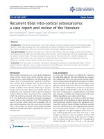
Reconstructed computed tomography coronal scan of the abdomenFigure 3
Reconstructed computed tomography coronal scan
of the abdomen. This image shows small bowel obstruc-
tion as a result of a stricture in the terminal ileum. A postop-
erative review suggested a Meckel's diverticulum could be
described.
Meckel’s diverticulum ?
Collapsed large bowel
Small bowel dilatation
Publish with BioMed Central and every
scientist can read your work free of charge
"BioMed Central will be the most significant development for
disseminating the results of biomedical research in our lifetime."
Sir Paul Nurse, Cancer Research UK
Your research papers will be:
available free of charge to the entire biomedical community
peer reviewed and published immediately upon acceptance
cited in PubMed and archived on PubMed Central
yours — you keep the copyright
Submit your manuscript here:
/>BioMedcentral
Journal of Medical Case Reports 2007, 1:8 />Page 5 of 5
(page number not for citation purposes)
Conclusion
This report therefore highlights the importance of consid-
ering a Meckel's diverticulum as a cause of small bowel
obstruction in individuals from all age groups and espe-
cially in a person with no previous abdominal pathology
or surgery.
Competing interests
The author(s) declare that they have no competing inter-
ests.
Authors' contributions
All authors have read and approved the final manuscript.
AST (Surgical House Officer): Involved in the conception
of the report, literature review, manuscript preparation,
manuscript editing and manuscript submission.
SSL (Surgical Registrar): Involved in the manuscript edit-
ing and manuscript review.
DOR (Consultant Surgeon): Involved in the manuscript
editing and manuscript review.
Acknowledgements
The authors would like to thank Dr. Watson with her help in the interpre-
tation and reconstruction of the computerised tomographic images used in
this case report.
Consent was obtained from the patient for the publication of this study.
References
1. Foster NM, McGory ML, Zingmond DS, Ko CY: Small bowel
obstruction: a population-based appraisal. J Am Coll Surg 2006,
203:170-176.
2. Park JJ, Wolff BG, Tollefson MK, Walsh EE, Larson DR: Meckel
diverticulum: the Mayo Clinic experience with 1476 patients
(1950-2002). Ann Surg 2005, 241:529-533.
3. Ishigami S, Baba K, Kato K, Nakame K, Okumura H, Matsumoto M,
Natsugoe S, Aikou T: Small bowel obstruction secondary to
meckel diverticulum detected and treated laparoscopically-
case report. Surg Laparosc Endosc Percutan Tech 2006, 16:344-346.
4. Nath DS, Morris TA: Small bowel obstruction in an adolescent:
a case of Meckel's diverticulum. Minn Med 2004, 87:46-48.
5. Prall RT, Bannon MP, Bharucha AE: Meckel's diverticulum caus-
ing intestinal obstruction. Am J Gastroenterol 2001, 96:3426-3427.
6. Tashjian DB, Moriarty KP: Laparoscopy for treating a small
bowel obstruction due to a Meckel's diverticulum. JSLS 2003,
7:253-255.
7. Suter M, Zermatten P, Halkic N, Martinet O, Bettschart V: Laparo-
scopic management of mechanical small bowel obstruction:
are there predictors of success or failure? Surg Endosc 2000,
14:478-483.
8. Kirshtein B, Roy-Shapira A, Lantsberg L, Avinoach E, Mizrahi S:
Laparoscopic management of acute small bowel obstruc-
tion. Surg Endosc 2005, 19:464-467.
9. Tomikawa M, Taomoto J, Saku M, Takeshita M, Yoshida K, Sugimachi
K: A loop formation of Meckel's diverticulum: a case with
obstruction of the ileum. Ulus Travma Acil Cerrahi Derg 2003,
9:134-136.