Báo cáo y học: "An impinging remnant meniscus causing early polyethylene failure in total knee arthroplasty: a case report" pot
Bạn đang xem bản rút gọn của tài liệu. Xem và tải ngay bản đầy đủ của tài liệu tại đây (740.36 KB, 3 trang )
BioMed Central
Page 1 of 3
(page number not for citation purposes)
Journal of Medical Case Reports
Open Access
Case report
An impinging remnant meniscus causing early polyethylene failure
in total knee arthroplasty: a case report
Rachid Saouti*, Barend J van Royen and Christiaan M Fortanier
Address: Department of Orthopaedic Surgery, VU University Medical Center, Amsterdam, The Netherlands
Email: Rachid Saouti* - ; Barend J van Royen - ; Christiaan M Fortanier -
* Corresponding author
Abstract
The management of patients with an apparently normal functional total knee arthroplasty (TKA)
suffering from unexplained persistent pain and swelling is a challenging issue. The usual causes of
pain after total knee replacement are well known, but there are a small number of patients in whom
its aetiology is obscure. Malfunction due to soft tissue impingement has rarely been reported. A
patient with an unusual case of posterior soft tissue impingement secondary to a trapped posterior
horn of a remnant medial meniscus after TKA and responsible for severe early polyethylene wear,
is reported. The diagnosis was confirmed by arthroscopy. Treatment was performed by
arthrotomy. The meniscus remnant was removed followed by total synovectomy and isolated
exchange of the polyethylene insert. To our knowledge, this is the first well-documented case
reporting this association.
Case presentation
A 63-year old male patient with a history of symptomatic
osteoarthritis of the left knee underwent a Total Knee
Arthroplasty (TKA) of posterior cruciate ligament retain-
ing design (Kinemax, Stryker, Mahwah, New Jersey, USA)
without a patella component. The postoperative course
was uneventful. Three weeks later he presented to our out
patient clinic with sudden swelling and discomfort of his
left knee. Clinical examination demonstrated medial joint
line tenderness and confirmed the patient's impression of
joint effusion. Radiographs demonstrated a well-aligned
TKA. All complaints, with exception of the knee effusion,
declined progressively over a period of months. Two years
postoperatively, the patient developed increasing pain
and complained of "catching" of the knee. Physical exam-
ination showed a stable knee with a normal range of
motion of 130 degrees flexion with no extension deficit.
There was a moderate swelling and joint line tenderness
medially. Standard radiographs showed a well-aligned
TKA with no signs of loosening or polyethylene wear (Fig-
ure 1). Laboratory analysis including a complete blood
count with differential, erythrocyte sedimentation rate, C-
reactive protein and knee aspiration for cell count and cul-
ture excluded infection. A technetium 99m diphospho-
nate bone scintigraphy showed an increased perfusion in
the early phase and increased uptake in the static phase at
the medial side of the femoral and tibial component and
in the patella of the left knee (Figure 2).
A diagnostic arthroscopy was performed to differentiate
between a mechanical and a soft tissue related problem.
Arthroscopy revealed a remnant of the posterior horn of
the medial meniscus impinging between the posterior
part of the femoral component and the polyethylene
insert. There was also an important delamination of the
anteromedial part of the insert with a crack at the ventral
Published: 13 July 2007
Journal of Medical Case Reports 2007, 1:48 doi:10.1186/1752-1947-1-48
Received: 17 January 2007
Accepted: 13 July 2007
This article is available from: />© 2007 Saouti et al; licensee BioMed Central Ltd.
This is an Open Access article distributed under the terms of the Creative Commons Attribution License ( />),
which permits unrestricted use, distribution, and reproduction in any medium, provided the original work is properly cited.
Journal of Medical Case Reports 2007, 1:48 />Page 2 of 3
(page number not for citation purposes)
side associated with substantial synovitis and polyethyl-
ene debris scattered all around the joint. Slight delamina-
tion of the posterolateral part of the insert was visible. An
arthrotomy showed no loosening of the femoral and tib-
ial components of the TKA. There was no malrotation of
both components. The tibial slope was not excessive
(almost neutral). The trapped posteromedial meniscus
remnant was removed (Figure 3) and a total synovectomy
with an isolated exchange of the polyethylene insert was
performed. Intraoperative cultures from both the fluid
aspiration and the remnant meniscus yielded no micro-
organisms. Postoperatively there were no complications
with a complete resolution of all complaints and symp-
toms. At 3 years follow-up he remains complete symptom
free with an unrestricted range of motion.
Discussion
TKA is a successful procedure with a satisfactory outcome
in patients with primary and secondary osteoarthritis of
the knee. Unfortunately, a small group of patients com-
plain about pain, recurrent knee effusion and limited
range of motion postoperatively. Acute and low grade
infection, "overstuffed knee", prosthetic loosening, rota-
tional component malpositioning and flexion instabili-
ties are the most recognised articular causes. Chronic
synovitis from soft tissue impingement has rarely been
reported [1-5]. Our patient had a remnant posterior horn
of the medial meniscus trapped between the femur com-
ponent and the polyethylene insert. This was responsible
for the catching sensation of the knee and the recurrent
pain. Because of the posteromedial impingement of the
remnant meniscus, the contact stresses at the anterome-
dial side, and in lesser extend at the posterolateral side
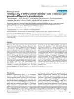
Photographs show a trapped posteromedial meniscus and severe damage of the polyethylene insert with polyethylene debrisFigure 3
Photographs show a trapped posteromedial meniscus and
severe damage of the polyethylene insert with polyethylene
debris.
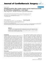
Radiographs of the prosthesis three years after total knee arthroplasty show normal alignment without evidence of looseningFigure 1
Radiographs of the prosthesis three years after total knee
arthroplasty show normal alignment without evidence of
loosening.
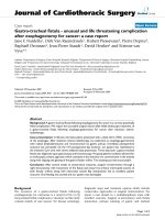
Triphasic bone scintigraphy shows an increased perfusion in the early phase and increased uptake in the static phase at the medial side of the femoral and tibial component and in the patella of the left kneeFigure 2
Triphasic bone scintigraphy shows an increased perfusion in
the early phase and increased uptake in the static phase at
the medial side of the femoral and tibial component and in
the patella of the left knee.
Publish with BioMed Central and every
scientist can read your work free of charge
"BioMed Central will be the most significant development for
disseminating the results of biomedical research in our lifetime."
Sir Paul Nurse, Cancer Research UK
Your research papers will be:
available free of charge to the entire biomedical community
peer reviewed and published immediately upon acceptance
cited in PubMed and archived on PubMed Central
yours — you keep the copyright
Submit your manuscript here:
/>BioMedcentral
Journal of Medical Case Reports 2007, 1:48 />Page 3 of 3
(page number not for citation purposes)
were probably higher and responsible for the severe poly-
ethylene wear with delamination.
Since there was no real symptom free interval between the
complaints and the index operation, we considered the
impinging remnant meniscus a result of incomplete
removal of the meniscus during the TKA procedure [2],
rather than regenerated after surgical removal [1,3]. We
consider the lack of a symptom free interval an important
finding related to the impinging remnant meniscus in
contrast to early polyethylene failure caused by other
mechanisms. The value of bone scintigraphy in the diag-
nosis of prosthesis loosening is limited. Bone scintigraphy
typically provides high sensitivity but exhibit variable spe-
cificity. An increased uptake can be seen many years after
the implantation of TKA but the tracer uptake is generally
mild or moderate and decreasing over time [6,7].
The diagnostic value of arthroscopy after TKA is controver-
sial. It has been suggested that several complications of
TKA, for example soft tissue-related problems, can suc-
cessfully be managed by arthroscopy [4,9,10]. However,
Van Mourik et al [8] stated that the indications for a diag-
nostic arthroscopy in painful TKA are, without any preop-
erative diagnosis, very limited. In our case, arthroscopy
clearly highlighted the problem of localised polyethylene
wear caused by a remnant meniscal part, warranting
arthrotomy to perform a polyethylene insert replacement
and resection of the remnant meniscus.
Early isolated polyethylene insert exchange in aseptic TKA
is a rare procedure. Generally, most indications are associ-
ated with varied forms of soft tissue complications need-
ing additional synovectomy, arthrolysis and ligament
release. The lack of a symptom free interval may suggest
an immediate postoperative problem caused by an
impinging remnant meniscus. To our best knowledge, this
is the first well-documented case reporting early polyeth-
ylene failure in TKA caused by an impinging remnant
meniscus.
Conclusion
Based on this report we emphasise on the importance of
meticulous resection of the menisci during TKA and the
diagnostic value of arthroscopy in unexplained pain and
swelling after TKA with no signs of infection.
Abbreviations
TKA – total knee arthroplasty
Competing interests
The author(s) declare that they have no competing inter-
ests.
Authors' contributions
RS conceived the study, participated in its design and
coordination and helped to draft the manuscript.
BVR revised the article for intellectual content details.
CMF conducted the literature review and carried out the
review of the patient's medical record in order to collect
all the available information.
All the authors read and approved the final manuscript.
Acknowledgements
A written informed consent was obtained for publication of this case
report.
References
1. Scher DM, Paumier JC, Di Cesare PE: Pseudomeniscus following
total knee arthroplasty as a cause of persistent knee pain. J
Arthroplasty 1997, 12:114-8.
2. Martini F, Kremling E, Kunz W: Symptomatic bucket handle tear
of the lateral meniscus after knee arthroplasty. Int Orthop
1999, 23:310-1.
3. Wigren A, Kolstad K, Brunk V: Formation of new menisci after
polycentric knee arthroplasty. Report of four cases, one with
a bucket handle tear. Acta Orthop Scand 1978, 49:615-7.
4. Takahashi M, Miyamoto , Nagano : Arthroscopic treatment of
soft-tissue impingement under the patella after total knee
arthroplasty. Arthroscopy 2002, 18(4):E20.
5. Allardyce TJ, Scuderi GR, Insall JN: Arthroscopic treatment of
popliteus tendon dysfunction following total knee arthro-
plasty. J Arthroplasty 1997, 12:353-5.
6. Rubello D, Carricasulo D, Borsato N, Chierichetti F, Zanco P, Ferlin
G: Three-phase bone scan pattern in asymptomatic unce-
mented total knee arthroplasty. Eur J Nucl Med 1996, 23:1400-3.
7. Hofmann AA, Wyatt RW, Daniels AU, Armstrong L, Alazraki N, Tay-
lor A Jr: Bone scans after total knee arthroplasty in asympto-
matic patients. Cemented versus cementless. Clin Orthop
1990, 251:183-8.
8. Van Mourik JBA, Verhaar JAN, Heijboer RP, Van Kampen A: Limited
value of arthroscopic evaluation and treatment of painful
knee prosthesis of 27 cases. Arthroscopy 1998, 14:877-79.
9. Klinger HM, Baums MH, Spahn G, Ernstberger T: A study of effec-
tiveness of knee arthroscopy after knee arthroplasty. Arthros-
copy 2005, 21(6):731-738.
10. Bocell JR, Thorpe CD, Tullos HS: Arthroscopic treatment of
symptomatic total knee arthroplasty. Clin Orthop 1991,
271:125-34.