Báo cáo y học: "Intussusception of the appendix secondary to endometriosis: a case report" docx
Bạn đang xem bản rút gọn của tài liệu. Xem và tải ngay bản đầy đủ của tài liệu tại đây (303.56 KB, 3 trang )
BioMed Central
Page 1 of 4
(page number not for citation purposes)
Journal of Medical Case Reports
Open Access
Case report
Intussusception of the appendix secondary to endometriosis: a case
report
Samia Ijaz*, Surjit Lidder, Waria Mohamid, Martyn Carter and
Hilary Thompson
Address: Department of General Surgery, Lister Hospital, Coreys Mill Lane, Stevenage, Hertfordshire, SG1 4AB UK
Email: Samia Ijaz* - ; Surjit Lidder - ; Waria Mohamid - ;
Martyn Carter - ; Hilary Thompson -
* Corresponding author
Abstract
Introduction: Intussusception of the appendix is an extremely rare condition that ranges from
partial invagination of the appendix to involvement of the entire colon. Endometriosis is an
exceptionally rare cause of appendiceal intussusception and only very few cases have been reported
in the literature to date.
Case presentation: A 40 year-old woman presented to clinic with a long history of lower
abdominal pain, loose motions and painful, heavy periods. Subsequent colonoscopy revealed
submucosal endometriotic nodules in the sigmoid as well as a polyp thought to be arising from the
appendix, which had inverted itself. She was referred to a colorectal surgeon because the polyp
could not be removed endoscopically despite several attempts. At laparotomy, the appendix had
intussuscepted but it was possible to reduce it and therefore a simple appendicectomy was carried
out. On histology, there were widespread endometrial deposits within the wall of the appendix and
this was thought to be the basis for the intussusception.
Conclusion: Histological evidence of the lead point is of crucial importance in cases of appendiceal
intussusception, in order to exclude an underlying neoplastic process. Consequently, surgical
resection is necessary either through an open or a laparoscopic approach. Gastrointestinal
endometriosis should be considered as a cause of appendiceal intussusception in post-menarchal
women with episodic symptoms and proven disease.
Introduction
Intussusception of the appendix is an extremely unusual
clinical entity. A study by Collins [1] described an inci-
dence of 0.01% based on 71,000 appendiceal specimens.
The condition ranges from partial invagination of the
appendix to involvement of the whole colon where the
appendix may protrude from the anus [2]. It occurs pre-
dominantly in the first decade of life, with a 4:1 male to
female ratio, and may be more common than tradition-
ally believed because transient appendiceal intussuscep-
tion has been reported on barium enema in
asymptomatic patients [3].
The coincidence of endometriosis and intussusception is
even more rare with few cases reported in the literature.
Published: 22 January 2008
Journal of Medical Case Reports 2008, 2:12 doi:10.1186/1752-1947-2-12
Received: 11 November 2007
Accepted: 22 January 2008
This article is available from: />© 2008 Ijaz et al; licensee BioMed Central Ltd.
This is an Open Access article distributed under the terms of the Creative Commons Attribution License ( />),
which permits unrestricted use, distribution, and reproduction in any medium, provided the original work is properly cited.
Journal of Medical Case Reports 2008, 2:12 />Page 2 of 4
(page number not for citation purposes)
Case presentation
A 40-year-old woman presented to gastroenterology out-
patients clinic with a several month history of right iliac
fossa pain and loose motions. Apart from longstanding
dysmenorrhoea and menorrhagia, she did not have any
other symptoms. There was no past medical history to
note and no family history of endometriosis. A clinical
examination of the patient, including a full gynaecologi-
cal examination, was within normal limits. Preliminary
investigations revealed an iron deficiency anaemia with a
haemoglobin level of 11.1 g/dl, a mean corpuscular vol-
ume of 71 fl and a low ferritin level of 8.4 ng/ml. A colon-
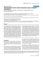
oscopy was duly organised which showed a sessile 1 cm
polyp in the caecum [see figure 1]. On biopsy, this proved
to be a metaplastic polyp. A subsequent attempted
polypectomy was unsuccessful so the patient was referred
to a tertiary centre where another attempt at polypectomy
was carried out. At this point, the polyp looked to be aris-
ing from the appendix, which itself was inverted. In addi-
tion, submucosal nodules in the sigmoid were noted and
these were thought to be endometrial in origin as the
patient had a long history of painful and heavy periods.
The polyp was not removed and the patient was referred
to the colorectal surgeons and gynaecologists for a possi-
ble right hemicolectomy, total abdominal hysterectomy
and bilateral salpingo-oophorectomy.
A preoperative CT scan of her abdomen and pelvis did not
reveal any firm evidence of endometriosis and only noted
small cysts on both ovaries.
At the time of the operation, the appendix had intussus-
cepted and a simple appendicectomy, rather than a right
hemicolectomy, was carried out in the absence of any
other findings at laparotomy.
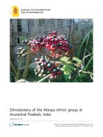
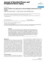
On histology, the wall of the appendix had widespread
endometrial deposits [see Figures 2 and 3] and there was
no evidence of malignancy. In addition, the cervix and fal-
lopian tubes were within normal limits and the ovaries
both had multiple follicular cysts and germinal inclusion
cysts and there were leiomyomas within the myometrium.
Discussion
Appendiceal intussusception is uncommon and typically
found at the time of operation. An incidence rate of 0.01%
has been reported in the literature [1]. Usually associated
with males in the first decade, patients tend to present
with symptoms of vague colicky lower abdominal pain
with or without symptoms of small bowel obstruction.
Endometriosis is defined as the proliferation and function
of endometrial tissue outside the endometrial cavity. The
reported incidence in pre-menopausal women is in the
order of 8–15%. Although the disease classically involves
the pelvic organs and pelvic peritoneum, seeding has been
observed in surgical scars, around the umbilicus, in the
inguinal canal, intestines, bladder, heart and lungs. The
exact aetiology of endometriosis is unknown but there are
two main theories on its pathogenesis. The transportation
theory presumes that endometrial cells are transported to
distant sites through surgical manipulation, menstrual
shedding via the fallopian tubes or through lymphatic or
vascular spread. Alternatively, the metaplastic theory sug-
gests that embryonic coelomic mesothelium dedifferenti-
ates into endometrial tissue in response to inflammation
or trauma [4,5]. The most common symptoms of
endometriosis are dysmenorrhoea, pelvic pain and infer-
tility but patients can also be asymptomatic.
Colonoscopy view of suspected caecal polypFigure 1
Colonoscopy view of suspected caecal polyp.
Low power (5 × 10) view of caecal wall showing endometri-otic glands and stroma within the submucosaFigure 2
Low power (5 × 10) view of caecal wall showing endometri-
otic glands and stroma within the submucosa. Haematoxylin
and eosin stain.
Publish with BioMed Central and every
scientist can read your work free of charge
"BioMed Central will be the most significant development for
disseminating the results of biomedical research in our lifetime."
Sir Paul Nurse, Cancer Research UK
Your research papers will be:
available free of charge to the entire biomedical community
peer reviewed and published immediately upon acceptance
cited in PubMed and archived on PubMed Central
yours — you keep the copyright
Submit your manuscript here:
/>BioMedcentral
Journal of Medical Case Reports 2008, 2:12 />Page 3 of 4
(page number not for citation purposes)
The incidence of gastrointestinal endometriosis varies
between 3–37% of those women who have proven dis-
ease. The rectum and sigmoid colon are most commonly
involved, followed by the rectovaginal septum, small
intestine, caecum and appendix. It usually takes the form
of asymptomatic, small, serosal deposits. Under cyclical
hormonal influences these deposits may proliferate and
infiltrate the bowel wall. Cyclical haemorrhage from the
endometrioma then leads to an intense, localised fibrosis
within the bowel wall that can result in the formation of
strictures. In addition, serosal deposits can lead to the for-
mation of adhesions between neighbouring pelvic struc-
tures or bowel loops [6].
Appendiceal endometriosis is usually asymptomatic.
When symptomatic it frequently presents as appendicitis.
Acute appendiceal inflammation arises due to partial or
complete luminal occlusion by the endometrioma [6].
Appendiceal intussusception secondary to endometriosis
is extremely rare with fewer than 30 cases reported in the
literature during the last fifty years. Endometrial involve-
ment of the appendix is usually accompanied by chronic
fibrosis, inflammation and hyperplasia or hypertrophy of
the muscularis propria. This hypertrophic segment serves
as a lead point for hyperperistalsis hence making it prone
to intussusception particularly when combined with a
fully mobile appendix that has a wide proximal lumen
and a fat free mesoappendix. CT abdominal scans may
demonstrate a soft tissue mass in the region of the cae-
cum, although in this particular case the CT scan did not
point towards the diagnosis.
Conclusion
As in all cases of intussusception, the index of suspicion
must be high as 90% of all intussusceptions in adults are
due to an underlying neoplastic process. Intestinal
endometriosis should be considered as a differential diag-
nosis in post-menarchal women who present with epi-
sodic gastrointestinal symptoms particularly in
conjunction with gynaecological symptoms. The gold
standard in the investigation of similar cases would
appear to be laparoscopy or laparotomy followed by sur-
gical resection in order to obtain histological evidence of
the lead point.
Competing interests
The author(s) declare that they have no competing interests.
Authors' contributions
All of the named authors were involved in the preparation
of this manuscript.
Consent
Written informed consent was obtained from the patient
for publication of this case report and any accompanying
images. A copy of the written consent is available for
review by the Editor-in-Chief of this journal.
Acknowledgements
The authors would like to express their thanks to both the gynaecology and
radiology departments for their help in this case. No funding was required
for this study.
References
1. Collins D: Seventy one thousand human appendix specimens.
A final report summarising forty years' study. Am J Proctol
1963, 14:356-381.
2. Burghard F: Intussusception of the vermiform appendix, the
intussusceptum protruding from the anus. Br J Surj 1914, 1:721.
3. Bachman AL, Clemett AR: Roentgen aspects of primary appen-
diceal intussusception. Radiology 1971, 101:531-538.
4. Igawa HH, Ohura T, Sugihara T, Hosokawa M, Kawamura K, Kaneko
Y: Umbilical endometriosis. Ann Plast Surg 1992, 29:266.
5. Hasegawa T, Yoshida K, Matsui K: Endometriosis of the appendix
resulting in perforated appendicitis. Case Rep Gastroenterol
2007, 1:27-31.
6. Cameron IC, Rogers S, Collins MC, Reed MWR: Intestinal
endometriosis. Int J Colorect Dis 1995, 10:83-86.
Low power (5 × 10) view of appendix wall showing foci of endometriosis within the muscle layerFigure 3
Low power (5 × 10) view of appendix wall showing foci of
endometriosis within the muscle layer. Haematoxylin and
eosin stain.
Publish with BioMed Central and every
scientist can read your work free of charge
"BioMed Central will be the most significant development for
disseminating the results of biomedical research in our lifetime."
Sir Paul Nurse, Cancer Research UK
Your research papers will be:
available free of charge to the entire biomedical community
peer reviewed and published immediately upon acceptance
cited in PubMed and archived on PubMed Central
yours — you keep the copyright
Submit your manuscript here:
/>BioMedcentral