báo cáo khoa học: " New insight into silica deposition in horsetail (Equisetum arvense)" doc
Bạn đang xem bản rút gọn của tài liệu. Xem và tải ngay bản đầy đủ của tài liệu tại đây (6.69 MB, 9 trang )
RESEA R C H ART I C L E Open Access
New insight into silica deposition in horsetail
(Equisetum arvense )
Chinnoi Law and Christopher Exley
*
Abstract
Background: The horsetails (Equisetum sp) are known biosilicifiers though the mechanism underlying silica
deposition in these plants remains larg ely unknown. Tissue extracts from horsetails grown hydroponically and also
collected from the wild were acid-digested in a microwave oven and their silica ‘skeletons’ visualised using the
fluor, PDMPO, and fluorescence microscopy.
Results: Silica deposits were observed in all plant regions from the rhizome through to the stem, leaf and spores.
Numerous structures were silicified including cell walls, cell plates, plasmodesmata, and guard cells and stomata at
varying stages of differentiation. All of the major sites of silica deposition in horsetail mimicked sites and structures
where the hemicellulose, callose is known to be found and these serendipitous observations of the coincidence of
silica and callose raised the possibility that callose might be templating silica deposition in horsetail. Hydroponic
culture of horsetail in the absence of silicic acid resulted in normal healthy plants which, following acid digestion,
showed no deposition of silica anywhere in their tissues. To test the hypothesis that callose might be templating
silica deposition in horsetail commercially available callose was mixed with undersaturated and saturated solutions
of silicic acid and the formation of silica was demonstrated by fluorimetry and fluorescence microscopy.
Conclusions: The initiation of silica formation by callose is the first example whereby any biomolecule has been
shown to induce, as compared to catalyse, the formation of silica in an undersaturated solution of silicic acid. This
novel discovery allowed us to specu late that callose and its associated biochemical machinery could be a missing
link in our unders tanding of biosilicification.
Keywords: Biosilicification, biogenic silica, silicic acid, horsetails, callose, PDMPO, fluorescence, acid digestion.
Background
Silicon is the second most abundant element of the
Earth’s crust after oxygen and, perhaps surprisingly, its
essentiality in biota remains equivocal [1]. The difficulty
in ascribing true biochemical essentiality to silicon prob-
ably emanates from a lack of demonstration of any sili-
con-requiring biochemistry and specifically Si-C, Si-O-
C, Si-N, et c. bonds in any form of extant life [2]. How-
ever, in spite of such limitations the essentiality of sili-
con in plants remains the subject of rigorous debate
[3,4] as do elaborations of the underlying mechanisms.
Biosilicification was recently defined as ’the movement of
silicic acid from environments in which its concentrat ion
does not exceed its solubility (< 2 mM) to intracellular
or systemic compartments in which it is accumulated for
subsequent deposition as amorphous hydrated silica’ [5]
and a number of plant s are known biosilicifiers [4]. One
of the best known of these are the horsetails, Equisetum
sp., and silica deposition in the tissues of these plants
has been studied extensively [6-12], perhaps the seminal
work in the field being carried out by Perry and Fraser
[13]. In this work scanning and transmission electron
microscopy was used to illuminate the elaborate and
detailed micromorphology and ultrastructure of silicas
extracted from different regions of the horsetail, Equise-
tum arvense. The images of silicified stomata and other
silica sculptures are truly breathtaking and the level of
organisation of silica in the tissues prompted the
authors to speculate that ’the silica acts as an in vivo
stain, faithfully reproducing the organic matrix skeleton
at the microscopic and macroscopic levels without stain-
ing’ . Perry and Lu (1992) suggested that the organic
* Correspondence:
The Birchall Centre, Lennard-Jones Laboratories, Keele University,
Staffordshire, ST5 5BG, UK
Law and Exley BMC Plant Biology 2011, 11:112
/>© 2011 Law and Exley; licensee Bi oMed Central Ltd. This is an Open Access article distributed under the terms of the Creative
Commons Attribution License ( which pe rmits unrestricted use, distribution, and
reproduction in any medium, provided the original work is properly cited.
matrix in question might be made from polymers of car-
bohydrates, for example, cellulose [14], and this sugges-
tion was reinforced recently by Fry and colleagues who
speculated that the hemicellulose,callose,inhorsetail
cell walls might be a potential site of silica deposition
[15]. Many different biomolecules, often having origin-
ally been extracted from biogenic silica, have been
shown to accelerate or catalyse silica deposition in satu-
rated solutions of silicic acid [16]. However, biosilicifiers,
such as horsetails, harvest silicic acid from solutions
whicharefarfromsaturationanddeposititasamor-
phous hydrated silica and it is the elucidation of this
mechanism which remains the ‘Holy Grail’ of biological
silicification research [5].
Herein we have taken inspiration from the work of
Perry and Fraser [13] on horsetail and we have used
fluorescence microscopy to investigate biosilicification in
horsetail and to identify the organic matrix involved in
templating silica deposition in this plant.
Results
PDMPO as a fluorescent marker of biosilicification
Microwave-assisted acid digestion of horsetail, either
grown hydroponically in the presence of silicic acid or
in plants coll ected from the wild, resulted in silica
depo sits and ‘skeletons’ which were successfully labelled
with the fluor PDMPO. Silica was identified in acid
digests of all areas of the plant from the rhizome
through to spores in the cone. There were no structu-
rally-distinct silica skeletons in the root, only what
appeared as diffuse deposits of siliceous materials (Fig-
ure 1a). Silica skeletons of basal stem showed epider-
mal-like cells, 30-40 μm wide and 100-300 μmlong,
with heavily silicified cell wall s and approximately equi-
distant punctate deposits of silica within the walls which
were suggestive of the expected locations of plasmodes-
mata. Each ‘silica cell’ included an amorphous, spherical
silica deposit between 10 and 20 μmindiameterwhich
had the appearance of a nucleus or vesicle. There were
also occasional heavily silicified (as indicated by an
enhanced fluorescence) skeletons of stomata, approxi-
mately 40 μmwideand70μm long, which appeared to
be at various stages of differentiation (Figure 1b). In
other silica skeletons of basal stem the sections were
characterised by many small punctate deposits of silica,
<1 μm across, while the stomata, ca 40-50 μmindia-
meter, were more numerous, only lightly silicified and
many appeared to be linked in pairs. Adjacent epider-
mal-like cells were ca 100-200 μm in length and 40-50
μm wide and included highly fluorescent silica deposits
which, concomitant with their parent silica cells
appeared to be in the process of division (Figure 1c).
Some sections of silicified stem showed silica cells
which were 100-400 μm in length but without the
intracellular, nucleus/vesicle-like deposits seen in other
stem sections. The silicified cell walls were heavily inva-
ginated and, again, included punctate and equidistant
deposits of silica which as suggested previously may be
indicative of the positions of plasmodesmata (Figure 1d).
Silica skeletons of distal stem sections were quite differ-
ent from basal sections in that they were characterised
by rosette-like accumulations of silica deposits approxi-
mately 20-30 μm in diameter as well as guard cells of
stomata studded with silica deposits of ca 1-2 μm across
and resembling ‘teeth’ where they extended into the sto-
matal pore (Figure 1e). These silica rosettes appear ed to
be further elaborated in nodal regions where they
formed doughnut-like structures, up to ca 40 μm in dia-
meter, which gave the distinct impression of being silici-
fied pores (Figure 1f). Other nodal regions showed long,
ca 200-500 μm, epidermal-like cells in which their
jagged-in-appearance cell walls were heavily silicified.
There were neither punctate silica deposits nor intracel-
lular silica inclusions evident in these structures (Figure
1g). The leaves showed silica skeletons which were very
similar to those of the nodal regions though perhaps
showing higher d ensities of the rosette-like silica struc-
tures (Figure 1h). Stomata were heavily silicified in some
sections of leaf and showed clear anatomical details
including an anular ring between the pore-forming
guard cells. Again stomata often appeared as pairs con-
nected by silicified threads of varying diameters (Figure
1i). Spores were found to be heavily silicified, being
associated with spore walls and present as sub-micron
punctate deposits of silica upon individ ual silicified
spores which were between 20 and 40 μm in diameter
(Figure 1j). Horsetail grown from rhizomes collected
from the wild under hydroponic conditions in the
absence of silicic acid grew normally without any
obvious requirement for silicon. Acid digestion of tissues
from these plants revealed no silica deposits or
skeletons.
PDMPO as a fluorescent indicator of silica formation in
vitro
Buffer solutions at pH 7 and including 0.125 μM
PDMPO showed no green fluorescence indicative of
silica and only occasional pa rticles of blue fluorescence
probably due to dust or insoluble contaminants in the
buffer (Figure 2a). Buffer solutions a t pH 7 and includ-
ing 5% w/v callose and PDMPO, but n ot Si(OH)
4
,
showed no green fluorescence while callose was indi-
cated as amorphous blue fluorescence (Figure 2b). Buf-
fer solutions at pH 7 and including 1 mM Si(OH)
4
(undersaturated) and 5% w/v callose showed significant
green fluorescence in the presence of PDMPO (Figure
2c). The fluorescent material was primarily made up of
aggregates of sub micron-sized particles (Figure 2c
Law and Exley BMC Plant Biology 2011, 11:112
/>Page 2 of 9
200 m
200 m
100 m
100 m100 m
100 m
100 m
100 m
200 m
a) b)
c) d)
e) f)
g) h)
i) j)
200 m
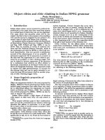
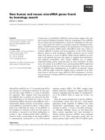
Figure 1 PDMPO-labelled silica deposition in horsetail. a.Rhizome;b. Basal stem, arrows (main and insert) indicate punctate deposits of
silica associated with cell walls; c. Basal stem, arrow (insert) indicates silica deposition at cell plate between dividing cells; d. Basal stem, arrow
(insert) indicates punctate deposits of silica associated with highly invaginated cell walls; e. Distal stem, showing (main and insert) rosette-like
silica structures and heavily silicified stomata; f. Node, showing high density of silicified structures including doughnut-like pore (insert); g. Node,
showing jagged appearance of silica-rich cell walls; h. Leaf, showing high densities of rosette-like silica structures; i. Leaf, demonstrating the
intimate association of silica with stomata (insert); j. Spores, showing heavily silicified spores including (insert) punctate deposits of silica on the
spore surfaces. Scale bars; 100 μm - d,e,f,g,h,i; 200 μm - a,b,c,j.
Law and Exley BMC Plant Biology 2011, 11:112
/>Page 3 of 9
insert and arrow) and these appeared to be associated
with or occluded within the blue fluorescent callose.
Identical solutions in the absence of callose showed no
green fluorescence and were similar to image Figure 2a.
In buffer solutions at pH 7 which included 2 mM Si
(OH)
4
and 5% w/v callose the PDMPO-positive green
fluorescence was more extens ive than at 1 mM Si (OH)
4
and included diffuse and particulat e materials, the latter
again being composed primarily of sub micron-sized
particles (Figure 2d). Identical solutions in the absence
of callose showed a significantly lesser amount of
PDMPO-positive green fluorescence and the fluorescent
material wa s similar in appear ance and size to that
observed in the presence of callose (Figure 2e). In buffer
solutions in which the concentration of Si(OH)
4
was 4
mM (saturated) there were significant flocs of PDMPO-
positive materials and particularly so in those prep ara-
tions which included 5% w/v callose (Figure 2f).
The presence of silica in an undersaturated (2 mM)
solution of Si(O H)
4
at pH 7 and including 5% w/v cal-
lose was further supported by fluorescence spectrometry
which demonstrated a callose-dependent shift in emis-
sion maximum from 450 to 510 nm (Figu re 3a,b). That
this shift was due to the formation of silica was con-
firmed in a saturated (7 mM) solution of Si(OH)
4
under
the identical solution conditions (Figure 3c). The silica-
dependent shift was significantly more pronounced in
the presence than absence of callose.
Discussion
When fresh or dried samples of horsetail were digested
in concentrated acid using a microwave oven all the
organic materials associated with the plants were com-
pletely dissolved leaving behind elaborate and detailed
silica ‘skeletons’ of the d ifferent plant regio ns. The sus-
pension of these silica remains in buffered solutions at
pH 7 which contained the fluorescent probe, PDMPO,
enabled their detailed structures to be viewed by fluores-
cence microscopy (Figure 1). It was of note that horse-
tail grown hydroponically in the complete absence of
added silicic acid grew normally for 10 weeks though
without leaving any trace of silica following tissue diges-
tion. While there was no immediate evidence that
horsetail required silicon for normal growth it was
observed that after 10 we eks of hydroponic culture in
the absence of added silicic acid some plants showed
wilting and blackening of d istal branch tips similar to
symptoms of ‘silicon-deficiency’ observed by Chen and
Lewin [17]. However, herein these symptoms appeared
simultaneously in parts of the plants where there was
evidence of infection by powdery mildew fungus and so
it was not clear as to whether they were the result of
silicon deficiency or fungal infection [18]. There was no
evidence of fungal infection in plants grown in the pre-
sence of a dded silicic acid. While it was clear in horse-
tail collected locally or gr own in sil icon-replete
hydroponic media that silica was deposited extensively
a) b) c)
d) e) f)
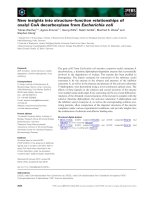
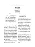
Figure 2 PDMPO-labelled silica in vitro. All [PDMPO] are 0.125 μM; All solutions are 20 mM PIPES at pH 7. All [callose] are 5% w/v. a. PDMPO
only; b. PDMPO + callose; c. PDMPO + callose + 1 mM Si(OH)
4
; the insert shows a close-up of one of the silica clusters; d. PDMPO + callose + 2
mM Si(OH)
4
; the insert shows a close-up of the precipitated silica; e. PDMPO + 2 mM Si(OH)
4
; the insert shows a close-up of silica; f. PDMPO +
callose + 4 mM Si(OH)
4
; the insert shows an example of silica formed in this treatment. Scale bars; 100 μm - b-f; 200 μm-a.
Law and Exley BMC Plant Biology 2011, 11:112
/>Page 4 of 9
throughout the stem and leaf certain structures showed
intense fluorescence which suggested significant silica
deposits in these regions. Stomata were often intensley
fluorescent (Figure 1) and it was noted that silicification
of stomata in horsetail appeared to mirror the known
deposition of the hemicellulose, callose, in guard cell
differentiation and stomatal pore formation in the
related fern, Asplenium nidus [19-21]. The observed
similarities between the deposition in stomatal struc-
tures of callose in A. nidus and silica in E. arvense were
remarkable. For example, in early post cytokinetic guard
cells the nascent ventral wall was silicified (Figure 4a).
In later examples, the ventral, dorsal and periclinal walls
as well as the wall thickenings were are all silicified (Fig-
ure 4b). I n some stomata silicification was reduced at
the centre of the ventral wall as stomatal pore formation
was iniated (Figure 4c). Thereafter in further differen-
tiated example s of stomata radial fibrillar arrays of silica
were observed on the periclinal wall where stomatal
pore formation takes place (Figure 4d). Finally in more
mature stomata the wall thickenings were silicified and
punctate deposits of silica were observed associated with
cell walls (Figure 4e). Annular rings of silica were also
observed lining the stomatal pore in more mature sto-
mata (Fig ure 1i). All of these observations of silica
deposition in E. arvense have been identified as sites of
callose deposition in A. nidus (Figure 4) in the recent
seminal and detailed studies of Apostolakos and collea-
gues [19-21]. These very close associations between the
known deposition of callose in differentiating stomata
and the presence of silica now strongly implicate callose,
or possibly, callose in conjunction with an underlying
microtubule array, in directing the silicification of sto-
mata in horsetail. Further strong evidence that callose
was i nvolved in templating the deposition of silica else-
where in horsetail was observed in silica skeletons of
cells undergoing cytokinesis (Figure 5). Again silica
deposition at phragmoplasts and eventually at cell plates
and young cell walls dividing daughter cells mirrored
the known deposition of callose in cytokinesis [22-24].
In some cells which were at an early stage of division, in
some cases before there was any evidence of silica
deposition at the phragmoplast, the cytosolic (and per-
haps nuclear) fragments of the emerging daughter cells
were found to be heavily silicified (Figure 1c). The iden-
tity of these silica ‘nuclei/vesicles’ is a mystery though
they may provide evidence for a role for callose in the
partitioning of cytosolic and nuclear materials during
cell division? The significant deposits of silica within cell
walls is supported by the known presence of c allose in
cell walls of horsetails [12,15, 25,26] . In addition, equidi -
stant punctate deposits of silica associated with cell
wallsmaybeindicativeof,again,theknowndeposition
of callose in plasmodesmata (Figure 1 b,d) [27,28].
Finally, the heavily silicified s pores (Figure 1j) may also
be evidence of the role which is known to be played by
callose deposition in plant reproduction [24,29]. Other
silica deposits observed in horsetail may also be related
to callose deposition. For example, the punctate deposits
of silica, sometimes singular and sometimes organised
into rosette-like structures, which could be found
throughout stem and leaf tissues were identical to those
found associated with mature stomata where they are
known to mimic callose deposition [30]. In addition the
silicified pores of internal diameter 3-5 μmwhichwere
identified in leaf tissues (Figure 1f) are not dissimilar t o
5% Callose +
Buffer/PDMPO
Buffer/PDMPO only
510 nm
450 nm
71.
2
5
-2
52.2
0.8
61.4
400
nm
Fluorescence (AU)Fluorescence (AU)Fluorescence (AU)
65
0
400 nm 65
0
4
00
nm
65
0
2mMSi(OH)
4
+
Buffer/PDMPO
2mMSi(OH)
4
+
5% Callose +
Buffer/PDMPO
7mMSi(OH)
4
+
5% Callose +
Buffer/PDMPO
7mMSi(OH)
4
+
Buffer/PDMPO
a)
b)
c)
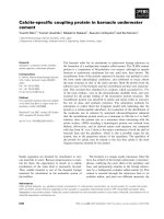
Figure 3 Emission spectra (Perkin-Elmer LS50B; Ex; 338 nm;
Em: 400-650 nm) of 0.125 μM PDMPO in 20 mM PIPES
solutions at pH 7 and; a. with or without 5% w/v callose; b.2mM
Si(OH)
4
with or without 5% w/v callose; c. 7 mM Si(OH)
4
with or
without 5% w/v callose.
Law and Exley BMC Plant Biology 2011, 11:112
/>Page 5 of 9
call ose lined sieve pores found, for exampl e, in A. thali-
ana [31]. We have successfully applied the fluor
PDMPO to demonstrate the deposition of silica in
horsetail and in doing so we have identified several
novel aspects of biosilicification in horsetail and in parti-
cular we have highlighted a potential role for callose in
templating silica deposition. Callose biochemistry is, of
course, essential in horsetail [15,25], as in many other
plants such as the ferns [19-21], and so it is not imme-
diately e vident as to how to test whether callose is ulti-
mately required for silica deposition. For example,
horsetail is unlikely to grow and/or prosper if the callose
synthase gene is knocked out. However, we have been
able to support our microscopy evidence linking silica
and callose deposition by demonstrating that an under-
saturated solution of Si(OH)
4
(i.e. a solution where the
[Si(OH)
4
] ≤ 2 mM) can be induced to form silica in the
presence of callose. The formation of silica was con-
firmed by both fluorescence microscopy (Figure 2) and
fluorimetry (Figure 3) and within the usual constraints
of such original results we believe that this is the first
time that an undersaturated solutio n of Si(OH)
4
at
room temperature and pressure has been induced to
form silica simply by the addition of a biomolecule.
When silica extracted from horsetail was added to a 20
mM PIPES-buffered solution at pH 7 which included
0.125 μM PDMPO t he emission spect rum changed to
give a single emission maximum at ca 510 nm. This
positive control confirmed the known silica-induced
shift in the emission spectrum of the fluor PDMPO. A
similar shift was also seen for solut ions under the same
conditions but including 5% w/v callose and either 2 or
4mMSi(OH)
4
(Figure 3). The former represents an
undersaturated solution of Si(OH)
4
and offered up the
first evidence that callose could induce Si(OH)
4
to auto-
condense and form silica. However, the in vitro evidence
was most compelling in preparations containing only 1
mM Si(OH)
4
when viewed by fluorescence microscopy
(Figure 2c). In the absence of callose no silica could be
identified by fluorescence microscopy in such
a) b) c) d) e)
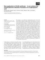
Figure 4 The deposition of callose (diagrams) and silica (fluorescent images) in th e differentiation of stomata in E. arvense. a. Callose
(yellow) and silica (arrow) deposition at the nascent ventral wall (VW) of post-cytokinetic guard cells; b. Deposition of callose (yellow) and silica
(arrows) in the periclinal wall and dorsal wall (DW) and callose/silica remaining in the ventral wall; c. Callose (yellow) and silica (arrow) disappear
from the centre of the ventral wall during pore initiation; d. Callose (yellow) and silica (arrows) appears as a radial fibrillar array as the stomatal
pore is formed; e. Upon stomatal pore formation callose (yellow) and silica (arrows) remain as punctate deposits upon the guard cell walls. All
stomata are ca 40 μm in diameter. Information on deposition of callose taken from [19-21].
Law and Exley BMC Plant Biology 2011, 11:112
/>Page 6 of 9
preparations while in the presence of callose there were
clear and numerous deposits of silica some of which
were spherical and approximately 0.5 - 1.0 μmindia-
meter. Intriguingly the silica bodies were intimately
associated with the polymer network of the callose,
identified as blue fluorescence, which suggest ed that the
constrained environment generated by the gel-like cal-
lose provided the cond itions under which an undersatu-
rated solution of Si(OH)
4
(1 mM) could be ‘tricked’ into
undergoing autocondensation and subsequent growth
towards stable aggregates of silica. Callose is a linear
homopolymer made up primarily of b-1,3-linked glucose
residues which at the c oncentration used herein, ca 5%
w/v, will form a viscoelastic gel [32] within which the
orientation of hydroxyl groups on the glucose mono-
mers may be such that they are able to iniate the first
steps in the autocondensation of silicic acid as i t slowly
diffuses within the callose matrix. The hydroxyl groups
on the polymer network of callose in some way enable
the energy barrier to the autocondensation of Si(OH)
4
to be ov ercome and once the first Si-O-Si linkages have
been made further condensation reactions can proceed
much more easily to eventually build the silica aggre-
gates observed, for example, in Figure 2c. While further
experiments will be required to delineate the range of
conditions under which callose i nduces silica formation
in undersaturated solutions of Si(OH)
4
and the exact
mechanism by which this is achieved we now have a
long sought after biomolecule which can act as a tem-
plate for silica formation and deposition in vitro.Ifthis
is also the basis for the mechanism of silica deposition
in horsetail then it may also be significant in other cal-
lose producing biosilicifiers such as diatoms [33]. If cal-
lose is the key then associated biochemistry including
enzymes such as callose synthase (potentially catalysing
Si-O-Si bond formation) and b-1,3-glucanases (poten-
tially cleaving Si-O-Si bonds) [25] will play a pivotal role
in the modelling and remodelling of silica frameworks.
The deposition of silica in horsetail has been studied for
many decades an d we now have a possible mechanism
of silica deposition in this plant which could also be a
general mechanism of biosilicification.
Conclusion
The fluor PDMPO has been used to identify silica
deposition in horsetail and to provide new insight into
silicification in this plant. It was observed that silica
deposition in horsetail exactly mirrored the known
deposition of callose in the related fern and other plants.
Callose was shown to induce the formation and precipi-
tation of silica in undersaturated solutions of silicic acid.
This was the first time that this had been demonst rated
for any biomolecule and it suggested that callose and
perhaps other similar carbohydrates might be key mole-
cules in biological silicification.
Methods
Hydroponic culture of horsetail
Horsetail (Equisetum arvense) rhizomes were collected
locally, washed in ultrapure water (conductivity < 0.067
μS/cm) and subjected to hydroponic culture in 1/6
th
MS
mediuminthepresence(2mM)orabsenceofadded
silicic acid. The latter media included an additional 8
mM Na
+
to account for Si addition as Na
4
SiO
4
.After
10-12 weeks of a 14 h light/10 h dark cycle at 25°C
healthy horsetail plants had grown under both sets of
conditions.
Digestion of horsetail materials
Horsetail plants, either collected locally or grown hydro-
ponically, were washed in ultrapure water, allowed to
air-dry, cut into discrete 1 cm sections of rhizome/root,
basal stem, distal stem, nodal regions and leaves and ca
0.5 g of each placed in acid-washed 20 mL PFA teflon
©
vessels. The samples were then digested in a 1:1 mixture
of 15.8M HNO
3
and 18.4M H
2
SO
4
using a Mars Xpress
microwave oven (CEM Microwave Technology Ltd.).
The acid digests were clear and, upon dilution with 8
mL of ultrapure water, were filtered and the residues
washed several times with further volumes of ultrapure
water. Filters were then placed in plastic Petri d ishes in
an incubator at 37°C to achieve dryness over several
days. Dry residues off each filter were then collected
into Bijoux tubes and stored in a dry, sealed, perspex
cabinet.
a)
b)
Figure 5 a,b PDMPO-labelling of silica deposition of cell plates
and young cell walls (arrows) forming in cytokinetic cells.
Law and Exley BMC Plant Biology 2011, 11:112
/>Page 7 of 9
PDMPO labelling of horsetail silica
Silica residues collected from filters were suspended in
20 mM PIPES at pH 7 and containing 0.125 μM2-(4-
pyridyl)-5-((4-(2-dimethylaminoethylaminocarbamoyl)
-methoxy)phenyl)oxazole (PDMPO; LysoSensor Yellow/
Blue DND-160, 1 mM in DMSO). This intracellular pH
probe [34] has been shown to be bound by silica (but
not silicic acid) and to emit ‘green’ fluorescence upon
excitation at 338 nm [35-38]. Suspensions were left for
24 h to allow the reaction between silica surfaces and
PDMPO to achieve completion after which 50 μLsam-
ples were transferred to a cavity slide and viewed using
an Olympus BX50 fitted with a BXFLA fluorescent
attachment using a U-MWU filter cube (Ex: 333-385
nm; Em: 400-700 nm). A ColourView III digital camera
(OSIS FireWire Camera 3.0 digitizer) was used to cap-
ture images in conjunction with CELL* Imaging soft-
ware (Olympus Cell* family, Olympus Soft Imaging
solutions GmbH 3.0).
In vitro preparations of callose and silicic acid
Callose (b-D Glucan, Barley, Sigma, UK) was dissolved
at 5% w/v in 20 mM PIPES buffer solutions at pH 7 and
containing 0, 1, 2, 4 and 7 mM Si(OH)
4
by warming
each preparation in a water bath at 100°C for 60 sec-
onds. Upon cooling to room temperature PDMPO was
added to a concentration of 0.125 μM. Equivalent con-
trol solutions to which no callose had been added were
treated in an identical manner. All solutions were then
incubated at room temperature in the dark for 5 days
before being examined by fluorescence microscopy, see
above, or their emission spectra were determined by
fluorimetry (Perkin-Elmer LS50B; Ex; 338 nm; Em: 400-
650 nm) as previously described [35].
Acknowledgements
CL was in receipt of a NERC studentship.
Authors’ contributions
CE designed the study and provided training and guidance in experimental
methods. CE wrote and prepared the first draft of the manuscript. CL carried
out the majority of the experimental work and helped with writing the
manuscript.
Both authors have read and approved this manuscript.
Competing interests
The authors declare that they have no competing interests.
Received: 15 April 2011 Accepted: 29 July 2011 Published: 29 July 2011
References
1. Exley C: Silicon in life: a bioinorganic solution to bioorganic essentiality. J
Inorg Biochem 1998, 69:139-144.
2. Exley C: Darwin, natural selection and the biological essentiality of
aluminium and silicon. Trends Biochem Sci 2009, 34:589-593.
3. Epstein E: The anomaly of silicon in plant biology. Proc Natl Acad Sci USA
1994, 91:11-17.
4. Currie HA, Perry CC: Silica in plants: biological, biochemical and chemical
studies. Ann Bot 2007, 100:1383-1389.
5. Exley C: Silicon in life: whither biological silicification? In Biosilica in
Evolution, Morphogenesis and Nano-biotechnology. Edited by: Muller WEG,
Grachev MA. Springer; 2009:173-184.
6. Page CN: An assessment of inter-specific relationships in Equisetum
subgenus Equisetum. New Phytol 1972, 71:355-369.
7. Kaufman PB, LaCroix JD, Dayanandan P, Allard LF, Rosen JJ, Bigelow WC:
Silicification of developing internodes in the perennial scouring rush
(Equisetum hyemale var. affine). Developmental Biol 1973, 31:124-135.
8. Laroche J, Guervin C, Le Coq C, Robert D: Activités pétrogénétiques chez
Equisetum arvense L. (Ptéridophytes). Bulletin de la Société Botanique de
France 1992, 139:47-55.
9. Holzhüter G, Narayanan K, Gerber T: Structure of silica in Equisetum
arvense. Analyt Bioanalyt Chem 2003, 376:512-517.
10. Sapei L, Nöske R, Strauch P, Paris O: Isolation of mesoporous biogenic
silica from the perennial plant Equisetum hyemale. Chem Mat 2008,
20:2020-2025.
11. Gierlinger N, Sapei L, Paris O: Insights into the chemical composition of
Equisetum hyemale by high resolution Raman imaging. Planta 2008,
227:969-980.
12. Currie HA, Perry CC: Chemical evidence for intrinsic ‘Si’ within Equisetum
cell walls. Phytochemistry 2009, 70:2089-2095.
13. Perry CC, Fraser MA:
Silica deposition and ultrastructure in the cell wall of
Equisetum arvense: the importance of cell wall structures and flow
control in biosilicification? Phil Trans Roy Soc London B 1991, 334:149-157.
14. Perry CC, Lu Y: Preparation of silicas from silicon complexes: Role of
cellulose in polymerisation and aggregation control. J Chem Soc Faraday
Trans 1992, 88:2915-2921.
15. Fry SC, Nesselrode BHWA, Miller JG, Mewburn BR: Mixed linkage (1→3,1→4)-
β-glucan is a major hemicellulose of Equisetum (horsetail) cell walls. New
Phytol 2008, 179:104-115.
16. Perry CC: An overview of silica in biology: Its chemistry and recent
technological advances. In Biosilica in Evolution, Morphogenesis and Nano-
biotechnology. Edited by: Muller WEG, Grachev MA. Springer; 2009:295-313.
17. Chen CH, Lewin J: Silicon as a nutrient element for Equisetum arvense.
Can J Bot 1969, 47:125-131.
18. Fauteux F, Chain F, Belzile F, Menzies JG, Belanger RR: The protective role
of silicon in the Arabidopsis-powdery mildew pathosystem. Proc Natl
Acad Sci USA 2006, 103:17554-17559.
19. Apostolakos P, Livanos P, Galatis B: Microtubule involvement in the
deposition of radial fibrillar callose arrays in stomata of the fern
Asplenium nidus L. Cell Motility Cytoskeleton 2009, 66:342-349.
20. Apostolakos P, Livanos P, Nikolakopoulou TL, Galatis B: The role of callose
in guard-cell wall differentiation and stomatal pore formation in the fern
Asplenium nidus. Ann Bot 2009, 104:1373-1387.
21. Apostolakos P, Livanos P, Nikolakopoulou TL, Galatis B: Callose implication
in stomatal opening and closure in the fern Asplenium nidus. New Phytol
2010, 186:623-635.
22. Scherp P, Grotha R, Kutschera U: Occurrence and phylogenetic
significance of cytokinesis-related callose in green alga, bryophytes,
ferns and seed plants. Plant Cell Rep 2001, 20:143-149.
23. Thiele K, Wanner G, Kindzierski V, Jürgens G, Mayer U, Pachl F, Assaad FF:
The timely deposition of callose is essential for cytokinesis in
Aribodopsis. The Plant J 2009, 58:13-26.
24. Chen XY, Kim JY: Callose synthesis in higher plants. Plant Signall Behav
2009, 4:489-492.
25. Fry SC, Mohler KE, Nesselrode BHWA, Franková L:
Mixed linkage β-glucan:
xyloglucan endotransgluocosylase, a novel wall remodelling enzyme
from Equisetum (horsetail) and charophytic algae. The Plant J 2008,
55:240-252.
26. Sørensen I, Pettolino FA, Wilson SM, Doblin MS, Johansen B, Bacic A,
Willats WGT: Mixed linkage (1→3, 1→4)- β-D-glucan is not unique to the
Poalesm and is an abundant component of Equisetum arvense cell walls.
The Plant J 2008, 54:510-521.
27. Xu XM, Jackson D: Lights at the end of the tunnel: new views of
plasmodesmal structure and function. Current Opin Plant Biol 2010,
13:684-692.
28. Zavaliev R, Ueki S, Epel BL, Citovsky V: Biology of callose (β-1,3-glucan)
turnover at plasmodesmata. Protoplasma 2011, 248:117-130.
29. Nishikawa SI, Zinkl GM, Swanson RJ, Maruyama D, Preuss D: Callose (beta-
1,3-glucan) is essential for Aribodopsis pollen wall patterning, but not
tube growth. BMC Plant Biol 2005, 5:art 22.
Law and Exley BMC Plant Biology 2011, 11:112
/>Page 8 of 9
30. Majewska-Sawka A, Münster A, Rodríguez-García MI: Guard cell wall:
immunocytochemical detection of polysaccahride components. Journal
of Experimental Botany 2002, 53:1067-1079.
31. Thompson MV, Wolniak SM: A plasma membrane-anchored fluorescent
protein fusion illuminates sieve element plasma membranes in
Aribodopsis and tobacco. Plant Physiol 2008, 146:1599-1610.
32. Cui SW, Wang Q: Cell wall polysaccharides in cereals: chemical structures
and functional properties. Struct Chem 2009, 20:291-297.
33. Størseth TR, Kirkvold S, Skjermo J, Reitan KI: A branched β-D-(1→3, 1→6)-
glucan from the marine diatom Chaetoceros dibilis (Bacillariophyceae)
charcaterised by NMR. Carbohydrate Res 2006, 341:2108-2114.
34. Diwu Z, Chen CS, Zhang C, Klaubert DH, Haugland RP: A novel acidotropic
pH indicator and its potential application in labeling acidic organelles of
live cells. Chem and Biol 1999, 6:411-418.
35. Shimizu K, Del Amo Y, Brzezinski MA, Stucky GD, Morse DE: A novel
fluorescent silica tracer for biological silicification studies. Chem Biol
2001, 8:1051-1060.
36. Leblanc K, Hutchins DA: New applications of a biogenic silica deposition
fluorophore in the study of eceanic diatoms. Limnol Oceanography::
Methods 2005, 3:462-476.
37. Hazelaar S, van der Strate HJ, Gieskes WWC, Vrieling EG: Monitoring rapid
valve formation in the pennate diatom Navicula salinarum
(Bacillariophyceae). J Phycol 2005, 41:354-358.
38. Ogane K, Tuji A, Suzuki N, Kurihara T, Matsuoka A: First application of
PDMPO to examine silicification in polycystine Radiolaria. Plankton
Benthos Res 2009, 4:89-94.
doi:10.1186/1471-2229-11-112
Cite this article as: Law and Exley: New insight into silica deposition in
horsetail (Equisetum arvense). BMC Plant Biology 2011 11:112.
Submit your next manuscript to BioMed Central
and take full advantage of:
• Convenient online submission
• Thorough peer review
• No space constraints or color figure charges
• Immediate publication on acceptance
• Inclusion in PubMed, CAS, Scopus and Google Scholar
• Research which is freely available for redistribution
Submit your manuscript at
www.biomedcentral.com/submit
Law and Exley BMC Plant Biology 2011, 11:112
/>Page 9 of 9