báo cáo khoa học: " Induction of stromule formation by extracellular sucrose and glucose in epidermal leaf tissue of Arabidopsis thaliana" pot
Bạn đang xem bản rút gọn của tài liệu. Xem và tải ngay bản đầy đủ của tài liệu tại đây (4.54 MB, 10 trang )
RESEARC H ARTIC L E Open Access
Induction of stromule formation by extracellular
sucrose and glucose in epidermal leaf tissue of
Arabidopsis thaliana
Martin Hartmut Schattat
1*
and Ralf Bernd Klösgen
2
Abstract
Background: Stromules are dynamic tubular structures emerging from the surface of plastids that are filled with
stroma. Despite considerable progress in understanding the importance of certain cytoskeleton elements and
motor proteins for stromule maintenance, their function within the plant cell is still unknown. It has been
suggested that stromules facilitate the exchange of metabolites and/or signals between plastids and other cell
compartments by increasing the cytosolically exposed plastid surface area but experimental evidence for the
involvement of stromules in metabolic processes is not available. The frequent occurrence of stromules in both
sink tissues and heterotrophic cell cultures suggests that the presence of carbohydrates in the extracellular space is
a possible trigger of stromule formation. We have examined this hypothesis with induction experiments using the
upper epidermis from rosette leaves of Arabidopsis th aliana as a model system.
Results: We found that the stromule frequency rises significantly if either sucrose or glucose is applied to the
apoplast by vacuum infiltration. In contrast, neither fructose nor sorbitol or mannitol are capable of inducing
stromule formation which rules out the hypothesis that stromule induction is merely the result of changes in the
osmotic conditions. Stromule formation depends on translational activity in the cytosol, whereas protein synthesis
within the plastids is not required. Lastly, stromule induction is not restricted to the plastids of the upper epidermis
but is similarly observed also with chloroplasts of the palisade parenchyma.
Conclusions: The establishment of an experimental system allowing the reproducible induction of stromules by
vacuum infiltration of leaf tissue provides a suitable tool for the systematic analysis of conditions and requirements
leading to the formation of these dynamic organelle structures. The applicability of the approach is demonstrated
here by analyzing the influence of apoplastic sugar solutions on stromule formation. We found that only a subset
of sugars generated in the primary metabolism of plants induce stromule formation, which is furthermore
dependent on cytosolic translational activity. This suggests regulation of stromule formation by sugar sensing
mechanisms and a possible role of stromules in carbohydrate metabolism and metabolite exchange.
Background
Stromules (stroma filled tubules) [1] are protrusions of the
plastid envelope with a diameter of usually less than 1 μm
[2]. These filament-like structures are highly dynamic and
can extend and retract within seconds [3]. Although
tubules extending from the plastid surface had been
described in a monograph about plastids in 1908 (see [4]),
their significance and morphological relevance w as
recognized only after development and improvement of
suitable fluorescence microscopy techniques. In particular,
the generation o f transgenic plants expressing chimeric
proteins consisting of green fluorescent protein (GFP)
fused to chloroplast targeting transit peptides allowed the
first detailed analy sis demonstrating the presence of stro-
mal proteins within these structures [1]. Over the past
years, stromules were found in a variety of vascular plant
species, non-vascular plant species and green algae (as
summarized in [5]) which suggests evolutionary conserva-
tion of these structures and implies that they might play
an important role in all members of the Viridiplantae.
* Correspondence:
1
Laboratory of Plant Development and Interactions; Department of
Molecular and Cellular Biology; University of Guelph; Guelph, ON Canada
Full list of author information is available at the end of the article
Schattat and Klösgen BMC Plant Biology 2011, 11:115
/>© 2011 Schattat and Klösgen; licensee BioMed Central Ltd. This is an Open Access article distributed under the terms of the Creative
Commons Attribution License ( 2.0), which permits unrestricted use, distribution, and
reproduction in any medium, provide d the original work is properly cited.
Despite significant progress in understanding the i mpor-
tance of certain cytoskeleton elements and motor proteins
for stromule maintenance [6-8], the function of stromules
remains elusive. One way to ap proximate their r ole i n
plant cells is to search for physiological conditions wh ich
lead to the induction of stromules.
Stromules are fo und at relatively high frequency, for
example, in sink tissues like ripening tomatoes [9], in leaf
samples placed on sucrose-rich medium as well as in BY2
cell cultu res [10]. In all these instances, the cells showing
high stromule frequency are exposed to a relatively high
concentration of carbohydrates which suggests a link
between the presence of sugars and stromule formation.
Here, we have tried to elucidate th is potential correlation
by measuring the influence of exposure to different sugar
solutions on stromule frequency in a model plant tissue,
notably the upper leaf epidermis of Arabidopsis thaliana.
We found in our experiments that stromule formation
is strongly induced in epidermal plastid s after appl ication
of sucrose and glucose. The specificity of this induction
is confirmed by the inability of either fructose, sorbitol
and mannitol to induce the same reacti ons. Fu rthermore,
stromule induction by sucrose and glucose is not
restricted to epidermal plastids but can likewise be
observed in chloroplasts of the palisade parenchyma.
Results
Upper leaf epidermis as suitable model tissue for
stromule induction
For the intended experiments, a suitable model system is
required that allows both controlled exposure to sugar
solutions and easy analysis of s ufficiently high numbers
of plastids. We chose Arabidopsis thaliana as model
plant,notablyatransgenicline(FNR/EGFP-7-4)which
constitutively expresses FNR/EGFP, a chimeric protein
composed of the chloroplast targeting transit peptide of
ferredoxin-NADP-oxidoreductase (FNR) fused to EGFP,
a derivative of green fluoresce nt protein with enhanced
fluorescence properties [11,12]. Due to the resulting
green fluorescent s troma, plastids and stromules can
easily be visualized in this transgenic line by conventional
epifluorescence microscopy (Figure 1A). In order to
avoid exposure of plant material to external sugars dur-
ing cultivation which might interfere with the intended
sugar induction experiments soil grown plants were used.
The upper epidermal tissue of rosette leaves was chosen
for these studies because it facilitates relatively easy data
acquisition. Due to the flat shape of these cells and the
comparably low number of plastids (usually below 20), all
plastids and stromules within a given cell can be m oni-
tored by epifluorescence microscopy in a z-stack co nsist-
ing of less than 20 focal planes (Figure 1A). Furthermore,
itssurfaceisnotastexturedasthatofthelower
epidermis with its emersed vascular veins, which w ould
impede microscopic imaging.
Stromule frequency is not influenced by cell size
To identify changes in the chosen model tissue a suitable
stromule parameter is needed. Stromule frequency (SF),
i.e. the proportion of plastids showing at least one stro-
mule, has been previously used for quantification of
changes induced by expe rimental treatments to epider-
mal cells of Nicotiana benthamiana [13] and occurring
during ripening of tomato fruit [9]. Because SF is a pro-
portion, Waters and colleagues [9] introduced the ‘stro-
mule index’ for stati stical a nalysis and gra phical display,
which represents the arcsin transformed SF (arcsin √ x,
where as × is the proportion). SF, as it has been used
before, is b ased on the comparison of SF estimated for
individual cells. This necessitates the cell context to be
considered and requires imaging of plastids and cell
boundaries. Not having to consider the cell context
wouldallowforstreamlinedimaginganddataanalysis
but would presuppose that cells of different size do not
differ in SF. O ur measurement of t he size of 1023 cells
from 3 leaves (as measure of cell size the surface area
bound by the lateral cell wall was used) shows that the
upper epidermis of Arabidopsis t haliana is compos ed of
cells of very different size (ranging in our measurements
from 15 μm
2
to 7044 μm
2
) with small cells dominating
the epidermis by number (Figure 1B and additional fil e 1
panel A). As illustrated in Figure 1C, the number of plas-
tids per cell (ranging in our measurements from 1 to 20)
is positively correlated with cell size (coefficient of deter-
mination r
2
0.8016 and p < 0.0001). In order to test if
stromule fre quency changes with cell size, cells were
pooled into size cla sses i ncrementing by 497 μm
2
.The
respective SF was calculated by dividing the total number
of plastids exhibiting stromules by the number of all plas-
tids within a given size class. As Figure 1D shows, a cor-
relation of cell size and SF is unlikely in t his tissue (r
2
0.009; p 0.1939; because of unequal size of size class
‘>4437’ this class has not been considered for reg ression
analysis). Taking this into account, stromule frequency in
the upper epidermis can be estimated without consider-
ing the cell context, which makes imaging cell boundaries
unnecessary.
Stromule frequency varies in untreated leaves
In order to determine the variability of SF in untreated tis-
sue, SF of 109 leaves from different plants was estimated.
The box plot shown in Figure 1E illustrates that 50% of
the samples had a SF ranging from 0.13 to 0.23 with a
median of 0.18, while in few cases (< 20 plants) leaves
showed markedly deviating frequencies (< 0.09 or > 0.27).
By using different leaves for different time points in a time
Schattat and Klösgen BMC Plant Biology 2011, 11:115
/>Page 2 of 10
course experiment, such variation could mask potential
inducing effects. Therefore, we used l eaf squa res from a
single leaf for a given time course experiment.
Stromule formation is induced by extracellular sucrose
The most prominent transport sugar present in the
phloemsapofplantsissucrose[14]whichisusually
unloaded from the phloem into the apoplast of the sin k
tissue. Sucrose is also the main carbon source in media
used for plant tissue culture. In both types of cells, stro-
mules have been observed in high abundance [9,10]
which suggests that the presence of sucrose in the apo-
plast of plant cells might support stromule formation. In
order to experimentally test this hypothesis, we infil-
trat ed leaves of Arabidopsis thaliana with a buffer solu-
tion (APW) supplemented with 40 mM sucrose, a
concentration routinely used in Arabidopsis thaliana tis-
sue culture medium.
We used vacuum infiltration of the leaf tissue to ensure
fast and uniform exposure of cells to the solution. At dis-
tinct time points (0, 60, 120, and 180 min), single samples
of a given leaf were analyzed independently from each
other by f luorescence microsco py (for a scheme of the
infiltration and incuba tion procedure se e ad ditional file 1
panel B). After image processing, the number of plastids
with or without stromules was counted for each sample
allowing determination of SF. Each treatment was carried
out three times. The resulting changes in SF are shown
as mean values along with the 99% confidence intervals
in Figure 2A (for absolute values of stromule index and
stromule frequency see additional file 1 panel C and D).
As illustrated by the graphs in Figure 2A and the micro-
scopic images depicted in Figure 2B and 2C, SF increases
dramatically during the first 60 min of exposure to
40 mM sucrose (for complete image series see additio nal
file 2). After 120 min, maximal SF is observed and further
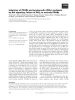
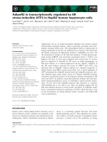
Figure 1 characteristics of the upper leaf epidermis. A)In‘Stacked’ images of upper epidermal cells of the Arab idopsis thalian a line FNR/
EGFP-7-4, all plastids in a given cell are visible (cell boundaries given as grey line). For better display of stromules, the image has been gray
scaled and inverted. Therefore, epidermal plastids are visible in dark grey (arrow) and the larger plastids from the palisade parenchyma appear
light grey (arrowhead). Size bar corresponds to 10 μ m. B) Histogram of cell size in the upper epidermis, illustrating the huge variety of cell sizes
and the predominance of small cells in this tissue. Values given on x-axes are upper limits of size classes. The visible surface area, defined by the
lateral cell walls, was used as a measure of cell size, (see A). C) Scatter plot and linear regression line of plastid number vs. cell size showing the
strong linear correlation between both variables (r
2
0.8016; p value < 0.0001). This underlines that cells of the epidermis can be very different in
cell size and plastid number. D) Plot and linear regression line of stromule frequency vs. cell size class suggesting no significant correlation
between the two parameters. Because of unequal class size, ‘ > 44376’ has not been considered in the regression test. E) Box plot of stromule
frequency found in 109 independent samples of untreated upper leaf epidermis. Specific parameters: maximum = 0.34, 90% percentile = 0.26,
75% percentile = 0.21, median 0.18, 25% percentile = 0.13, 10% percentile = 0.08, minimum 0.02.
Schattat and Klösgen BMC Plant Biology 2011, 11:115
/>Page 3 of 10
exposure to the sucrose solution leads to slowly decreas-
ing SF. Control leaf samples were treated as described
above except that APW lacking sucrose was used for
infiltration. In these samples, only marginal increase in
SF compar ed to non-infiltrated samples was observed
(Figure 2A) demonstrating that the presence of sucrose,
and not the vacuum treatment of the l eaf tissue, is
responsible for stromule induction.
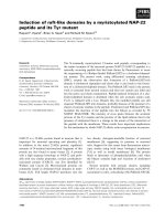
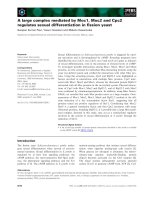
Figure 2 change in stromule frequency in response t o sugar exposure. A) Line plots illustrating changes in SF over time after vacuum
infiltration of either 40 mM sugar solution (sucrose, mannitol, sorbitol, fructose or glucose; depicted as black line), or buffer control (APW,
depicted as grey line). Error bars represent the 99% confidence intervals. For better comparison, values of the buffer control were plotted along
with the sugar treatments. For absolute SF values see additional file 1C. B-C) ‘Stacked’ and inverted epifluorescence images showing leaf
epidermal plastids (black) either 0 h (B) or 2 h (C) after infiltration of 40 mM glucose solution. Note the significantly higher number of plastids
having stromules in the image taken at 2 h (C). Size bar corresponds to 10 μm. D) Line plot depicting changes of stromule frequency induced
by different sucrose concentrations (1 mM, 10 mM, 40 mM, and 80 mM) as well as by the buffer control (APW). The plots show that increase of
sucrose concentration above 40 mM does not result in stronger stromule induction. Error bars represent the 99% confidence intervals. Values for
40 mM and APW have been taken from previously shown experiments (A). E) Line plots with 99% confidence intervals showing the time-course
of increase in SF in the presence or absence of translational inhibitors. Leaf samples were pretreated for 1 h (-1 h to 0 h) with APW
supplemented with either cycloheximide, DMSO, streptomycin, or spectinomycin. At time point “0h”, all buffers were additionally supplemented
with 40 mM sucrose and incubated for additional 3 h. For further details see the legend to A of the same figure.
Schattat and Klösgen BMC Plant Biology 2011, 11:115
/>Page 4 of 10
Like other sucrose-induced physiological reactions, e.g.
anthocyanin accumulation in Arabidopsis thaliana seed-
lings or alp ha-amylase induction in barley embryos
[15,16], the increase in SF is concentration dependent.
Infiltrating tissue sampleswithsolutionsof1mM,
10 mM or 40 mM sucrose suggests correlation of SF
and sucrose concentration of the infiltration medium
(Figure2D;forabsolutevaluesofstromuleindexand
stromule frequency see additional file 1 panel E and F).
However, a further increase in sucrose concentration
(> 40 mM) did not lead to an additional increase in SF
suggesting a kind of saturation effect. Furthermore, we
have never observed SF exceeding 60%, i.e. even under
“optimal” inducing conditions approximately 40% of all
plastids in a sample are yet devoid of visible stromules.
Induction of stromule formation is not an osmotic effect
In order to elucidate whether s ucrose induction of stro-
mule formation is merely an osmotic effect, the expe ri-
ments were repeated with solutions of mannitol and
sorbitol, which are both not part of the primary carbon
metabolism and are routinely used to apply osmotic stress
[17-20]. In neither case did vacuum infiltration result in
significant increa se in SF (Figure 2A). Instead, infiltration
of sorbitol led even to a mild inhibition of stromule forma-
tion demonstrating that changes in osmotic conditions
cannot be the reason for the stromule induction observed
in the presence of sucrose. This suggests that stromule
formation can be elicited specifically by sugars present
during primary carbon metabolism.
However,notevenallofthesesugarsarecapableof
inducing stromule formatio n. If sucro se is replaced by
either glucose or fructose in the infiltration experiments
only glucose was able to induce stromule formation,
whereas fructose treatment did not le ad to any change in
SF (Figure 2A). Although the process is not well under-
stood, it i s widely accepted (based on s upporting experi-
mental evidence, summarized in [21]) that extracellular
fructose generated by sucrose-cleavage in the apopl ast is
imported into the cell. Considering that intracellular fruc-
tose can be converted to phosphorylated glucose, stromule
induction is probably not caused by an overall increase in
cell metabolic activity but likely depends on specific meta-
bolic and/or signaling pathways.
Stromule formation requires de novo protein synthesis in
the cytosol
The apparent influence of metabolic activity on stromule
formation was further analyzed by sucrose induction
experiments performed in the presence of inhibitors of
protein biosynthesis. If the sucrose solution is addition-
ally supplemented with cycloheximide (CHX), which
inhibits the activity of 80S ribosomes and thus is used to
prevent translation in the cytosol [22,23], we observed
complete inhibition of stromule formation (Figure 2E). In
contrast, control experiments performed with sucrose
solutions supplemented with 0.03% DMSO, the solvent
of CHX, did not show any inhibitory or inducing effect
(Figure 2E) confirming the specificity of this reaction. On
the other ha nd, neither streptomycin nor spectinomycin,
which are used to prevent translation within plastids by
inhibition of the 70S ribosomes [22,24-26], affected
sucrose-triggered stromule induction (Figure 2E). This
indicates that de novo synthesis of nuclear encoded pro-
teins but not of those encoded by the plastid genome is
required to mediate the signal from apoplastic sucrose
accumulation to stromule formation.
Sucrose-dependent stromule formation is observed also
in the palisade parenchyma
In order to examine if fully developed chloroplasts are also
compe tent for stromule formation following sucrose and
glucose treatment, the image stacks obtained in the experi-
ments shown in Figure 2A were additionally screened for
the presenc e of palisade parenchyma cells. This photo-
synthetically act ive tissue carries fully developed chloro-
plasts. Indeed, we detected in the images taken for sucrose,
glucose, APW and sorbitol treatments not only sufficient
amounts of chloroplasts but found that those chloroplasts
showed stromu le induction characteristics i ndistinguish-
able from those of the epidermal plastids. While solutions
of sucrose and glucose led to pronounced induction of
stromule formation, neither sorbitol nor the buffer control
had any stimu latory effect (Figure 3A; for absolute values
of stromule index and stromule frequency see additi onal
file 3 panel A and B). Even the maximal SF determined for
epidermal cells after sucrose induction (60% in single treat-
ments, for the mean of absolute SF values see additional
file 1 panel D and F and compare with additional file 3
panel B) was observed for the ch loroplasts of the photo-
synthetically active cells. It should be noted though that
the detection of stromules in these cells was complicated
by the dense packing o f most chloroplasts. Furthermore,
many chloroplasts of parenchyma cells in the field of view
were not captured in the images and could thus not be
considered for estimating stromule frequency. Both factors
might hav e contribut ed to the relatively large confidence
intervals. However, our data still clearly demonstrate that
stromu le induction by selected sugars is not restricted to
the plastids found in the upper leaf epidermis and suggests
that the mechanism leading to stromule formation is con-
served amon g diverse plastid types.
Discussion
In the present study, we aimed to establish an experi-
mental system facilitating the repro ducible induction o f
stromule formation in living plant tissue in order to
make these enigmatic structures better accessible to
Schattat and Klösgen BMC Plant Biology 2011, 11:115
/>Page 5 of 10
sys tematic investigation. A possible connection between
extracellular sugars and st romule formation has be en
suggested by several reports concerning high stromule
frequency in heterotrophic cell cultures and sink tissues.
Using the upper leaf epidermis of a transgenic Arabi-
dopsis t halia na line harboring green f luorescent plastid
stroma as model tissue, we addressed the influence of
extracellular sugars on stromule formation.
Stromule formation is specifically induced by sucrose and
glucose
We found that formation of stromules is specifically
induced only by a subset of sugars generated in plants.
While vacuum infiltration of either sucrose or glucose
leads to a significant increase i n SF, an inducing effect of
fructose or mannitol cannot be observed. Infiltration of
sorbitol leads even to a mild inhibition of stromule forma-
tion. Thus, our data suggest that stromule formation is
most likely due to neither osmotic effects nor the result of
the presence of metabolizable sugars in general. Instead, it
seems that specific signaling pathways involving sucrose
and/or glucose play a role in the induction process.
The role of sucrose in signal transduction is difficult to
evaluate despite the fact that there is strong evidence for
sucrose-specific intracellular and extracellular s ensing
mechanisms operating in plants [15,27]. Since sucrose is
efficiently cleaved into fructose and glucose, both in the
apoplast and in the cytosol by invertases and sucrose
synthase, the signaling function of sucrose is difficult to
distinguish from that of its cleavage products - glucose or
UDP-glucose. Glucose sensing, on the other hand, is
already understood in some detail. In particular, the
intracellular enzyme hexokinase1 (HXK1) has been iden-
tified as a key player in this process. Beside its enzymatic
activity, HXK1 is an important glucose sensor. Isoforms
of this enzyme are present within plastids as well as
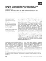
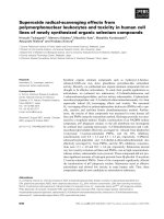
Figure 3 response of palisade parenchyma plastids to sugar exposure . A) Increase of stromule frequency in mesophyl l cells after vacuum
infiltration of either APW or APW supplemented with 40 mM glucose, 40 mM sucrose, or 40 mM sorbitol. Error bars indicate the 99%
confidence intervals. For absolute values of SF see additional file 2 panel B. B-E)’Stacked’ and inverted epifluorescence images showing leaf
tissue either at 0 h (B), 1 h (C), 2 h (D) or 3 h (E) after infiltration of 40 mM glucose solution. The plastids of epidermal cells appear in dark, while
the larger mesophyll chloroplasts appear brighter. Note the increasing proportion of plastids in both tissues that form stromules. The asterisk
highlights mesophyll chloroplasts with stromules. Size bar corresponds to 10 μm.
Schattat and Klösgen BMC Plant Biology 2011, 11:115
/>Page 6 of 10
associated with mitochondria. The latter isoform is also
found in the nucleus where it is part of a protein complex
involved in gene regulation [28]. In addition to HXK1,
further potential glucose sensors have been reported,
which alternatively or additionally might be involved in
glucose induced stromule formation [27].
At this stage, it is not known if sucrose and glucose can
act as independent signals for stromule formation or if the
sucrose induction observed is caused by the release of glu-
cose after sucrose cleavage. Likewise, the question remains
to be answered as to whether the stromule inducing signal
is sensed extracellularly or intracellularly.
It s hould be kept in mind that changes in extracellular
sugar levels might not o nly influe nce the carbohydrate
metabolism of a cell but may be a cause of stress for the
plant cells potentially leading to the induction of stress sig-
naling pathways [29]. Further experimental evidence is
the refore required to substantiate the presu med interde-
pendence of st romule formation and carbohydrate meta-
bolism. Hence, our next experiments will address the
question if glucose and sucrose generate independent stro-
mule inducing signals and if internal changes in sugar
levels are sufficient to change stromule frequency (making
use, for example, of non-metabolizable glucose and
sucrose analogues as well as mutants with altered intracel-
lular sucrose and glucose levels).
Stromules may support metabolite exchange
Although several possible functions for stromules have
been suggested and discussed [5], the final role of stro-
mules in plant cells re mains still enigmatic. The observa-
tion of stromules or other envelope protrusions being in
direct contact with mitochondria and peroxisomes led to
the suggestion that formation of envelope protrusions, like
stromules, supports photorespiration [30-33] by increasing
the interactive surface between the organelles and, in turn,
facilitating efficient metabolite exchange. However, experi-
mental evidence that these organellar connections become
more fre quent under photorespiratory conditions, which
would support this assumption, is yet missing.
Alternatively, the i ncrease in interactive plastid surface
by stromule formation may have more gener al c onse-
quences on the int eraction of plastids with the cytosol or
other organelles, which might be particularly relevant
under conditions of increased demand of metabolite
import or exp ort across the plastid envelope membranes
[2,5]. Indeed, our results demonstrating stromule induc-
tion by sucrose and glucose seem to support this hypoth-
esis. Apoplastic glucose and/or sucrose are particularly
prominent in sink tissue and heterotrophic cell cultures.
The non- or less-photosynthetically active plastids of these
cells import large amounts of glucose-6 phosphate from
the cytosol in order to generate the ATP and NADPH
needed to fulfill their metabolic functions, which in turn
originates from extracellular sucrose or glucose pools. On
the other hand, triose phosphates, which are simulta-
neously produced in this process, are exported back into
the cytoso l. This continuous need for import and export
of metabolites in heterotrophic tissue might explain the
high stromule abundance in BY2 cells as well as in non-
green tissue like ripening tomato fruits and dark grown
seedlings. Furthermore, it could explain why chloroplasts,
which generate ATP and NADPH by photosynthetic pro-
cesses, are reported to show generally lower stromule fre-
quencies than non-green plastids [2]. The fact that
chloroplasts develop stromules t o a similar extent as epi-
dermal plastids after vacuum infiltration of glucose or
sucrose seems to be contradictory at first glance. However,
it is well established that under high sugar conditions
source activity is suppressed and sink activities are trig-
gered [34]. Naturally, this change occurs during fruit
development [35], a process that in tomato fruits goes
along w ith an increase in stromule frequency and length
[9]. Furthermore exposure to extracellular glucose and
sucrose induces major changes in gene expression [36,37].
Such a change might thu s take place also by our sucrose
and glucose treatments, since the cycloheximide expe ri-
ments demonstrate the requirement of de nov o protein
synthesis for stromule induction.
Conclusions
While u p to now only speculations about stromule
related processes w ere possible, the present study pro-
vides experimental evidence, which suggests a possible
involvement of stromules in carbohy drate me tabolism.
This supports the idea of stromules being involved in
optimizing metabolite exchange. The stromule inducing
capacity of glucose and sucrose, important metabolites
and signal molec ules, provides experimental evidence for
the involvement of a typical sugar sensing m echanism in
stromule regulation. However, the sugar sensing mechan-
ism and signaling cascades involved remain still unknown
and require further investigation. Our model system, th e
upper leaf epidermis of Arabidopsis thaliana,maypro-
vide a useful tool for solving these questions.
Methods
Chemicals and solutions
All chemicals were purchased from Sigma-Aldrich (Dei-
senhofen, Germany), Roth (Karlsruhe, Germany), or
Serva (Heidelberg, Germany). As buffer for dissolving
and diluting sugars and inhibitors, artificial pond water
(APW) [38] was used. All solutions were prepared imme-
diately before use.
Microscopy, hardware and software
For imaging of EGFP fluorescence, an Axioscop 2 upright
microscope (Carl Zeiss, Jena, Germany) operating in
Schattat and Klösgen BMC Plant Biology 2011, 11:115
/>Page 7 of 10
epifluorescence mode (fluorescence filter ‘ endowGFP’
F41-017 purchased from AHF Analysetechnik, Tübingen,
Germany) was used. Image s were captured using either
an Axiocam HRc camera (Carl Zeiss, Jena , Germany) or
a KY-F75 camera (JVC, Japan ). Microscope, camera and
piezo stepper were controlled by either of the frame
grapping software packages Ax ioVision (Carl Zeiss, Jena,
Germany) or DISKUS (Hilgers, Königswinter, Germany).
Plant material, sample preparation and drug treatments
Transgenic Arabidopsis th aliana plants constitutively
expressing the chimeric protein FNR/EGFP, which con-
sists of the chloroplast targeting transit peptide of ferre-
doxin-NADPH-oxidoreductase (FNR) fused to an
enhanced derivative of the green fluorescent protein
(EGFP), were grown on soil at 120 μEinstein m
-2
s
-1
and
60% relative air humidity under a short-day ligh t regim e
(8 h light/16 h dark). For vacuum infiltration, expanding
leaves from 10 - 12 week old plants, which had reached
approximately 75% of the size of mature leaves, were har-
vested. After removing the mid vein, the leaves were cut
into four squares and vacuum infiltrated using a 5 ml or
10 ml syring e and a 2 ml rea ction tu be. The tu be was
filled with 1.5 ml of the respective solution and a 10 ml
syringe was placed on top of the tube. By pulling the
plunger of the syringe, vacuum was applied for not longer
than 2s. Upon release, the resulting negative pressure in
the tissue caused the liquid to flood the intercellular
space. The infiltrated leaves were immediately analyzed
or further incubated. For treatment of leaf samples with
inhibitors of translation, samples were infiltrated with
APW supplemented with either 100 μM cycloheximide,
100 μgml
-1
spectinomycin, or 100 μgml
-1
streptomycin
and incubated for one hour in darkness. Then the solu-
tions were replaced by APW supplemented with 40 mM
sucrose in addition to the respective inhibitor. As a sol-
vent control for cycloheximide treatment, APW was sup-
plemented with DMSO at 0.0 3%. Each experiment was
performed at least three times with leaves of different
plants.
Imaging and data processing
After vacuum infiltration leaf squares were either immedi-
ately analyzed (time point 0 h) or incubated at room tem-
perature in the dark for the given time periods (1, 2, or
3 h). For each time point, epidermal plastids of 6 indivi-
dual leaf sectors were imaged by capturing an image series
along the z-axes. The resulting image stac k was further
processed using the software package AxioVision (Carl
Zeiss, Jena, Germany). Image stacks were processed into
one ‘stacked’ 2D image with the help of CombineZP [39]
as described previously [40]. After import of the stacked
images into the ImageBrowser package (Carl Zeiss, Jena,
Germany), plastids with and without stromules were
marked following a color code. The resulting image layer,
which con sisted solely of mark ings, w as exported as an
image file. Markings in these images were automatically
counted u sing the Phot oshop plug-in F ovea Pro 4 (Rein-
deer Graphics, Asheville, USA). Data files produced with
FoveaPro 4 were analyzed with Excel (Microsoft, Red-
mond, Washington, USA).
Calculating stromule frequency
The values for SF were calculated as follo wed. For a time
point of a time course experiment image stacks at 6 differ-
ent spots per leaf square were taken as described (captur-
ing approx. 250 epiderm al plastids per spot, i.e. approx.
1500 plastids per leaf square). For each leaf square stro-
mule frequencies of the six spots were calculated resulting
in six SF values for each leaf square (for estimating the SF
in the palisade parenchyma, only chloroplasts which were
completely visible in the taken image stacks were consid-
ered). Afterwards SF values were arcsin transformed (arc-
sin √ SF) according t o Waters et al. [9] resulting in
stromule index (SI) values. To summarize the data of
experimental repeats, for each experiment the arithmetic
average and the 99% confidence intervals wer e calculated
using SI values (additional file 1 panel C, E and additional
file 3 panel A).
These average SI values and confidence intervals have
been converted back into SF values by calculating the
square of the sinus of the SI values ((sin SI)
2
)foreaseof
conveyance. Bar charts of stromule index as well as back-
transformed data are shown in additional file 1 panel C-F
and additional file 3 panel A and B. For better comparison
of the effect of different treatments, the increase or
decrease in relation to the initial stromu le frequency was
plotted in the graphs presented in Figure 2A, D, E and 3A.
Additional material
Additional file 1: experimental procedure and absolute values of
stromule index as well as stromule frequency in epidermal cells. A)
Depiction of epidermal cell outlines which illustrates the large variety of
cell sizes found in the epidermises of Arabidopsis thaliana. Epidermal cells
were colored according to the respective size class. Stomata that are
shown in gray were not considered. Size bar corresponds to 50 μm. B)
Schematic depiction of the experimental procedure showing sample
preparation, infiltration and data acquisition. C) Bar charts showing upper
epidermal ‘stromule index’ mean values for 40 mM sugar (sucrose,
sorbitol, mannitol, glucose, or fructose) and buffer control (APW)
treatments calculate d as described in Material and Methods. Scale
maximum of y-axes was set to 1.57, which corresponds to a stromule
frequency of 1 (or 100%). Error bars show the 99% confidence intervals
and therefore represent the likelihood of the calculated mean value. D)
By doing the opposite of the mathematical function used for
transforming stromule frequencies into ‘stromule index’, ‘stromule index’
mean values were back-transformed into stromule frequency values. The
same procedure was applied to the 99% confidence intervals. Bar charts
showing both values for each time point are depicted in C. To illustrate
the relation of stromule frequencies to a ‘stromule saturated’ tissue, the
maxima of the y-axes were set to 1 (or 100%). E-F) Absolute stromule
Schattat and Klösgen BMC Plant Biology 2011, 11:115
/>Page 8 of 10
indices and back-transformed stromule frequency values for 1 mM, 10
mM and 80 mM sucrose treatments.
Additional file 2: image series for a sucrose induction experiment.
‘Stacked’, inverted, gray scaled images of time points 0 h (A), 1 h (B), 2 h
(C), 3 h (D) of a 40 mM sucrose induction experiment. Scale bar
corresponds to 10 μm.
Additional file 3: absolute values of stromule index as well as
stromule frequency in palisade parenchyma cells. A and B) Absolute
stromule index and back-transformed stromule frequency values for the
40 mM sorbitol, 40 mM sucrose, 40 mM glucose and APW treatments
based on chloroplasts in palisade parenchyma cells.
Acknowledgements
We thank Jaideep Mathur and Sebastian Schornack for helpful discussions
and comments on the manuscript; Naomi Marty and Michael Wozny for
helping with English wording; Martin Paulmann, Max Paulmann and Armin
Danziger for their kind support in marking plastids. This work was supported
by grants from the state Sachsen-Anhalt (Exzellenznetzwerk
Biowissenschaften).
Author details
1
Laboratory of Plant Development and Interactions; Department of
Molecular and Cellular Biology; University of Guelph; Guelph, ON Canada.
2
Institute of Biology - Plant Physiology, Martin-Luther-University Halle-
Wittenberg, Weinbergweg 10, 06120 Halle (Saale), Germany.
Authors’ contributions
MHS designed and carried out all the experiments and wrote the
manuscript. RBK participated in the experimental design and helped to draft
and write the manuscript. Both authors read and approved the final
manuscript.
Received: 25 April 2011 Accepted: 16 August 2011
Published: 16 August 2011
References
1. Köhler RH, Cao J, Zipfel WR, Webb WW, Hanson MR: Exchange of protein
molecules through connections between higher plant plastids. Science
1997, 276(5321):2039-2042.
2. Hanson MR, Sattarzadeh A: Stromules: Recent Insights into a Long
Neglected Feature of Plastid Morphology and Function. Plant Physiology
2011, 155(4):1486-1492.
3. Gunning BE: Plastid stromules: video microscopy of their outgrowth,
retraction, tensioning, anchoring, branching, bridging, and tip-shedding.
Protoplasma 2005, 225(1-2):33-42.
4. Gray JC, Sullivan JA, Hibberd JM, Hansen MR: Stromules: Mobile
Protrusions and Interconnections Between Plastids. Plant Biol 2001,
3(3):223-233.
5. Hanson MR, Sattarzadeh A: Dynamic morphology of plastids and
stromules in angiosperm plants. Plant Cell Environ 2008, 31(5):646-657.
6. Kwok EY, Hanson MR: In vivo analysis of interactions between GFP-
labeled microfilaments and plastid stromules. BMC Plant Biol 2004, 4:2.
7. Sattarzadeh A, Krahmer J, Germain AD, Hanson MR: A Myosin XI Tail
Domain Homologous to the Yeast Myosin Vacuole-Binding Domain
Interacts with Plastids and Stromules in Nicotiana benthamiana. Mol
Plant 2009, 2(6):1351-1358.
8. Kwok EY, Hanson MR: Microfilaments and microtubules control the
morphology and movement of non-green plastids and stromules in
Nicotiana tabacum. Plant J 2003, 35(1):16-26.
9. Waters MT, Fray RG, Pyke KA: Stromule formation is dependent upon
plastid size, plastid differentiation status and the density of plastids
within the cell. Plant J 2004, 39(4):655-667.
10. Köhler RH, Hanson MR: Plastid tubules of higher plants are tissue-specific
and developmentally regulated. J Cell Sci 2000, 113(Pt 1):81-89.
11. Marques JP, Dudeck I, Klosgen RB: Targeting of EGFP chimeras within
chloroplasts. Mol Genet Genomics 2003, 269(3):381-387.
12. Marques JP, Schattat MH, Hause G, Dudeck I, Klosgen RB: In vivo transport
of folded EGFP by the DeltapH/TAT-dependent pathway in chloroplasts
of Arabidopsis thaliana. J Exp Bot 2004, 55(403):1697-1706.
13. Natesan SKA, Sullivan JA, Gray JC: Myosin XI Is Required for Actin-
Associated Movement of Plastid Stromules. Mol Plant 2009,
2(6):1262-1272.
14. Dinant S, Lemoine R: The phloem pathway: New issues and old debates.
Comptes Rendus Biologies 2010, 333(4)
:307-319.
15.
Solfanelli C, Poggi A, Loreti E, Alpi A, Perata P: Sucrose-specific induction
of the anthocyanin biosynthetic pathway in Arabidopsis. Plant Physiol
2006, 140(2):637-646.
16. Loreti E, Alpi A, Perata P: Glucose and disaccharide-sensing mechanisms
modulate the expression of alpha-amylase in barley embryos. Plant
Physiol 2000, 123(3):939-948.
17. Luo Y, Liu YB, Dong YX, Gao XQ, Zhang XS: Expression of a putative
alfalfa helicase increases tolerance to abiotic stress in Arabidopsis by
enhancing the capacities for ROS scavenging and osmotic adjustment.
J Plant Physiol 2009, 166(4):385-394.
18. Davletova S, Schlauch K, Coutu J, Mittler R: The zinc-finger protein Zat12
plays a central role in reactive oxygen and abiotic stress signaling in
Arabidopsis. Plant Physiology 2005, 139(2):847-856.
19. Arce DP, Godoy AV, Tsuda K, Yamazaki K, Valle EM, Iglesias MJ, Di
Mauro MF, Casalongue CA: The analysis of an Arabidopsis triple knock-
down mutant reveals functions for MBF1 genes under oxidative stress
conditions. J Plant Physiol 2010, 167(3):194-200.
20. Zhu JH, Lee BH, Dellinger M, Cui XP, Zhang CQ, Wu S, Nothnagel EA,
Zhu JK: A cellulose synthase-like protein is required for osmotic stress
tolerance in Arabidopsis. Plant Journal 2010, 63(1):128-140.
21. Büttner M: The monosaccharide transporter(-like) gene family in
Arabidopsis. Febs Letters 2007, 581(12):2318-2324.
22. Mulo P, Pursiheimo S, Hou CX, Tyystjarvi T, Aro EM: Multiple effects of
antibiotics on chloroplast and nuclear gene expression. Functional Plant
Biology 2003, 30(11):1097 1103.
23. Boulter D: Protein Synthesis in Plants. Annual Review of Plant Physiology
1970, 21:91.
24. Burns DJW, Cundliff E: Bacterial Protein Synthesis - Novel System for
Studying Antibiotic Action in-Vivo. European Journal of Biochemistry 1973,
37(3):570-574.
25. Moazed D, Noller HF: Interaction of antibiotics with functional sites in
16S ribosomal RNA. Nature 1987, 327(6121):389-394.
26. Etzold T, Fritz CC, Schell J, Schreier PH: A Point Mutation in the
Chloroplast 16 S Rna Gene of a Streptomycin Resistant Nicotiana-
Tabacum. Febs Letters 1987, 219(2):343-346.
27. Hanson J, Smeekens S: Sugar perception and signaling-an update. Curr
Opin Plant Biol 2009, 12(5):562-567.
28. Smeekens S, Ma JK, Hanson J, Rolland F: Sugar signals and molecular
networks controlling plant growth. Current Opinion in Plant Biology 2010,
13(3):274-279.
29.
Wingler A, Roitsch T: Metabolic regulation of leaf senescence: interactions
of sugar signalling with biotic and abiotic stress responses. Plant Biology
2008, 10:50-62.
30. Sage TL, Sage RF: The functional anatomy of rice leaves: implications for
refixation of photorespiratory CO2 and efforts to engineer C4
photosynthesis into rice. Plant Cell Physiol 2009, 50(4):756-772.
31. Hanson MR, Sattarzadeh A: Stromules: recent insights into a long
neglected feature of plastid morphology and function. Plant Physiol 2011.
32. Holzinger A, Wasteneys GO, Lutz C: Investigating cytoskeletal function in
chloroplast protrusion formation in the arctic-alpine plant Oxyria digyna.
Plant Biol (Stuttg) 2007, 9(3):400-410.
33. Buchner O, Holzinger A, Lütz C: Effects of temperature and light on the
formation of chloroplast protrusions in leaf mesophyll cells of high
alpine plants. Plant Cell Environ 2007, 30(11):1347-1356.
34. Rolland F, Baena-Gonzalez E, Sheen J: Sugar sensing and signaling in
plants: conserved and novel mechanisms. Annu Rev Plant Biol 2006,
57:675-709.
35. Neuhaus HE, Emes MJ: Nonphotosynthetic metabolism in plastids. Annual
Review of Plant Physiology and Plant Molecular Biology 2000, 51:111-140.
36. Gonzali S, Loreti E, Solfanelli C, Novi G, Alpi A, Perata P: Identification of
sugar-modulated genes and evidence for in vivo sugar sensing in
Arabidopsis. Journal of Plant Research 2006, 119(2):115-123.
Schattat and Klösgen BMC Plant Biology 2011, 11:115
/>Page 9 of 10
37. Price J, Laxmi A, St Martin SK, Jang JC: Global transcription profiling
reveals multiple sugar signal transduction mechanisms in Arabidopsis.
Plant Cell 2004, 16(8):2128-2150.
38. Yamada SY, Sonobe S, Shimmen T: Synthesis of a callosic substance
during rhizoid differentiation in Spirogyra. Plant Cell Physiol 2003,
44(11):1225-1228.
39. Hadley A: My software to combine pictures to increase Depth of Field.
2006 [ />40. Schattat MH, Klösgen RB: Improvement of plant cell microscope images
by use of “Depth of Field” - extending software. Endocytobiosis Cell Res
2009, 19:11-19.
doi:10.1186/1471-2229-11-115
Cite this article as: Schattat and Klösgen: Induction of stromule
formation by extracellular sucrose and glucose in epidermal leaf tissue
of Arabidopsis thaliana. BMC Plant Biology 2011 11:115.
Submit your next manuscript to BioMed Central
and take full advantage of:
• Convenient online submission
• Thorough peer review
• No space constraints or color figure charges
• Immediate publication on acceptance
• Inclusion in PubMed, CAS, Scopus and Google Scholar
• Research which is freely available for redistribution
Submit your manuscript at
www.biomedcentral.com/submit
Schattat and Klösgen BMC Plant Biology 2011, 11:115
/>Page 10 of 10