báo cáo khoa học: " Identification and characterization of the Nonrace specific Disease Resistance 1 (NDR1) orthologous protein in coffee" doc
Bạn đang xem bản rút gọn của tài liệu. Xem và tải ngay bản đầy đủ của tài liệu tại đây (1.13 MB, 17 trang )
RESEARC H ARTIC L E Open Access
Identification and characterization of the Non-
race specific Disease Resistance 1 (NDR1)
orthologous protein in coffee
Jean-Luc Cacas
1,4
, Anne-Sophie Petitot
1
, Louis Bernier
2
, Joan Estevan
1
, Geneviève Conejero
3
, Sébastien Mongrand
4
and Diana Fernandez
1*
Abstract
Background: Leaf rust, which is caused by the fungus Hemileia vastatrix (Pucciniales), is a devastating disease that
affects coffee plants (Coffea arabica L.). Disadvantages that are associated with currently developed phytoprotection
approaches have recently led to the search for alternative strategies. These include genetic manipulations that
constitutively activate disease resistance signaling pathways. However, molecular actors of such pathways still
remain unknown in C. arabica. In this study, we have isolated and characterized the coffee NDR1 gene, whose
Arabidopsis ortholog is a well-known master regulator of the hypersensitive response that is dependent on coiled-
coil type R-proteins.
Results: Two highly homologous cDNAs coding for putative NDR1 proteins were identified and cloned from leaves
of coffee plants. One of the candidate coding sequences was then expressed in the Arabidopsis knock-out null
mutant ndr1-1. Upon a challenge with a specific strain of the bacterium Pseudomonas syringae (DC3000::AvrRpt2),
analysis of both macroscopic symptoms and in planta microbial growth showed that the coffee cDNA was able to
restore the resistance phenotype in the mutant genetic background. Thus, the cDNA was dubbed CaNDR1a
(standing for Coffea arabica Non-race specific Disease Resistance 1a ). Finally, biochemical and microscopy data were
obtained that strongly suggest the mechanistic conservation of the NDR1-driven function within coffee and
Arabidopsis plants. Using a transient expression system, it was indeed shown that the CaNDR1a protein, like its
Arabidopsis counterpart, is localized to the plasma membrane, where it is possibly tethered by means of a GPI anchor.
Conclusions: Our data provide molecular and genetic evidence for the identification of a novel functional NDR1
homolog in plants. As a key regulator initiating hypersensitive signalling pathways, CaNDR1 gene(s) might be target
(s) of choice for manipulating the coffee innate immune system and achieving broad spectrum resistance to
pathogens. Given the potential conse rvation of NDR1-depende nt defense mechanisms between Arabidopsis and
coffee plants, our work also suggests new ways to isolate the as-yet-unidentified R-gene(s) responsible for
resistance to H. vastatrix.
Background
The genus Coffea includes about 120 species of subtropi-
cal/tropical woody perennial trees and shrubs (family
Rubiaceae), of which at lea st two species are of world-
wide agr o-economic interest. Nearly 75% of world coffee
production originates from Coffea arabica L., while
about 20% comes from C. canephora Pierre ex A. Froeh-
ner (= C. robusta). Orange coffee leaf rust is considered
to be one of the major plagues affecting C. arabica [1].
The fungus responsible for the disease, Hemileia vasta-
trix Berkeley & Broome, is widely spread throughout cof-
fee-growing countries and can cause severe def oliation,
resulting in substantial berry y ield losses [1,2]. Further-
more, the two current approaches for restricting patho-
gen infection offer limited advantages. First, fungicide
application, although c ost-effective, does not always
result in adequate disease contr ol and, moreover, it has a
* Correspondence:
1
UMR 186 - IRD/CIRAD/UM2 Résistance des Plantes aux Bio-agresseurs,
Institut de Recherche pour le Développement (IRD), BP64501, 34394
Montpellier Cedex 5, France
Full list of author information is available at the end of the article
Cacas et al. BMC Plant Biology 2011, 11:144
/>© 2011 Cacas et al; licensee BioMed Central Ltd. This is an Open Access article distributed under the terms of the Creative Commons
Attribution License ( es/b y/2.0), which permits unrestricted use, distribution, and reproduction in
any medium, provided the origin al work is properly cited.
negative environmental impact. Second, even though sev-
eral varieties of coffee that are resistant to H. vastatrix
have been used for introgression purposes [3,4], such
alternatives are time-consuming and do not provide dur-
able resistance due to the rapid co-evolution of races of
the fungus that harbor new virulence genes [5]. There-
fore, additional met hods to co ntrol leaf rus t in the fields
are required.
H. vastatrix is an obligate biotrophic parasite belonging
to the division Basidiomycetes, order Pucciniales [6].
Following urediospore germination on the abaxial leaf
surface, hyphae grow and penetrate intercellular spaces
of the mesophyll tissue through stomatal openings before
differentiating intra-cellular feeding structures, or haus-
toria. In susceptible coffee plants, the successful pathogen
can complete its dikaryotic cycle within three weeks fol-
lowing infection and reach the ultimate stage, which is
characterized by the formation of a sporulating sorus. In
resistant plants, hyphal invasion is rapidly sensed and
arrested within 2-3 days [7,8]. Based on quantitative and
Mendelian genetic studies [3,4], the occurrence of at least
nine dominant resistance (R) genes in Coffea spp., and a
similar number of fungal virulence genes, have been
inferred. It is thus commonly accepted that the outcome
of coffee/rust interactions, whether the plant resists
pathogen attack (incompatibility) or develops disease
(compatibility), relies on the gene-for-gene model [9],
which has been recently amended [10]. Once delivered
into coffee cells, H. vastatrix effector proteins, and the
intracel lular perturbations that they trigger, are supposed
tobeperceivedbyspecificR-proteins.Therecognition
step promotes the launching of signaling defense path-
way(s) and subsequent resistance. Alternatively, virulent
rust races a re believed to secrete effectors that escape or
even counterac t the host surv eillance system, which
allow for the highjacking of coffee cell metabolism and
tissue colonization [11].
During incompatible interactions with biotrophic patho-
gens, the plant resistance phenotype results from the onset
of a complex and multilayered-defense response, which is
the so-called hypersensitive response or HR [12,13].
Although little is still known about the molecular mechan-
isms that govern resistance to H. vastatrix, several studies
have advanced the case for the existence of a HR-like phe-
nomenon in coffee plants. Resistant varieties that were
inoculated with avirulent fungal strains displayed a mor-
photype that exhibits many HR character istics. These
include rapid host cell death, which is localized at the
infection site and that is associated with fungal hyphae col-
lapse [7,8], callose encasement of haustori a and subsequent
cell wall lignification [8], early oxidative burst [14,15], and
the activation of ty pical defense-related genes [16-18].
In previous work, we performed a suppression subtrac-
tive hybridization-based screening in C. arabica that had
been challenged with H. vastatrix and identified a series
of Expressed Sequence Tags (ESTs) that were regulated
during compatible or incompatible interactions [16,19].
One of these ESTs shared a significant identity with the
coding sequence of the NON-RACE-SPECIFIC DISEASE
RESISTANCE 1 (NDR1) gene. Originally isolated in
Arabidopsis thaliana, NDR1 encodes a small p lasma
membrane-resident protein, the deficiency of which was
found to abolish HR and confer susceptibility to some
fungal and bacterial pathogens carrying specifi c effector
genes [20-22]. Notably, it has been established that
NDR1-driven resistance is dependent on a specific subset
of R-proteins (such as RPM1, RPS2 and RPS5) that are
defined by the presence of a coiled- coil (CC) structure
within their N -terminal parts [23]. Fro m a mechanistic
perspective, the best characterized example illustrating
NDR1 function is the pathosystem involving strain
DC3000::AvrRpt2 of the plant pathogenic bacterium
Pseudomonas syringae pv. tomato (Pst). In this model,
under resting conditions, AtNDR1 indirectly retains the
RPS2 protein on the cyto solic side of the plasma mem-
brane through its interaction with the RPM1-INTER-
ACTING PROTEIN 4 (RIN4), thereby preve nting HR
activation [24]. Upon infection with Pst,thebacterial
protease AvrRpt2 is secreted into the cytoplasm where it
can cleave RIN4, releasing RPS2 and initiating a disease
resistance signaling pathway [25].
In this study, we cloned two C. arabica candidate
cDNAs for NDR1 andanalyzedthededucedprimary
amino-acid sequences. Domain conservation and the
high degree of homology between the coffee proteins and
AtNDR1 led us to undertake a genetic complementation
approach. Using the Arabidopsis ndr1-1 null mutant, we
obtained genetic and molecular evidence that at least one
of our candidate genes is a functiona l NDR1 ortholog.
Both laser-confocal microscopy and biochemical analyses
further suggested that the protein is likely to be attached
to the plasma membrane via a glycosylphosphatidylinosi-
tol-anchor. Based on these data, the possibility that a
NDR1-contingent me chanism could be invoked in R-
gene-mediated resistance to H. vastatrix is discussed.
The impact this result could have in the context of resis-
tance
improvement
is also outlined.
Results
Cloning and analysis of a novel NDR1 sequence homolog
from Coffea arabica
In previous work [19], we used a subtractive hybridiza-
tion approach to identify genes involved in defense/resis-
tanceofcoffeeplants(C. arabica L.)totheorangerust
fungus H. vastatrix. Of the 9 ESTs which were signifi-
cantly up-regulated during HR, one displayed 43% iden-
tity with the canonical NDR1 coding sequence from
A. thaliana. In this study, we focused our efforts on the
Cacas et al. BMC Plant Biology 2011, 11:144
/>Page 2 of 17
coffee candidate for NDR1 gene and isolated two distinct
full-length transcripts by nested RACE-PCR. The corre-
sponding cDNA s were cloned as described in the ‘Meth-
ods’ section (CaNDR1a [GenBank:DQ335596], CaNDR1b
[GenBank:DQ335597]). Open reading frames differed
from one another by only 3 single nucleotide positions
with one of the substitutions being non-silent (F69L).
Both sequences were predicted to encode proteins that
were 214 amino acids long, which shared a calculated
molecular weight of 23.8 kDa and an isoelectric point of
9.58.
Searching for Arabidopsis relatives of our proteins, we
screened the GenBank database by means of the BLAST P
algorithm [26] and retrieved 15 non-redundant hits.
As expected, t he best match appeared to be NDR1 with
42/61% identity/homology. Apart from an unknown
sequence, all identified homologs had been previously
described as members of the NDR1/HIN1-like (NHL) pro-
tein superfamily [27]. NHLs account for a vast class of
plant defense-associated proteins that, within their N-
terminal halves, contain two highly conserved peptide pat-
terns (motifs 2 and 3) and a less conserved one (motif 1)
[28]. Alignment of the proteins, along with the tobacco
HIN1 for c omparison, revealed the position of t he three
motifs within sequences (Figure 1; see also additional file 1
for full length sequence alignment). A phylogenetic analy-
sis using solely the conserved region that is presented i n
Figure 1, and which encompasses the three NHL motifs,
showed that CaNDR1a/b, NDR1, NHL38 and NHL16
formed a group that was distinct from other NHLs (Figure
2a,b). These data indicate that NDR1, NHL38 and NHL16
are the closest Arabidopsis relatives of CaNDR1a/b.
Ectopic expression of CaNDR1a in Arabidopsis ndr1-1 null
mutant restores specific resistance to Pseudomonas
syringae pv. tomato (DC3000::AvrRpt2)
From our in silico analysis, the question arises as to
whether the two identified coffee genes are functional
homologs of AtNDR1 or code for distinct NHL counter-
parts. To answer this question, a genetic complementa-
tion approach was undertaken. Given the high degree of
identity between the two predicted CaNDR1 amino-acid
sequences, we decided to stud y CaNDR1a and expressed
the corresponding ORF under the control of the
CaMV35S promoter in the Arabidopsis ndr1-1 null
mutant. Segregation analysis on a selective medium
allowed for the isolation of single-locus, homozygous
insertion lines (see additional file 2 for segregation
results). T3 lines were then screened by RT-qPCR for
high steady-state levels of transgene transcripts and three
of them were selected for further experiments. The
expression level of CaNDR1a in these lines, designated
T3-1, T3-2 and T3-3, was respectively 92-, 190-, and
714-fold higher than that of the endogenous AtNDR1
gene,whencomparedtoWTCol-0 plants grown under
the exact same conditions.
Previous work has shown that the ndr1-1 null mutant
is incapable of HR activation in response to Pst strain
AtNHL22 38 FLVWII-LQPKNPEFILQDTTVYAFNLS QPNLLTSKFQITIASRNRNSNIGIYYDHLHAYASYR NQQITLASDLPPTYQRHKE 119
AtNHL11 38 FLVSII-LQPKKPEFILQDTTVYAFNLS QPNLLTSKFQITIASRNRNSNIGIYYDHLHAYASYR NQQITLASDLPPTYQRHKE 119
AtNHL12 37 FLVWII-LQPTKPRFILQDATVYAFNLS QPNLLTSNFQITIASRNRNSRIGIYYDRLHVYATYR NQQITLRTAIPPTYQGHKE 118
AtNHL18 35 FLVWVI-LRPTKPRFVLQDATVYAFNLS QPNLLTSNFQVTIASRNPNSKIGIYYDRLHVYATYM NQQITLRTAIPPTYQGHKE 116
AtNHL1 35 LLIWAI-LQPSKPRFILQDATVYAFNVSGN-PPNLLTSNFQITLSSRNPNNKIGIYYDRLDVYATYR SQQITFPTSIPPTYQGHKD 117
AtNHL23 37 LLVWAI-LQPSKPRFVLQDATVFNFNVSGN-PPNLLTSNFQFTLSSRNPNDKIGIYYDRLDVYASYR SQQITLPSPMLTTYQGHKE 119
unknown 7 PIDCAI-LLPSKPRFIFQDVTVFNFNVSGN-PSDLNTPVVQFNLSFRNPNANIRIYYDTLDVYAFYGNG SQQIIIPTPMPSTYQGHKE 92
AtNHL26 44 FLVWLI-LHPERPEFSLTEADIYSLNLTTS-STHLLNSSVQLTLFSKNPNKKVGIYYDKLLVYAAYR GQQITSEASLPPFYQSHEE 126
AtNHL2 72 LILWLI-FRPNAVKFYVADANLNRFSFDP NN-NLHYSLDLNFTIRNPNQRVGVYYDEFSVSGYYG DQRFGSANVSSFYQGHKN 151
NtHin1 62 LVLWLV-LRPNKVKFYVTDATLTQFDLST TNNTIFYDLALNMTIRNPNKRIGIYYDSIEARALYQ GERFDSTNLEPFYQGHKN 142
AtNDR1 31 LFLWLS-LRADKPKCSIQNFFIPALGKDP NSRDNTTLNFMVRCDNPNKDKGIYYDDVHLNFSTINTTKINSSALVLVGNYTVPKFYQGH-K 118
AtNHL38 31 LILWLS-LRAKKPKCSIQNFYIPALSKNL SSRDNTTLNFMVRCDNPNKDKGIYYDDVHLTFSTINTTTTNSSDLVLVANYTVPKFYQGH-K 118
AtNHL16 30 LCLWLSTLVHHIPRCSIHYFYIPALNKSL ISSDNTTLNFMVRLKNINAKQGIYYEDLHLSFSTRINNSS LLVANYTVPRFYQGH-E 113
CaNDR1a 28 LFMWLS-LRGSKPSCSIEDFYVPSLNATDNSTTTRSNHTLYFDFRFKNEMKDKGVGYDDLNLTFFYVQNGS LGIANYTVPSFYQGH-D 112
AtNHL21 63 FILWLS-LRPHRPRFHIQDFVVQGLDQPT GVENARIAFNVTILNPNQHMGVYFDSMEGSIYYKDQR VGLIPLLNPFFQQPT-N 142
AtNHL5 61 FILWIS-LQPHRPRVHIRGFSISGLSRPD GFETSHISFKITAHNPNQNVGIYYDSMEGSVYYKEKR IGSTKLTNPFYQDPK-N 140
AtNHL6 85 IGILYLVFKPKLPDYSIDRLQLTRFALNQD SSLTTAFNVTITAKNPNEKIGIYYEDGSKITVWY MEHQLSNGSLPKFYQGHEN 166
NPNKRIGIYYD
LILWLILRPXKPKFXVQDATV
Motif 1 Motif 2 Motif 3
PFYQGHKN
Ύ
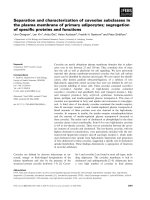
Figure 1 The two coffee candidates for NDR1 protein belong to the NHL family.PutativeArabidopsis orthologs of CaNDR1a/b proteins
were identified by means of the BLAST algorithm using as queries the two deduced coffee amino-acid sequences. The retrieved sequences
were aligned using version 2.0.10 of the Clustal X program [59] and the resulting alignment was then processed online at the BoxShade server
( The conserved region containing the three NHL motifs is presented. The position of the
motifs is indicated with red lines and numbers. An asterik shows the position of the substituted amino-acid residue between the two coffee
proteins (F69L). The full length sequence alignment can be found in Additional file 1. Accession numbers of the genes coding for the
Arabidopsis proteins are as follows: NDR1 [AGI:At3g20600]; NHL1 [AGI:At3g11660]; NHL2 [AGI:At3g11650]; NHL5 [AGI:At1g61760]; NHL6, [AGI:
At1g65690]; NHL11 [AGI:At2g35970]; NHL12 [AGI:At2g35960; NHL16 [AGI:At3g20610]; NHL18 [AGI:At3g52470]; NHL21 [AGI:At4g05220]; NHL22 [AGI:
At4g09590]; NHL23 [AGI:At5g06330]; NHL26 [AGI:At5g53730]; NHL38 [AGI:At3g20590]; unknown, [AGI:At5g05657]. The accession number of the
Nicotiana tabacum Hin1 coding sequence is GenBank: AB091429.1.
Cacas et al. BMC Plant Biology 2011, 11:144
/>Page 3 of 17
AtNDR1
CaNDR1a/b
NtHin1
Class 1
Class 3
AtNDR1
CaNDR1a/b
NtHin1
Class 1
Class 3
(a)
(b)
AtNDR1 1 MNNQNEDTEGGRNCCTCCLSFIFTAGLTSLFLWLS-LRADKPKCSIQNFFIPALGKDP
AtNHL38 1 MTKIDPEEELGRKCCTCFFKFIFTTRLGALILWLS-LRAKKPKCSIQNFYIPALSKNL
AtNHL16 1 -MDRDDAWEWFVTIVGSLMTLLYVSFLLALCLWLSTLVHHIPRCSIHYFYIPALNKSL
CaNDR1a 1 MSDPSSSAGGCCRCCCSFILTSGLTALFMWLS-LRGSKPSCSIEDFYVPSLNATDNS
CaNDR1b 1 MSDPSSSAGGCCRCCCSFILTSGLTALFMWLS-LRGSKPSCSIEDFYVPSLNATDNS
AtNDR1 58 -NSRDNTTLNFMVRCDNPNKDKGIYYDDVHLNFSTINTTKINSSALVLVGNYTVPKFYQG
AtNHL38 58 -SSRDNTTLNFMVRCDNPNKDKGIYYDDVHLTFSTINTTTTNSSDLVLVANYTVPKFYQG
AtNHL16 58 -ISSDNTTLNFMVRLKNINAKQGIYYEDLHLSFSTRINNSS LLVANYTVPRFYQG
CaNDR1a 57 TTTRSNHTLYFDFRFKNEMKDKGVGYDDLNLTFFYVQNGSLG IANYTVPSFYQG
CaNDR1b 57 TTTRSNHTLYFDLRFKNEMKDKGVGYDDLNLTFFYVQNGSLG IANYTVPSFYQG
AtNDR1 117 HKKKAKKWGQVKPLNN QTVLRAVLPNGSAVFRLDLKTQVRFKIVFWKTKRYG-VEVGA
AtNHL38 117 HKKKAKKWGQVWPLNN QTVLRAVLPNGSAVFRLDLKTHVRFKIVFWKTKWYRRIKVGA
AtNHL16 112 HEKKAKKWGQALPFNN QTVIQAVLPNGSAIFRVDLKMQVKYKVMSWKTKRYK-LKASV
CaNDR1a 111 HDKKARRKELVQTYGVPWEAAYRAVSNGSTVTFRVGLTTRVRYKILFWYTKRHG-LKVGA
CaNDR1b 111 HDKKARRKELVQTYGVPWEAAYRAVSNGSTVTFRVGLTTRVRYKILFWYTKRHG-LKVGA
AtNDR1 174 DVEVNGDGVKAQ KKGIKMKKSDSS-
AtNHL38 175 DVEVNGDGVKANEKEIKMEKSNFWKTHGYWSEFGFDDDVELTGDGAQKKGSKTKKSDSS-
AtNHL16 169 NLEVNEDGATKVKDK EDGIKMKISDSSP
CaNDR1a 170 NVDVNNSGKKVN KKGIRLKSGAPES
CaNDR1b 170 NVDVNNSGKKVN KKGIRLKSGAPES
AtNDR1 198 FPLRSSFPISVLMNLLVFFAIR
AtNHL38 234 LPLRSSFPIFVLMNLLVFFAIR
AtNHL16 197 QRLTFFQVCFSIICVLMNWLIFLAIR
CaNDR1a 195 VRCPGLVVISIALYFLVLLL
CaNDR1b 195 VRCPGLVVISIALYFLVLLL
*
Figure 2 NDR1, N HL16 and NHL3 8 are the closest Arabidopsis relatives of CaNDR1 proteins. (a) Phylogenetic relationships between
CaNDR1 proteins and their Arabidopsis relatives. The phylogenetic tree was built using the Phylowin freeware using the neighbor-joining
method [60]. Sequence alignment was previously obtained using version 2.0.10 of the Clustal X program [59]. (b) Full length sequence alignment
of CaNDR1a/b and the Arabidopsis protein NDR1, NHL16 and NHL38. Locations of the three NHL motifs within sequences are indicated with red
lines above the alignment. The star indicates the amino acid residue substituted between both coffee NDR1 sequences. For sequence accession
numbers, see legend of Figure 1.
Cacas et al. BMC Plant Biology 2011, 11:144
/>Page 4 of 17
DC3000::AvrRpt2 carrying an AvrRpt2 cassette-contain-
ing plasmid [20,21]. Conversely, a high overexpression
level of AtNDR1 in the Col-0 genetic background was
found to conf er enhanced disease resistance to strain
DC3000 [22]. The behavior of our overexpressor lines
was thus examined in response to the two isogenic bac-
terial strains (DC3000::AvrRpt2 and DC3000) by record-
ing macroscopic symptoms and following in planta
bacterial growth over a four-day period. Although Cop-
pinger et al. [22] had previously reported the occurrence
of HR-like lesions in non-inoculated Arabidopsis trans-
genic lines overexpressing AtNDR1,nosuchlesions
were observed in our non-inoculated T3 lines. Although
the three genotypes developed disease symptoms in
response to DC3000 (Figure 3a), T3-2 and T3-3 lines
were less susceptible than the ndr1-1 mutant plants, as
shown by the leaf bacterial contents at four days p ost-
inoculation (dpi) (Figure 3c). Upon a challenge with
DC3000::AvrRpt2, WT plants exhibited typical hypersen-
sitive lesions located within the infiltrated area, whereas
ndr1-1 mutants showed disease-like symptoms charac-
terized by tissue yellowing, which spread outside the
inoculated zone (Figure 3a). As expected, such striking
difference s between the WT and ndr1-1 genotypes were
closely correlated with lea f bacterial amoun ts. For
instance, as early as 2 dpi, mutant leaves already showed
a 10-fold increase in the concentration of bacteria com-
pared to WT leaves (Figure 3b). More importantly,
when inoculated with strain DC3000::AvrRpt2,allthree
CaNDR1a-expressing lines presented a HR-like pheno-
type (Figure 3a) that was associated with bacterial levels
statistically comparable to that of WT plants (Figure
3b). Furthermore, expression of the coffee transgene in
the Arabidopsis mutant had no significant impact on
the RPS4-coordinated HR that had been previously
shown to be independent of AtNDR1 [23] (Additional
file 3). Altogether, these results provide genetic evidence
that CaNDR1a functionally and specifically comple-
ments the ndr1-1 mutant.
The mature CaNDR1a protein is C-terminally processed
The Arabidopsis NDR1 protein undergoes several post-
translational modifications, including mult iple glycosyla-
tions and C-terminus processing. The latter cleavage
removes a small portion of the protein, thereby freeing an
amino-a cid residue known as a ω-site (Figure 4) that was
proposed to be modified by covalent binding to a glycosyl-
phosphatidyl-inositol (GPI)-anchor [22]. In accordance
with the cognate role of AtNDR1 in disease resistance sig-
nalling [23], GPI anchoring is usually encountered in
eukaryotic plasma membrane-resident proteins and allows
for the cell surface-tethering phenomenon [29]. Although
there is no established consensus sequence of GPI-anchor
attachment sites, prediction algorithms are available
online. Using the Big-Pi Plant Predictor [30,31], we identi-
fied two putative overlapping cleavage sites in the primary
amino-acid sequence of CaNDR1a (Figure 4), with resi-
dues S189 and G190 being strong ω-site candidates (with
P-values of 2.48 × 10
-6
and 2.76 × 10
-5
, respectivel y).
Furthermore, CaNDR1a and its Arabidopsis ortholog
share common structural fe atures that are believed to be
necessary for GPI attachment by the transamidase com-
plex in endoplasmic reticulum (ER) membranes [31].
Directly downstream of the potential ω-residuesisa
region predicted to encom pass a short polar spa cer, fol-
lowed by a hydrophobic tail. An uncleavable signal peptide
(1-39) comprising a potential transmembrane domain (16-
32) was also predicted with a high probability of occur-
rence (P = 0.867) using SignalP-3.0 software [32,33]. As
previously suggested [22], this N-terminal signal sequence
might be required for the protein to enter the ER network
and travel through the secretory pathway.
Based on this in silico analysis, we decided to investigate
the possibility of C-terminus processing for CaNDR1a. To
this end, a doubly-tagged CaNDR1a version (HA-CaN-
DR1a-His) was created (Figure 5a) and transiently
expressed in tobacco leaves. We reasoned that, if the CaN-
DR1a protein is cleaved in tobacco cells, the loss of its C-
terminus should be easily visualized upon immunoblotting
by the absence of a His-specific signal, whereas the proof
that the protein is synthesized would be provided by the
presence of a HA-specific signal.
Two to three days post-infiltration with an Agrobacter-
ium strain, which was dedicated to the expr ession of the
HA-CaNDR1a-His construct, protein extracts prepared
from fresh tissues were resolved by SDS-PAGE and immu-
noblotted using either HA- or His-specific antisera as
described in the ‘Methods’ section. Immunoblot conditions
were tested using a N-terminally HA-tagged CaNDR1a
(HA-CaNDR1; Figure 5a) and C-terminally His-tagged
Bax Inhibitor 1 (BI1-His) versions as controls. S ix inde-
pen dent experiments including independent Agrobacter-
ium infiltrations and protein extractions were carried out.
Using anti-HA antibody, only one major band was detect-
able in lanes loaded with NDR1 samples (Figure 5b, lanes
3-6), whereas no specific signal was visualized in lanes
loaded with negative control samples (Figure 5b, lanes 1, 2
& 7). Although the nucleotide sequences of HA-CaNDR1a
and HA-CaNDR1a-His code for proteins with predicted
molecular weights averaging 25-26 kDa, the detected pro-
teins migrated to approximately 45 kDa under denaturat-
ing conditions. Such an apparent discrepancy is not
surprising based on previous work. Coppinger and cowor-
kers [22], indeed, showed that the native AtNDR1 protein
resolved by SDS-PAGE displays a mass of about 48 kDa
instead of the predicted 24.6 kDa. These authors further
demonstrated that the protein regains its theoretical size
when translated in vitro without the machinery dedicated
Cacas et al. BMC Plant Biology 2011, 11:144
/>Page 5 of 17
ŶĚƌϭͲϭ͗͗ĂEZϭ
ŽůͲϬ
ϯϬϬϬ
ϯϬϬϬ͗͗
ǀƌZƉƚϮ
ŶĚƌϭͲϭ
ϭϬ
ϵ
ϴ
ϳ
ϲ
ϱ
ϰ
>ŽŐ;&hͬŵ>ͬŐ&tͿ
;ĂͿ
;ďͿ
ϭϬ
ϵ
ϴ
ϳ
ϲ
ϱ
ϰ
>ŽŐ;&hͬŵ>ͬŐ&tͿ
;ĐͿ
ϯϬϬϬ͗͗ǀƌZƉƚϮ
ϯϬϬϬ
A
BBBB
β
ββ
β
β
ββ
ββ
ββ
ββ
ββ
β
α
αα
α
α
αα
α
αβ
αβαβ
αβ
αβ
αβαβ
αβ
β
ββ
ββ
ββ
β
AAA AA
Figure 3 The coffee gene CaNDR1a functionally complements the Arabidopsis ndr1-1 null mutant. Bacterial solutions were hand-infiltrated
into leaves with syringes as described in the ‘Methods’ section. (a) Representative symptoms triggered by the virulent (DC3000) and avirulent
(DC3000::AvrRpt2) Pst strains. A 2 × 10
7
cfu mL
-1
inoculum was used for this experiment, which was conducted twice. Pictures were taken 7 days
after inoculation. (b) and (c) Bacterial growth was monitored in planta by assaying leaf samples 0, 2, and 4 days post-inoculation. CaNDR1a-
expressing lines (T3-1, T3-2 and T3-3), like the WT plants, are resistant to Pst DC3000::AvrRpt2, whereas ndr1-1 mutants are susceptible. Expressing
CaNDR1a in the ndr1-1 genetic background increased resistance to strain DC3000, as shown by significant reductions in leaf bacterial populations
in lines T3-2 and T3-3 at 4 dpi. A 2 × 10
5
cfu mL
-1
inoculum was used for this experiment and the experiment was conducted twice. Means and
standard errors (4 biological replicates) are shown for a representative experiment. Different letters indicate a significant difference at 2 dpi
(Roman letters) or 4 dpi (Greek letters), as determined by ANOVA of square-root transformed data followed by a Student-Newman-Keuls (SNK)
test (a < 5%). No significant difference in leaf bacterial concentration was observed among Arabidopsis genotypes at T0.
Cacas et al. BMC Plant Biology 2011, 11:144
/>Page 6 of 17
to glycosylation, indicating that the latter post-transla-
tional modification could account for the migration shi ft
of the mature proteins on polyacrylamide gels. Consis-
tently, the CaNDR1a protein, like its Arabidopsis ortholog,
exhibits a significant number of putative glycosylation sites
(Figure 4). Hence, one can assume that our protein
extracts (Figure 5b, lanes 3-6) are likely to contain glycosy-
lated forms of CaNDR1a, the migration behavior of which
is altered on polyacrylamide gels.
Finally, using the same set of samples and anti-His anti-
body, we were unable to de tect HA-NDR1-His protein
(Figure 5b, lanes 4-6), whereas BI1-His protein (31 kDa)
was clearly identified (Figure 5b, lane 7). The latter data
indicate that CaNDR1a is C-terminally processed in
tobacco leaves, which strongly suggests that the protein is
modified by addition of a GPI moiety. Further experiments
are nevertheless needed to confirm this assumption.
CaNDR1a is localized to the plasma membrane
Indirect data support the association of the CaNDR1a
protein with m embranes: (i) the potential post-transla-
tional modification by addition of a GPI-anchor; (ii) a
predicted transmembrane-spanning domain located
within the N-terminal signal peptide (Figure 4), and (iii)
the need of a detergent for the protein to be extracted
from tobacco leaf tissues when transiently expressed
(Additional file 4). Accordingly, the CaNDR1a protein
was predicted to be localized to the plasma membrane
(PM) using ChloroP1.1 and PSORTII software [34,35].
Therefore, in order to assess its subcellular localization, a
GFP6 translational fusion was created (Figure 6a), trans-
formed into leaf epidermal tobacco cells using Agrobac-
terium tumefaciens as the vector, and imaged by confocal
microscopy (as described in the ‘ Metho ds’ section). In
accordance with our working hypothesis, independent
experiments showed a consistent fluorescent pattern deli-
neating cellular contours (Figure 6b, panel i). Such a pat-
tern was also observed (Figure 6b, panel ii) with a PM-
resident protein fused to mCherry f luorophore [36]. In
addition, further experiments where both proteins were
simultaneously expressed in the same cells revealed a sig-
nificant overlap between the GFP6 and mCherry signal s
at the cell surface (Figure 6b, panels iv, v, vi). It is note-
worthy that a few GFP6-CaNDR1a-expressing cells dis-
played not only cell surface labeling, but also internal
fluorescence resembling an ER-like reticulated network
with brighter dots that could represent Golgi structures
(Figure 6b, panel iii).
Because leaf epidermal tobacco cells possess a large cen-
tral vacuole that presses the cytoplasmic compartment
against the PM and cell wall, it is difficult to conclude on
the subcellular localization of CaNDR1a based solely on
' W
ϭϱͲϭϴ ϭϵͲϮϬ ϭϱϲ ϲϭϲ
E,
Ϯ
KK,
dD ,LJĚƌŽƉ
ŚŽďĞ
,LJĚƌŽͲ
ƉŚŝůĞ
ƚEZϭ
'ůLJĐŽƐLJůĂƚĞĚƌĞŐŝŽŶ
ϭϲͲϭϳ ϭϲ ϭϱϲ ϲϭϲ
^ƉĂĐĞƌ
E,
Ϯ
KK,
ĂEZϭĂ
ůĞĂǀĂŐĞƐŝƚĞ
ƉсϬ͘ϴϵϮ
ƉсϬ͘ϴϲϳ
ϳƉƌĞĚŝĐƚĞĚƐŝƚĞƐ
ϴƉƌĞĚŝĐƚĞĚƐŝƚĞƐ
^ ^
ȦȦнϭȦнϮ
^ '
/ // ///
/ // ///
^ŝŐŶĂůƉĞƉƚŝĚĞ
Figure 4 Structural similarities between the Arabidopsis and coffee NDR1 proteins. Predicted structural domains and motifs of NDR1
proteins are represented. The overall structure of both proteins appears conserved; it is furthermore reminiscent of GPI-anchored proteins [29].
The C-terminus of NDR1 proteins exhibits putative cleavage sites, including the ω-site to which the glycolipid moiety of the anchor is attached.
Domains following the attachment site display the necessary features for proper transamidase activity, the enzyme complex involved in GPI
modifications of proteins and localized to the ER membrane. A putative uncleavable N-terminal signal peptide that might be implicated in ER
targetting is also present in both proteins. TMD indicates a predicted transmembrane domain. The size of each protein domain is indicated as
Arabic numbers. The number of predicted glycosylation sites (in the middle domain, shown in light grey) is also indicated above and below the
proteins. For convenience, the three conserved NHL motifs are shown as hatched regions I, II and III. Predictive models and methods used for
building this scheme are described in the ‘Methods’ section.
Cacas et al. BMC Plant Biology 2011, 11:144
/>Page 7 of 17
our microscopy data. In order to unambigously ascertain
the localization of CaNDR1a, the N-terminally HA-
tagged version of CaNDR1a (Figure 5a) was transiently
expressed in tobacco leaves and purified PM fractions
were directly tested for the presence of the protein by
immunoblotting using HA-specific antisera. Immuno-
blotting of crude extracts (CE) prepared by directly boil-
ing agroinfiltrated tissues in Laemmli buffer indicated
that HA-CaNDR1a proteins were succesfully expressed
in plant cells (Figure 7a). Most importantly, the tagged
version of CaNDR1a was significantly enriched in PM
fractions compared to microsomal ones, as also observed
for the endogenous PM-resident protein PMA2 (Figure
7b). In addition, while no signal was detected when 5, 10
and 15 μg proteins of the soluble fraction (100.000 × g
supernatant) was blotted, a HA-specific band, the inten-
sity of which increased with the amount of total proteins
loaded, was clearly visualized (Figure 7c). Altogether,
these results show that the mature CaNDR1a prote in is
targeted to PM in the tobacco heterologous system,
further suggesting a similar subcellular localization for
the protein in coffee cells.
Identification of a potential homologous RIN4 protein
from coffee plants
The Arabidopsis NDR1 protein has been demonstrated to
physically interact with RIN4 both in a yeast heterolo-
gous system and in pla nta [24]. Searching for RIN4
sequence homologs in the HarvEST
©
Coffea database
resulted in the identification o f a candidate contig from
Coffea cane phora [GenBank: DV705409.1]. The deduced
protein sequence shares a high percentage of identity/
homology (36/53%) w ith the begin ning of our query
sequence, AtRIN4. This region is also highly conserved
within the RIN4 family of proteins (Figure 8a). One of
the two clea vage sites that permit the hydrolysi s of RIN4
upon delivery of the bacterial protease AvrRpt2 into
Arabidopsis cells [25,37,38] is also conserved in the coffee
protein (Figure 8a). In line with our previous data (Fig-
ures 5, 6 and 7), this in silico analysis points to potential
mechanistic conservation of the NDR1 function in Arabi-
dopsis and coffee plants.
Discussion
The Arabidopsis ndr1 locus was identified in the late
1990’s using a forward genetic screen based on the loss
of resistance to the Pst strain DC3000::AvrRpt2 [20,21].
Since then, NDR1 homologous genes have been found by
sequence comparison in other plant species such as Bras-
sica napus [39] and Vitis vinifera [40]. Many sequence
homologs (around 19 non-redundant hits within 11 plant
species) can also be retrieved from the GenBank database
by means of the BLAST P algorithm (data not shown).
However, to our knowledge, our data constitute a novel
report on the identification a nd characterization of a
functi onal NDR1 homolog, despite the plethora of ortho-
logous candidates.
In this study, several lines of evidence indeed demon-
strated that ectopic expression of CaNDR1a coding
sequence was able to rescue the phenotype of the Arabi-
dopsis ndr1-1 null mu tant. Upon infection with DC3000::
AvrRpt2, the three mutant lines expressing the coffee
transgene were found to develop hypersensitive cell
death symptoms that were absent in mutant plants (Fig-
ure 3a). This macroscopic study was further corroborated
by two independent in planta bacterial growth assays
showing that leaf populations of the bacterial pathogen in
our transgenic lines were low and comparable to those of
WT plants (Figure 3b). In addition, high overexpression
level of the coffee CaNDR1a gene in the Col-0 genetic
background was also found to confer enhanced disease
resistance to the DC3000 strain, as previously reported
,Ϯdžϯϱ^ ŶŽƐƚ͘ĂEZϭĂ
Ϯdžϯϱ^ ŶŽƐƚ͘
ĂEZϭĂ
, ,ŝƐ
;ĂͿ
;ďͿ
ϭϮϯϰϱϲϳ
ϱϬ
ϯϳ
Ϯϱ
ďĮ ,
ďĮ ,ŝƐ
ϱϬ
ϯϳ
Ϯϱ
ƉDϯϮ
ǀĞĐƚŽƌ
ƉDϯϮ
ǀĞĐƚŽƌ
Figure 5 The C-terminal end of CaNDR1a is removed from the
mature protein in tobacco. (a) Constructs used for transiently
expressing HA- and HA-His-tagged CaNDR1a proteins in tobacco
leaves. (b) Detection of CaNDR1a-tagged proteins by
immunoblotting. The upper and lower panels show scanned films
corresponding to membranes blotted with anti-HA and anti-His
sera, respectively. For comparison, the same protein extracts were
resolved by SDS-PAGE and subsequently transferred onto both
membranes. Ten micrograms of proteins were loaded in each lane.
Samples contained the main insoluble proteins that were extracted
using SDS as described in the ‘Methods’ section. Lanes 1 & 2,
negative controls (samples prepared from leaves expressing a GUS
protein and non-infiltrated leaves, respectively); lane 3, HA-positive
control, His-negative control (sample prepared from tissues
expressing the N-terminally HA-tagged CaNDR1a protein); lanes 4-6,
samples prepared from tissues expressing the doubly-tagged
CaNDR1a protein (3 independent experiments); and lane 7, HA-
negative control, His-positive control (sample prepared from
Arabidopsis leaves constitutively expressing the C-terminally His-
tagged AtBI1 protein) [56].
Cacas et al. BMC Plant Biology 2011, 11:144
/>Page 8 of 17
when the AtNDR1 gene was overexpressed in A. thaliana
[22].
Importantly, NDR1-driven resistance in A. thaliana is
not restricted to bacter ial pathogen attacks. Two re ports
have demonstrated that the ndr1 mutation renders plants
susceptible to infection by the oomycete Hyaloperonospora
arabidopsidis [20] and the fungus Verticillium longis-
porum [41]. Therefore, given that (i) CaNDR1a is a func-
tional homolog of the Arabidopsis NDR1 gene, and (ii)
transcripts of the former accumulate in coffee leaves
undergoing HR in res ponse to the fung us H. vastatrix
[16,19], it would not be surprising if NDR1 proteins could
regulate the defense signaling pathway(s) leading to coffee
rust resistance. This hypothesis is currently under investi-
gation in our laboratory using a functional approach.
Recently, we also showed that A. thaliana Col-0 plants
display a rapid non-host response to H. vastatri x.This
response is reminiscent of HR in that it prevents haustor-
ium formation and hyphal spread in plant tissues [42].
This work raises the possibility of testing the role of NDR1
in response to the coffee leaf rust in the A. thaliana he t-
erologous system.
As predicted by our bioinformatic a nalysis, imaging of
GFP6-tagged CaNDR1a protein by confocal microscopy
revealed a fluorescent pattern that was consistent with a
plasma membrane localization (Figure 6b, (i)). Colocali-
zation experiments with a PM fluorescent protein marker
also supported this observation (Figure 6b, (iv-vi)).
Furthermore, the need of an anionic det ergent like
sodium dodecyl-sulfate for the HA-tagged CaNDR1a
proteins to be extracted from tob acco leaves (Additional
file 4) indicated an association with membranes. Finally,
our biochemical approach based on the purification of
PM by tw o-phase PEG/dextran partitioning (Figure 7b,c)
Ϯdžϯϱ^
ŶŽƐƚ͘
ĂEZϭĂ'&Wϲ
;ĂͿ
;ďͿ
ƉDϰϯ
ǀĞĐƚŽƌ
;ǀŝͿ;ǀͿ
;ŝǀͿ
;ŝͿ
;ŝŝͿ ;ŝŝŝͿ
ϱϬђŵ
ϭϬђŵ
Figure 6 The CaNDR1a protein is localized at the plasma membrane. (a) Scheme of the construct used for determining the subcellular
localization of CaNDR1a protein. (b) Confocal-laser microscopy pictures illustrating the plasma membrane localization of CaNDR1a: (i) GFP6-
CaNDR1a; (ii) mCherry-labeled protein targeted to the plasma membrane; (iii) GFP6-CaNDR1a, a close-up of the internal labeling observed in a
few cells; (iv), (v) and (vi), colocalization experiments where both the GFP6-CaNDR1a and mCherry-labeled plasma membrane marker were
simultaneously expressed in the same cells. Independent experiments were conducted five times.
Cacas et al. BMC Plant Biology 2011, 11:144
/>Page 9 of 17
clearly demonstr ated the presence of HA-CaNDR1a pro-
teins in tobacco PM fractions. Therefore, it is likely that
the mature CaNDR1a protein resides in the plasma
membrane of coffee cells.
No fluorescent labeling of the organelle corresponding
to a GFP6 spectrum was observed in chloroplasts,
although it had been reported previously for a tagged
version of AtNDR1 [27]. Instead, internal reticulated
labeling reminiscent of the ER network (Figure 6b, (iii))
was observed in a few cases and may correspond to
cells overloaded with the ectopic fluorescent proteins.
Thi s observation is consistent with our results, suggest-
ing that the CaNDR1a protein could be modified by
addition of a G PI moiety to its C-terminal part (Figure
5b). It has been well-described that prot eins tethered to
the cell surface by means of a GPI anchor undergo this
sort of post-translational modification in the ER before
being sorted via the secretory pathway to their final des-
tination, i.e., the plasma membrane.
Usually, GPI-anchored-proteins are also thought to
locate on the apoplasm side of the plasma membrane [43].
In A. thaliana, it has been clearly established that NDR1 is
attached to the plasma membrane through a C-terminal
GPI anchor [22]. It has also been inferred that the N-term-
inal portion of NDR1 lies within the cytoplasm because it
was found to interact with t he cytosolic protein R IN4 in
planta [24]. Since the C-terminal anchor of AtNDR1 is
resistant to cleavage by phospholipase C, these data
further led to the hypothesis that the protein possesses a
transmembrane-spanning domain as a second anchor site.
This was recently corroborated by a modelling study [ 44]
and, in fact, the coffee protein, like its Arabidopsis relative,
was predicted to present a single transmembrane domain
(Figure 4), suggesting a similar, but atypical topology o f
the two counterparts (Figure 8b).
Recently, a new mode of action of NDR1 was revealed
by Knepper et al. [44]. Based on structural homology with
mammalian integrins and the Arabidopsis late embryogen-
esis abundant (LEA) protein 14, known to be involved in
abiotic stress response [45], the aforementioned authors
investigated the possibility that AtNDR1 may control cell
integrity through PM-cell wall adhesions. Besides its well-
characteriz ed role as a key signaling component during
pathogen attack, a broader function for NDR1 is strongly
suggested by the data in mediating primary cellular func-
tions in Arabidospsis through maintenance of PM-cell wall
connections [44]. From these unexpected results, the ques-
tion arises as to whether or not CaNDR1a could perform a
similar function in C. arabica.
Interestingly, upon inoculat ion with DC3000::AvrRpt2,
successful activation of HR required NDR1-RIN4 physical
interaction. Further examination using an alanine-scan-
ning mutagenesis strategy revealed that two amino acid
residues within the N-terminal part of NDR1 were neces-
sary for the interaction [24]. Despite the apparent lack of
conservation of these two amino acid determinants within
the CaNDR1a end (Figure 8c), our results showing that
the coffee gene was able to restore RPS2-me diated resis -
tance in the ndr1-1 mutant tend to prove that CaNDR1a
does interact w ith AtRIN4 in our transgenic lines. Thus,
this raises the possibility that mechanism(s) whereby
NDR1 proteins exert their function could be conserved in
Arabidopsis and coffee plants.
Cons istent with this idea, searching for RIN4 sequence
homologs in the HarvEST
©
Coffea database resulted in
the identification of a candidate contig from Coffea cane-
phora. The deduced protein shows, within its N-terminal
portion, a highly conserved region with the m embers of
the RIN4 family, as well as a putative conserved canonical
AvrRpt2 cleavage site (Figure 8a). Nonetheless, further
experiments are needed to answ er the question as to
120
86
47
34
26
(a)
(b)
μ PM
PMA2
NDR1
CE
(c)
5 10 15
soluble
PM
(μg)
Figure 7 The CaNDR1a protein is enriched in plasma membrane
fraction. The HA-CaNDR1a construct (see Figure 5a) was used for
carrying out two independent experiments that consisted of two
independent agroinfiltrations and plasma membrane (PM) preparations.
A representative experiment is presented in this figure. Agroinfiltration
and immunoblot con ditions are described in the ‘Methods’ section. (a)
Detection of HA-CaNDR1a proteins in Agrobacterium-infiltrated leaf
tissues. Crude extract (CE) was prepared by directly incubating tissues at
95°C for 5 min in 1X Laemmli buffer [57]. (b) Detection of HA-CaNDR1a
proteins and endogenous PM-resident proteins PMA2 in microsomal
and PM fractions. PMA2 is a proton-ATPase pump previously shown to
be localized exclusively at the PM [61]. Membrane was probed using a
specific anti-PMA2 serum [58] in order to check for the purity of the PM
fraction. As expected, PMA2 proteins appeared to be significantly
enriched in the PM fraction versus the microsomal (μ) one, as also
observed for H A-CaNDR1a proteins upon stripping and reprobing of
the same blotting membrane with HA-specific antiserum (Middle
panel). Membranes were also stained with Ponceau S to show the
equal loading between both fractions, i.e. μ and PM (lower panel). (c)
HA-tagged CaNDR1a proteins are not detected in soluble fractions.
Distinct protein amounts of soluble (100.000 × g supernatant) and PM
fractions (5, 10 and 15 μg) were resolved by SDS-PAGE and
immunoblotted using a HA-specific antiserum.
Cacas et al. BMC Plant Biology 2011, 11:144
/>Page 10 of 17
whetherornotCaNDR1a,likeitsArabidopsis ortholog,
could serve as a PM anchor that indirectly recruits R-
protein(s) via its interaction with RIN4-like intermediates
(Figure 8b) [24,46]. Split-ubiquitin and yeast two-hybrid
systems, combined with bimo lecular fluorescence com-
plementation (BiFC), would be useful tools for tackling
this question. This might also be a faster and more
convenient strategy, as opposed to classical genetic
approaches, for the isolation of R-gene (s) conferring
resistance to H. vastatrix.Todate,nocoffeeR-gene( s)
have been isolated despite the efforts of the coffee
research community [3,4]. The reproductive barriers
affecting genetic exchanges between diploid coffee spe-
cies and the allopolyploid C. arabica have thus far pre-
vented the successful isolation of the loci responsible for
resistance to H. vastatrix through a map-b ased cloning
strategy [4].
Conclusions
The functional and biochemical characterization o f the
orthologous NDR1 prote in from C. arabica that we
have carried out represents a cruc ial step towards the
elucidation of the molecular events underpinning resis-
tance to coffee rust. It should help identify new players
in the coffee NDR1-dependent signaling pathway(s) in
the near future, and might thus be crucial for the engi-
neering of tr ansgenic coffee plants with broad spectrum
resistance to H. vastatrix races. The development of effi-
cient techniques to transform and propagate coffee vari-
eties renders these biotechnological approaches feasible
[47,48].
Methods
Plants and growth conditions
Tobacco plants (Nicotiana benthamiana) that were used
for transient expression experiments were grown in a
greenhouse, at 150 μmol/m
2
/s light radiance, with a 14/
10 h, 23/20°C light-dark cycle, and 60% relative
humidity.
Wild-type Arabidopsis thaliana ecotype Columbia
( Col-0), ndr 1-1 null mutants [20], and transgenic lines
expressing CaNDR1a were all grown in a growth cham-
ber under short-day conditions (10 h photoperiod,
100 μmol/m
2
/s light fluency), at 22/20°C day/night with
(a)
At 1 MARSNVPKFGNWEAEENVPYTAYFDKARKTRAPGSKIMNPNDPEYNSDS
Vv 1 MAQRSHVPKFGNWESEENVPYTAYFDKARKGRT-GTKIINPNDPQENPDM
Pt 1 MAQRSHVPKFGNWESEENVPYTAYFDKARKGRT-GGKMINPNDPQENPDL
Gm 1 MAQRSHVPKFGNWDSGENVPYTAYFDKARKGRT-GARIINPNDPEENADL
Mt 1 MTTQRSHVPKFGNWEGEDDVPYTVYFDKARKSRP-GSKMINPNDPEENPDL
Cc 1 LKASKGEFWKMAQRSQVPKFGNWESEEDVPYTVYFDNAMKGKK GSKMNTNDPQEDLDA
At 50 QSQ-APPHPPSS-RTKPEQVDT VRRSREHMRSREESELKQFGDAGG S
Vv 50 FSD-NASEARSPPRTRAEQEES IGQQVTHEHRR P
Pt 50 VSDYAAPDQAPPFRAKAPPEEAAGQGAVRQAHEHRTSREESDLKQFANSPARNENLNRRA
Gm 50 SLDNPSSDHLPPTRPRANSEDQSGKGSLPLED-DPKHFVDSPARHDNVSSRSGSRSHGVG
Mt 51 VLQNSSSDDVIPPKPRVSSENQSEKGTVRLTHNDLQKNKEEGDVKHSVNSPARPGGHGVG
Cc 59 ETK GQKRPEATRAKHVRRTSREDGDLRKSIDSPLHSDAMSQKSANESPHHKQGGLKHG
At 95 SNEAANKRQGRA SQNNSYDNKSPL
Vv 83 STESTHQRQG GKGSSYDS
Pt 110 SYEPAPQRYGGRGPSFGEAHKRPARYSIGSENSMEQSPIHNHARISGRNSGAPSPSWEGK
Gm 109 SAENRRRHSTQSTG SEYSIERSPLHRQARAPGRD SPQWEPK
Mt 111 SADSRRRPSRQSTAS SEYSVERSPLHRQAKTPGRD SPSWEGK
Cc 117 SRKPESEGSKGTDTVR PRHESREEGDLRRPTDSPLRN-ETGNRRTSHD
At 119 HKNSYDGTGKSRPKPTNLRADESPEKVTVVPKFGDWDENNPSSADGYTHIFNKVREER
Vv 101 SHGTPGRSRMKPT RGDESPDKGAAVPKFGDWDENNPSSADGYTHIFNKVREER
Pt 170 NSNDGSHGTPGRSRLRPK GDESPDKGAAVPKFGDWDENNPSSADGYTHIFNKVREEK
Gm 150 NSYDNSQGTPGRSRLRPVN-RGDETPDKGAAVPKFGDWDVNNPSSADGFTHIFNKVREER
Mt 153 STYDSSHGTPGRSRLRPVN-RDDEIPDKSAAVPKFGEWDESDPASADGYTHIFNKVREEK
Cc 164 SPHHRHGGLSAGETPKRVARQSVGSDRSIDQSPLHPHSQVRTGGRGSGVSSPSWERKGSS
At 177 SSGAN VSGSSRTPTHQSSRNPN-NTSS-CCCFGFGGK
Vv 154 QTGAATRVPGMASEPSYQTNRKHN-TSSSKSCCFPWGRK
Pt 227 QIGEG-KMPGMPTESSNAYVRKQTPSDSAKCCCFPWGRN
Gm 209 QGVPG-QVPGTPNERPQ-AIRGQSNDDKVQCCCFAWGGKK
Mt 212 HVAAG-NTPGTPNGRSY-VIRNQPANDKAQGCCFFWGRK
Cc 224 EGGLGLAPSTPGGSRLKSVTRGDETPDHSPAVXKSAIGMRLILH
AvrRpt cleavage site I
AvrRpt cleavage site II
(c)
R
I
N
4
(b)
apoplasm
RPS2
cytoplasm
RPM1
GPI
C-ter
N-ter
1- MSDPSSSAGGCCRCCCSF-18
Ca-NDR1a
At-NDR1
1- MNNQNEDTEGGRNCCTCC-18
1- MSDPSSSAGGCCRCCCSF-18
Ca-NDR1a
At-NDR1
1- MNNQNEDTEGGRNCCTCC-18
NDR1
Figure 8 Putati ve mechanistic conserva tion of NDR1 function. (a) Alignment of RIN4 homologous sequences. The closest sequence
homologs of AtRIN4 [AGI:At3g25070] were aligned with the putative coffee RIN4 protein [harvEST:Coffea:UG5351] using ClustalX [59]. The
positions of the two AvrRpt2 cleavage sites [37,38] are highlighted in red. Accession numbers of the genes coding for the proteins presented in
the figure are as follows: Glycine max Gm [GenBank:ADJ67468]; Medicago truncatula Mt [GenBank:ACJ83941]; Populus trichocarpa Pt [GenBank:
XP_002301798]; Vitis vinifera, Vv [GenBank:CBI33050]. (b) Scheme showing how NDR1 is anchored to the plasma membrane. AtNDR1 indirectly
retains both R-proteins, RPS2 and RPM1, at the plasma membrane via its interaction with RIN4 [24,48]. (c) Comparison of the N-terminal portions
of the two orthologous NDR1 proteins from A. thaliana and C. arabica. Amino-acid residues necessary for the interaction with AtRIN4 are
highlighted in red. Intriguingly, these residues do not seem to be conserved in the coffee sequence.
Cacas et al. BMC Plant Biology 2011, 11:144
/>Page 11 of 17
80% relative humidity. Pathogen challenge conditions are
described hereafter.
Isolation and cloning of CaNDR1a/b cDNA
As previously described [19], the 5’ end of the CaNDR1
cDNA that is referred to as DSS12 had alre ady been
sequenced (Genbank:CO773976). 3’-RACE PCR was thus
conducted to determine the sequence of the full-length
cDNA. Total RNA (1 μg) isolated from C. arabica cv.
caturra leaves that had been challenged with H. vastatrix
for 18 hours were first reverse-transcribed using the Smart
CDS primer and a co mbination of the Omniscript RT
(Qiagen, Courtaboeuf, France) and SMART PCR cDNA
synthesis kits (Clontech, Mountain View, CA, USA).
RACE assays were then performed using specific oligonu-
cleotides designed in the 5’ non-coding region (3R-NDR1,
5’ -CTACTTTGTTCACTGGTAGTCCCTC-3’ ;n3R-N
DR1, 5’-CATAATACTTCACCGGAGAACCACC-3’) and
the 5’PCR Smart primer (Clontech). The resulting 1-kb
PCR product was cloned into the pGEM-Teasy vector
(Promega, Charbonnières-les-bains, France) and finally
sequenced (Genome Express, Grenoble, France).
Constructs
To assess the complementation of the Arabidopsis null
mutant ndr1-1 [20], the open reading frame of CaNDR1a
was cloned into the binary vector pCAMBIA 1305.1
(Cambia, Brisbane, Australia) downstream of the strong
and constitutive 35S promoter of the cauliflower mosaic
virus. For this purpose, the iud gene was removed from
the vector by restriction digestion with BglII and BstEII
enzymes. The coding sequence of CaNDR1a was amplified
by PCR (DAP Goldstar DNA polymerase, Eurogentec,
Seraing, Belgium) using the corresponding pGEM-T clone
(GenBank:DQ335596) as a template and the following pri-
mers: CaNDR1-BglII 5’-TCAGATCTTATGGACA AAG-
GATGGGGC-3’,andCaNDR1-BstEII 5’-T AGGTCAC
CAAATTAATTCCCAGGAAA-3’.DigestedPCRpro-
ducts were t hen ligated into the binary vector to get the
final construct.
To test the hypothesis that the C-terminal part of CaN-
DR1a is removed from the mature protein, single- and
double -tag construct s were created (Figure 5). CaNDR1a
was amplified by PCR using a high fidelity DNA polymer-
ase according to the manufacturer’sinstructions(Pfu-
Turbo, Stratagene, La Jolla, CA, USA). The following
primer couple was used for directly adding haemaglutinin
(HA) and poly-histidine (His) sequences to the 5’- and 3’-
ends of the PCR products, respectively: CaNDR1-Forward
4, 5’-
CACC ATG TAT CCC TAC GAC GTA CCA GAT
TAT ATG TC AGA CCC CAG CAG CAG TGC-3’ and
CaNDR1-Reverse 3, 5’-CTA ATG GTG ATG GTG ATG
GTG CAA CAG CAG AAC CAA GAA A-3’. The primers
used for obtaining the single HA-tagged version were
CaNDR1-Forward 4 and CaNDR1-Reverse 2 (5’ -CTA
CAA CAG CAG AAC CAA GA-3’). PCR fragments were
then subcloned into the pENTR-D/TOPO vector (Invitro-
gen, Cergy Pontoise, France) and sequenced. To get direc-
tional cloning, the underlined nucleotide sequence was
added to the forward primers. Selected clones were
digested with Mlu-I restriction enzyme (R0198L, NEB,
OZYME, Saint Quentin Yvelines, France) before overnight
recombination with the binary vector pMDC32 [49] using
the LR Clonase II kit (Invitrogen).
The N-terminally GFP6-tagged version was produced to
examine the subcellular localization of CaNDR1a. PCR
products were amplified, subcloned, sequenced and
recombined with the b inary vector pMDC43 [49] as
described above. Prime rs used for the initial PCR step
were as follows: CaNDR1-Forward 1, 5’-
CACC ATG TCA
GAC CCC AGC A GC AGT-3’ and CaNDR1-Reve rse 2.
The vector used for in planta expression of the plasma
memb rane fluorescent marker (mCherry-tagged protein)
was purchased from the Arabidopsis Biol ogical Resource
Center (ABRC) at Ohio State University (Stock # CD3-
1008) [36].
Final binary constructs were all sequenced (Genome
Express) prior to transformation into Agrobacterium
tumefaciens by heat shock or electroporation methods.
Bacterial strain GV3101 was used for transformation of
Arabidopsis plants by floral dipping according to [50].
Strain LBA1119 was used for transient expression experi-
ments in tobacco plants.
Pathogen challenge and growth curve assays
The Pseudomonas syringae pv. tomato (Pst )strain
DC3000 and the isogenic strains expressing the bacterial
effector proteins AvrRpt2 or AvrRps4 were provided by
Dr. Jane Glazebrook (University of Minnesota ) [51]. For
pathogen challenges, bacteria were grown overnight at
28°C under mild shaking in liquid King B medium. Pst
DC3000 bacteria were selected with rifampicin (50 μg
mL
-1
); DC3000::AvrRpt2 and Pst DC3000::AvrRps4 with
rifampicin and tetracycline (10 μgmL
-1
). Bacteria were
collected by centrifugation a nd resuspended at 2 × 10
5
CFU mL
-1
in physiological water (9 g NaCl/L) prior to
inoculation.
Progeny of Arabidopsis ndr1-1 T0 plants (ndr1-1::
CaNDR1a) were screened on half-strength Murashige
and Skoog medium supplemented with 30 μgmL
-1
hygromycin. Transformation of individual resistant seed-
lings was confirmed by PCR using genomic DNA as the
template and CaNDR1a-specific primers (CaNDR1-BglII
and CaNDR1-BstEII). Homozygous single locus inser-
tion lines were then isolated by following segregation of
hygromycin-resistant plants in T2/T3 generations (Addi-
tional file 2). To a ssess ndr1-1 complementation, three
independent T3 lines displaying distinct expression
Cacas et al. BMC Plant Biology 2011, 11:144
/>Page 12 of 17
levels for CaNDR1a (designated T3-1, T3-2 and T3-3)
were challenged with Pst and in planta bacterial growth
was followed over a four-day period (0, 2 and 4 dpi).
Wild type (Columbia, Col-0)andndr1-1 plants were
also inoculated for comparison. Negative controls were
infiltrated with physiological water. Half-leaves (6 to 7-
week-old plants) were hand- infiltra ted with a 1-mL nee-
dleless syringe. Two independent experiments that gave
similar results were carried out. Each experiment com-
prised four replicates that were each performed by dif-
ferent individuals. In one replicate, each plant genotype
(5 plants/genotype) was infiltrated with water or suspen-
sions of Pst DC3000, Pst DC3000::AvrRpt2 or Pst
DC3000::AvrRps4. Upon infiltration, plants were imme-
diatel y placed in a tray covered with a plastic dome that
was removed at 24 hours post-inoculation. Bacterial
growth was monitored as follows. At each time point,
two leaves (per plant) were harvested and ground with a
mortar and pestle. The resulting mixture was serially
diluted in sterile physiological water and plated onto
solid King B medium supplemented with the appropriate
antibiotics. The bacterial population was scored two days
upon plating. Inoculation data were square-root trans-
formed prior to ANOVA and subsequently subjected to
the Student-Newman-Keuls multiple comparison test.
When transformation failed to satisfy assumptions of nor-
mality and homoscedasticity, the non-parametric Kruskal-
Wallis test was used.
Hypersensitive and disease symptoms were also visually
assessed in an independent experiment using higher con-
centrations of bacterial suspension (2 × 10
7
CFU mL
-1
)for
infiltration. Samples from this experiment were also
harvested for RT-qPCR analysis.
RNA extraction, reverse transcription and real time
quantitative-polymerase chain reaction
Expression of AtNDR1 an d CaNDR1a was measured as
previously described [15] with the specific primers (Addi-
tional file 5) that were previously used [42]. Each assay
was conducted in duplicate and included a negative con-
trol without template. The strong and constitutive actin
gene (At3g18780) was chosen as internal control for nor-
malization. Specificity of amplification was estimated by
analyzing melting-temperature curves. Calculations for
gene expression quantification were carried out using the
comparative cycle-threshold method, as described pre-
viously [16].
Agrobacterium tumefaciens-mediated transient expression
Ten mL Agrobacterium cultures were grown overnight
under mild shaking at 30°C in regular Luria-Bertani med-
ium containing 2 5 μgmL
-1
rifampicin, and 50 μgmL
-1
kanamycin when necessary. Bacteria were collected the
followin g day by centrifugation. Pellets were resuspended
in induction buffer (20 mM MES pH5.5, 10 mM MgSO4,
200 μM acet osyringone) so that OD
600 nm
of the solution
reaches 0.5-0.6. Upon incubation at room temperature
for 3 hours, the bacterial suspension was inf iltrated onto
the abaxial side of Nicotiana benthamiana leaves (4 to 6-
week-old plants) using a needleless syringe. Samples for
western blot analysis and microscopy studies were har-
vested 2-3 dpi. Each experiment included a transforma-
tion control that was carried out by infiltrating a bacterial
clone containing a 35S::uidA intron construct [52]. Histo-
chemical beta-glucuroni dase (GUS) staining was
performed according to [53] using X-Gluc (5-bromo-4-
chloro-3-indolyl-beta-D-glucuronic acid) as substrate.
Protein colocalization by confocal microscopy
Subcellular localization of CaNDR1a was assessed by
means of a transient expression system as described in
the above sections. Overnight grown bacterial suspen-
sions (GFP6-fused CaNDR1a and mCherry-fused mar-
ker) were individually induced and then mixed at 1:1
ratio before infiltration into tobacco leaves. Induction
buffer and individual bacterial suspensions were also
infiltrated as controls. Two to three days post infiltra-
tion, leaf disks (1.2 cm diameter) were punched from
the infiltrated area and directly observed with a LSM
510 Meta Zeiss upright laser scanning confocal micro-
scope (Objective C-Apochromat 40X/1,2 water, 488 nm
laser and 505-530 band-pass filter to GFP, 543 nm laser
and 585-615 band-pass filter to mCherry). Spectral ima-
ging was obtained with a 488 nm laser on the Meta
detector. After Lambda stack acquisition between 500
and 640 nm, the Linear Unmixing Function of confocal
microscope discriminates between the fluorescence of
GFP and mCherry in cells from reference spectra of
these molecules obtained on leaves from GFP or
mCherry plants (method of Emission Fingerprinting
from Zeiss). The autofluorescence of ch lorophyll was
detected via a 650-nm long pass f ilter. The images were
coded green (GFP) or red (mCherry). The experiment
was repeated five times (each replicate included at least
two infiltrated leaves per plant and three independent
plants).
Plasma membrane purification
To unambigously determine the subcellular localization
of CaNDR1a p roteins, the HA-tagged version of CaN-
DR1a was ectopically expressed in N. benthamiana
leaves under conditions described above. Plasma mem-
brane was prepared from infiltrated leaves at 2 dpi and
purified by two-phase PEG/dextran partitioning, as pre-
viously described [54]. The purity of PM fractions was
checked by assess ing the enrichment of the endogenous
PM-resident protein PMA2. Western blotting conditions
for PMA2 are described in the next section.
Cacas et al. BMC Plant Biology 2011, 11:144
/>Page 13 of 17
Protein extraction, SDS-PAGE and immunoblotting
Protein samples were isolated by a two-step extraction
protocol. Briefly, frozen leaf tissues (1 g fresh weight) were
ground in ice-cold buffer 100 mM, pH 8.0, Tris buffer
(50 mL) containing 1 mM ethylenediamine tetraacetic
acid, 1 mM dithiothreitol and a protease inhibitor cocktail
(1 tablet for 100 mL of buffer, Complete Mini, Roche
Diagnostics, Meylan, France). Mixtures w ere centrifuged
for 40 min at 12 ,000 × g at 4°C. Protein concentration of
supernatants was determined according to [55] using BSA
as a standard. Overnight acetone precipitation was per-
formed in order to concentrate samples. Upon western
blot analysis, these crude extracts comprisin g the main
soluble proteins appeared to contain neither of the two
HA-tagged CaNDR1 versions. Mono- and polytopic mem-
brane proteins were then e xtracted by resuspending the
pellet in 400 μL of extraction buffer in the presence of 2%
(w/v) sodium dodecyl sulphate (SDS) (Additional file 4).
Mixtures were warmed in a water bath at 70°C for 15 min
and centrifuged for 25 min at 18,000 × g at room tempera-
ture. Pellets were discarded. Concentration of supernatant
proteins was determined using the bicinchoninic acid
assay (B-9643/C-2284, S igma-Aldrich, Saint-Qu entin-
Fallavier, France) according to the manufacturer’s instruc-
tions. Protein samples were loaded onto 12.5% polyacryla-
mide gels to be separated by SDS-PAG E. Proteins were
transferred for immunoblot analysis by electroblotting
onto nitrocellulose membranes (0.45 μm, Hybond , GE
Healthcare, Saclay, France) using X Cell II™ Blot Modules
(Invitrogen). Successful trans fer of proteins was checked
by staining with a Ponceau S solution. Membranes were
then i ncubated overnight at 4°C under mild shaking in a
Tris-buffered saline solution containing 4% (w/v) dry milk
(Cat. # 170-6404, Bio-Rad, Marnes-la-Coquette, France)
and 0.2% (v/v) Tween 20. They were probed with anti-
HA-HRP (Cat.# A00169, GenScript Corporation, Paris,
France) or anti-(His)
5
-HRP (Cat.# 34460, Qiagen) antibo-
dies to detect epitope-tagged proteins. Both antibodies
were used at a 1:2000 dilution.
Protein samples used as positive controls for His blots
were prepared from Arabidopsis transgenic lines constitu-
tively overexpressing a C-terminally His-tagged version of
AtBI1 [56]. Seeds w ere kindly provided by Dr. Eric Lam
(Biotechnology Center for Agriculture and the Environ-
ment, Rutgers University, USA). Proteins were extracted
as described by the authors [56]. Freshly harvested leaves
were directly ground in Laemmli buffer [57], warmed at
95°C for 5 min and centrifuged. The resulting supernatant
was resolved by SDS-PAGE and blotted like other protein
samples. The expected size of epitope-tagged AtBI1 is
about 31 kDa.
Microsomal and plasma membrane samples were
resolved by SDS-PAGE and transferred onto PVDF mem-
branes for immunoblotting under the exact same
conditions as ot her protein samples. HA-CaNDR1a
detection was carried out as described above. When
membranes were probed with antibodies raised against
PMA2 (1:16.000 dilution) [58], a goat anti-rabbit anti-
body coupled to HRP (Cat.# 65 6120, Invitrogen) was
used as secondary antibody at a 1:2000 dilution. To test
for the presence of HA-CaN DR1a, membra nes probed
with the PMA2-specific antiserum were then stripped off
in the electrop horesis SDS-PAGE migration buffer in the
presence of b-mercaptoethanol (28 mM final concentra-
tion) at 50°C for 30 minutes. Membranes were then
blocked and reprobed with anti-HA-HRP antibodies.
Bioinformatic analysis
Searches for CaNDR1 sequence homologs in the Gen-
Bank database were performed by means of Basic Local
Alignment Search Tools, or BLAST [27], available online
at the National Center for Biotechnology Information
(http:// ncbi.nlm. nih.gov/). Sequences were aligned using
the ClustalX algorithm (version 2.0.10) [59] and further
process ed online at the BoxShade server (http ://www.ch.
embnet.org/software/BOX_form.html). The phylogenetic
tree was built using Phylowin freeware using the neigh-
bor-joining method [60]. Putative GPI-anchor attach-
ment sites were identified using the Big-Pi Plant
Predictor ( />server.html) [30,31]. The occurence of signal peptides
and transmembrane domains with in primary amino-acid
sequences was assessed using SignalP-3.0 (http://www.
cbs.dtu.dk/services/SignalP/) [32,33]; that of glycosylation
sites was predicted using NetNGlyc 1.0 (.
dtu.dk/services/NetNGlyc/). The freeware Mwcalc was
used for calculations of the theoretical protein molecular
weight and isoelectric point ( />search/?q=mwcalc). Subcellular localization of protein s
was predicted using the PSORTII program [34].
ChloroP1.1 [35] was also used for checking for the
absenceofputativechloroplast-targetingsequencesin
our proteins of interest. HarvEST
©
software that was
used to identify the coffee RIN4-like prot ein is available
online at />Additional material
Additional file 1: Full length alignment of CaNDR1a coding
sequence with its Arabidopsis relatives. Alignment was performed as
described in the legend of Figure 1. For sequence ID, see also the
legend of Figure 1. The positions of the three NHL motifs within
sequences are highlighted in red.
Additional file 2: T2 segregation results of CaNDR1a transgenic
lines used in this study. Table showing the segregation of Hyg
R
and
Hyg
S
phenotypes in T2 progeny from three T1 transgenic lines of
Arabidopsis thaliana expressing CaNDR1a. The T3 lines that were selected
for further work originated from T2 individuals that gave only Hyg
R
phenotypes upon selfing.
Cacas et al. BMC Plant Biology 2011, 11:144
/>Page 14 of 17
Additional file 3: Ectopic expression of CaNDR1a in Arabidopsis
ndr1-1 null mutant does not alter resistance to Pseudomonas
syringae pv. tomato (DC3000::AvrRps4). Inoculation experiments were
carried out as described in the ‘Methods’ section. A 2 × 10
-5
cfu mL
-1
inoculum was used for this experiment, and the experiment was
conducted twice. Bacterial growth was measured in planta over a four-
day period. Means and standard errors (4 biological replicates) are shown
for a representative experiment. Putative differences among leaf bacterial
concentrations at T0 and 4 dpi were statistically assessed by ANOVA of
square-root transformed data followed by a SNK test (a < 0.05). Data
measured at 2 dpi were analyzed using the non-parametric Kruskal-Wallis
test. No significant differences in leaf bacterial concentration were
observed among the Arabidopsis genotypes.
Additional file 4: Detergent is needed to extract CaNDR1a from
tobacco leaves. CaNDR1a-tagged proteins that were transiently
expressed in tobacco leaves were resolved by SDS-PAGE and
subsequently transferred onto membrane by immunoblotting. Panel
shows the scanned film corresponding to a representative membrane
blotted with anti-HA serum (3 independent experiments). Ten μgof
protein were loaded in each lane. Samples containing the main insoluble
proteins extracted using SDS were loaded in lanes 1-4; those containing
the main soluble proteins extracted without SDS were loaded in lanes 5-
8. Protein extracts were prepared as described in the ‘Methods’ section.
Lanes 1 & 5, samples prepared from tissues expressing the doubly-
tagged CaNDR1a protein; lanes 2 & 6, samples prepared from leaves
expressing the N-terminally HA-tagged CaNDR1a protein; lanes 3 & 7,
negative controls, samples prepared from leaves infiltrated with the
buffer that was used for resuspending Agrobacterium pellets; lanes 4 & 8,
negative controls, samples prepared from non-infiltrated leaves.
Additional file 5: Primers used for real-time quantitative PCR
approach of gene expression in 35S::CaNDR1 A. thaliana
transformed lines. Table with the name and sequence of primers used
for RT-qPCR.
List of abbreviations
BiFC: bimolecular fluorescence complementation; ER: endoplasmic reticulum;
EST: expressed sequence tag; GPI: glycosyl-phosphatidylinositol; HIN1:
Harpin-induced gene 1; HR: Hypersensitive Response; NDR1: Non-race
specific Disease Resistance 1; NHL: NDR1/HIN1-like; PCR: polymerase chain
reaction; Pst: Pseudomonas syringae pv. tomato; R-gene: Resistance-gene;
RACE: Rapid Amplification of cDNA ends; RIN4: RPM1-interacting protein 4.
Acknowledgements
The Arabidopsis null mutant ndr1-1 was a generous gift from Dr. Brian J.
Staskawicz (University of California, Berkeley, CA, USA). Arabidopsis His-tagged
Bax Inhibitor 1 lines were kindly provided by Dr. Eric Lam (Biotechnology
Center for Agriculture and the Environment, Rutgers University, New
Brunswick, NJ, USA). Construct CD3-1008 (mChe rry plasma membrane
marker) was purchased from the Arabidopsis Biological Resource Center
(Ohio State University, Columbus, OH, USA). We wish to thank Dr. Jane
Glazebrook (University of Minnesota, St. Paul, MN, USA) for providing the
virulent and avirulent strains of Pseudomonas syringae pv. tomato used in
this study (DC3000, DC3000::AvrRpt2 and DC3000::AvrRps4). We are grateful to
Drs. Patrick Moreau and Su Melser for their contributions to the microscopy
study. We also thank the Bordeaux Imaging Center (BIC, Université Bordeaux
2, UMS 3420 CNRS-US4 INSERM, Bordeaux, France). We also wish to warmly
thank Drs Mark Diamond, and Jean-Luc Montillet for critically reading the
manuscript, as well as Drs. Alison D Munson, Mark Diamond and William FJ
Parsons for English editing. This work was partially supported through a
bilateral cooperative agreement between France and Brazil (CAPES-COFECUB
n° Sv 555/07). We declare no conflicts of interest with any work cited in this
study.
Author details
1
UMR 186 - IRD/CIRAD/UM2 Résistance des Plantes aux Bio-agresseurs,
Institut de Recherche pour le Développement (IRD), BP64501, 34394
Montpellier Cedex 5, France.
2
Centre d’Étude de la Forêt, Université Laval,
Québec (QC), G1V 0A6, Canada.
3
Plate-forme d’Histocytologie et d’Imagerie
Cellulaire Végétale, Biochimie et Physiologie Moléculaire des Plantes-
Développement et Amélioration des Plantes, INRA-CNRS-CIRAD, TA96/02
Avenue Agropolis, 34398 Montpellier, France.
4
Laboratoire de Biogenèse
Membranaire (LBM), UMR 5200, CNRS-Université Victor Ségalen, Bordeaux 2,
Case 92, 146 Rue Léo Saignat, 33076 Bordeaux Cedex, France.
Authors’ contributions
JLC & ASP carried out the bioinformatic analysis; JLC, ASP & JE performed
the cloning experiments; JLC, SM & GC carried out the microscopy study;
JLC purified the plasma membrane and conducted the western blotting
approach; JLC, JE, LB & DF performed the pathogen inoculation and in
planta growth assay; ASP conducted the RT-qPCR experiments; LB
conducted the statistical analysis. JLC, LB & DF designed/interpreted the
experiments. JLC & DF wrote the manuscript. All authors read and approved
the final manuscript.
Received: 14 March 2011 Accepted: 24 October 2011
Published: 24 October 2011
References
1. Silva-Acuna R, Zambolim L, Berger RD: Incidence-severity relationships in the
pathosystem Coffea arabica-Hemileia vastatrix. Plant Dis 1998, 83:186-188.
2. Silva MC, Várzea V, Guerra-Guimarães L, Azinheira HG, Fernandez D, Petitot AS,
Bertrand B, Lashermes P, Nicole M: Coffee resistance to the main diseases:
leaf rust and coffee berry disease. Braz J Plant Physiol 2006, 18:119-147.
3. Rodrigues CJ Jr, Bettencourt AJ, Rijo L: Races of the pathogen and
resistance to coffee rust. Annu Rev Phytopathol 1975, 13:49-70.
4. Bettencourt AJ, Rodrigues CJ Jr: Principles and practice of coffee breeding
for resistance to rust and other diseases. In Coffee Agronomy. Volume 4.
Edited by: Clarke RJ, Macrae R. London and New York: Elsevier Applied
Science Publishers; 1988:199-234.
5. Várzea VMP, Marques DV: Population variability of Hemileia vastatrix vs
coffee durable resistance. In Durable resistance to coffee leaf rust. Edited by:
Zambolim L, Zambolim E, Várzea VMP. Viçosa: Universidade Federal de
Viçosa; 2005:53-74.
6. Mayne WW: Physiologic specialization of Hemileia vastatrix B. & Br. Nature
1932, 129:510.
7. Martins EMF, Moraes WBC: Development of Hemileia vastatrix in coffee
plants with genetic and induced resistance. J Phytopathol 1996,
144:519-526.
8. Silva MC, Nicole M, Guerra-Guimarães L, Rodrigues CJ Jr: Hypersensitive
cell death and post-haustorial defense responses arrest the orange rust
(Hemileia vastatrix) growth in resistant coffee leaves. Physiol Mol Plant
Pathol 2002, 60:169-183.
9. Flor HH: Inheritance of pathogenicity in Melampsora lini. Phytopathology
1942, 32:653-669.
10. Jones JD, Dangl JL: The plant immune system. Nature 2006, 444:323-329.
11. Fernandez D, Tisserant E, Talhinhas P, Azinheira H, Vieira A, Petitot AS,
Loureiro A, Poulain J, Silva MC, Da Silva C, Duplessis S: 454-pyrosequencing
of Coffea arabica leaves infected by the rust fungus Hemileia vastatrix
reveals pathogen-secreted proteins in planta and plant functions
triggered in a late compatible plant-rust interaction. Mol Plant Pathol
2011.
12. Morel JB, Dangl JL: The hypersensitive response and the induction of cell
death in plants. Cell Death Differ 1997, 4:671-683.
13. Cacas JL: Devil
inside:
does plant programmed cell death involve the
endomembrane system? Plant Cell Env 2010, 33:1453-1473.
14. Silva MC, Guerra-Guimaraes L, Loureiro A, Nicole M: Involvement of
peroxidases in the hypersensitive reaction of coffee plants to orange
rust (Hemileia vastatrix). Physiol Mol Plant Pathol 2008, 72:29-38.
15. Ramiro DA, Escoute J, Petitot AS, Nicole M, Maluf MP, Fernandez D:
Biphasic haustorial differentiation of coffee rust (Hemileia vastatrix race
II) associated with defence responses in resistant and susceptible coffee
cultivars. Plant Pathol 2009, 58:944-955.
16. Ganesh D, Petitot AS, Silva MC, Alary R, Lecouls AC, Fernandez D:
Monitoring of the early molecular resistance responses of coffee (Coffea
arabica L.) to the rust fungus (Hemileia vastatrix) using real-time
quantitative RT-PCR. Mol Plant Pathol 2006, 5:527-536.
17. Petitot AS, Lecouls AC, Fernandez D: Sub-genomic origin and regulation
patterns of a duplicated WRKY gene in the allotetraploid species Coffea
arabica. Tree Genet Genomes 2008, 3:379-390.
Cacas et al. BMC Plant Biology 2011, 11:144
/>Page 15 of 17
18. Ramiro D, Jalloul A, Petitot AS, Grossi-de-Sa MF, Maluf M, Fernandez D:
Identification of coffee WRKY transcription factor genes and expression
profiling in resistance responses to pathogens. Tree Genet Genomes 2010,
6:767-781.
19. Fernandez D, Santos P, Agostini C, Bon MC, Petitot AS, C Silva M, Guerra-
Guimarães L, Ribeiro A, Argout X, Nicole M: Coffee (Coffea arabica L.)
genes early expressed during infection by the rust fungus (Hemileia
vastatrix). Mol Plant Pathol 2004, 5:527-536.
20. Century KS, Holub EB, Staskawicz BJ: NDR1, a locus of Arabidopsis thaliana
that is required for disease resistance to both a bacterial and a fungal
pathogen. Proc Natl Acad Sci USA 1995, 92:6597-6601.
21. Century KS, Shapiro AD, Repetti PP, Dahlbeck D, Holub E, Staskawicz BJ:
NDR1, a pathogen-induced component required for Arabidopsis disease
resistance. Science 1997, 278:1963-1965.
22. Coppinger P, Repetti PP, Day B, Dahlbeck D, Mehlert A, Staskawicz BJ:
Overexpression of the plasma membrane-localized NDR1 protein results
in enhanced bacterial disease resistance. Arabidopsis thaliana Plant J 2004,
40:225-237.
23. Aarts N, Metz M, Holub E, Staskawicz BJ, Daniels MJ, Parker JE: Different
requirements for EDS1 and NDR1 by disease resistance genes define at
least two R gene-mediated signaling pathways in Arabidopsis. Proc Natl
Acad Sci USA 1998, 95:10306-10311.
24. Day B, Dahlbeck D, Staskawicz BJ: NDR1 interaction with RIN4 mediates
the differential activation of multiple disease resistance pathways in
Arabidopsis. Plant Cell 2006, 18:2782-2791.
25. Coaker G, Falick A, Staskawicz B: Activation of a phytopathogenic
bacterial effector protein by a eukaryotic cyclophilin. Science 2005,
308:548-550.
26. Altschul SF, Lipman DJ: Protein database searches for multiple
alignments. Proc Natl Acad Sci USA 1990, 87:5509-5513.
27. Zheng MS, Takahashi H, Miyazaki A, Hamamoto H, Shah J, Yamaguchi I,
Kusano T: Up-regulation of Arabidopsis thaliana NHL10 in the
hypersensitive response to Cucumber mosaic virus infection and in
senescing leaves is controlled by signalling pathways that differ in
salicylate involvement. Planta 2004, 218:740-750.
28. Dörmann P, Gopalan S, He SY, Benning C: A gene family in Arabidopsis
thaliana with sequence similarity to NDR1 and HIN1
. Plant
Physiol
Biochem 2000, 38:789-796.
29.
Schultz C, Gilson P, Oxley D, Youl J, Bacic A: GPI-anchors on
arabinogalactan-proteins: implications for signalling in plants. Trends
Plant Sci 1998, 3:426-431.
30. Eisenhaber B, Bork P, Eisenhaber F: Post-translational GPI lipid anchor
modification of proteins in kingdoms of life: analysis of protein
sequence data from complete genomes. Protein Eng 2001, 14:17-25.
31. Eisenhaber B, Wildpaner M, Schultz CJ, Borner GH, Dupree P, Eisenhaber F:
Glycosylphosphatidylinositol lipid anchoring of plant proteins. Sensitive
prediction from sequence- and genome-wide studies for Arabidopsis
and rice. Plant Physiol 2003, 133:1691-701.
32. Nielsen H, Krogh A: Prediction of signal peptides and signal anchors by a
hidden Markov model. In Proceedings of the Sixth International Conference
on Intelligent Systems for Molecular Biology: 28 June-1 July 1998; Montréal.
Edited by: Glasgow JI, Littlejohn TG, Major F, Lathrop RH, Sankoff D, Sensen
C. Menlo Park: AAAI Press; 1998:122-130.
33. Nielsen H, Engelbrecht J, Brunak S, von Heijne G: A neural network
method for identification of prokaryotic and eukaryotic signal peptides
and prediction of their cleavage sites. Int J Neural Syst 1997, 8:581-599.
34. Nakai K, Kanehisa M: A knowledge base for predicting protein localization
sites in eukaryotic cells. Genomics 1992, 14:897-911.
35. Emanuelsson O, Nielsen H, von Heijne G: ChloroP, a neural network-based
method for predicting chloroplast transit peptides and their cleavage
sites. Protein Sci 1999, 8:978-984.
36. Nelson BK, Cai X, Nebenführ A: A multiple-color set of in vivo organelle
markers for co-localization studies in Arabidopsis and other plants. Plant
J 2007, 51:1126-1136.
37. Kim HS, Desveaux D, Singer AU, Patel P, Sondek J, Dangl JL: The
Pseudomonas syringae effector AvrRpt2 cleaves its C-terminally acylated
target, RIN4, from Arabidopsis membranes to block RPM1 activation. Proc
Natl Acad Sci USA 2005, 102:6496-6501.
38. Takemoto D, Jones DA: Membrane release and destabilization of
Arabidopsis RIN4 following cleavage by Pseudomonas syringae AvrRpt2.
Mol Plant-Microbe Inter 2005, 18:1258-1268.
39. Lee SB, Ham BK, Park JM, Kim YJ, Paek KH: BnNHL18A shows a localization
change by stress-inducing chemical treatments. Biochem Biophys Res
Comm 2006, 339:399-406.
40. Chong J, Le Hananff G, Bertsch C, Walter B: Identification, expression
analysis and characterization of defense and signaling genes in Vitis
vinifera.
Plant Physiol Biochem 2008, 46:469-481.
41.
Johansson
A, Staal J, Dixelius C: Early responses in the Arabidopsis-
Verticillium longisporum pathosystem are dependent on NDR1, JA- and
ET-associated signals via cytosolic NPR1 and RFO. Mol Plant-Microbe Inter
2006, 19:958-969.
42. Azinheira HG, Silva MC, Medeira C, Maia I, Petitot AS, Fernandez D: Non-
host Resistance Responses of Arabidopsis thaliana to the Coffee Orange
Rust Fungus (Hemileia vastatrix). Botany 2010, 88:621-629.
43. Thompson GA Jr, Okuyama H: Lipid-linked proteins of plants. Prog Lipid
Res 2000, 39:19-39.
44. Knepper C, Savory E, Day B: Arabidopsis NDR1 is an integrin-like protein
with a role in fluid loss and plasma membrane-cell wall adhesion. Plant
Physiol 2011, 156:286-300.
45. Goyal K, Walton LJ, Tunnacliffe A: LEA proteins prevent protein
aggregation due to water stress. Biochem J 2005, 388:151-157.
46. Belkhadir Y, Nimchuck Z, Hubert DA, Mackey D, Dangl JL: Arabidopsis RIN4
negatively regulates disease resistance mediated by RPS2 and RPM1
downstream or independent of the NDR1 signal modulator and is not
required for the virulence functions of bacterial type III effectors
AvrRpt2 or AvrRpm1. Plant Cell 2004, 16:2822-2835.
47. Etienne H, Anthony F, Dussert S, Fernandez D, Lashermes P, Bertrand B:
Biotechnological applications for the improvement of coffee (Coffea
arabica L.). In Vitro Cell Dev-Plant 2002, 38:129-138.
48. Albuquerque EVS, Cunha WG, Barbosa AEAD, Teixeira JB, Vianna GR,
Cabral GB, Fernandez D, Grossi-de-Sa MF: Transgenic coffee fruits from
Coffea arabica genetically-modified by bombardement. In Vitro Cell Dev-
Plant 2009, 45:532-539.
49. Curtis MD, Grossniklaus U: A gateway cloning vector set for high-
throughput functional analysis of genes in planta. Plant Physiol 2003,
133:462-469.
50. Clough SJ, Bent AF: Floral dip: a simplified method for Agrobacterium-
mediated transformation of Arabidopsis thaliana. Plant J 1998, 16:735-43.
51. Whalen MC, Innes RW, Bent AF, Staskawicz BJ: Identification of
Pseudomonas syringae pathogens of Arabidopsis and a bacterial locus
determining avirulence on both Arabidopsis and
soybean. Plant
Cell 1991,
3:49-59.
52. Hood E, Gelvin SB, Melchers LS, Hoekema A: New Agrobacterium helper
plasmids for gene transfer to plants. Transgenic Res 1993, 2:208-218.
53. Jefferson RA, Kavanagh TA, Bevan MW: GUS fusions: beta-glucuronidase as
a sensitive and versatile gene fusion marker in higher plants. EMBO J
1987, 6:3901-3907.
54. Mongrand S, Morel J, Laroche J, Claverol S, Carde JP, Hartmann MA,
Bonneu M, Simon-Plas F, Lessire R, Bessoules JJ: Lipid rafts in higher
plants. Purification and characterization of triton X-100-insoluble
microdomains from tobacco plasma membrane. J Biol Chem 2004,
279:36277-36286.
55. Bradford WR: A rapid and sensitive method for the quantification of
microgram quantities of protein utilizing the principle of protein-dye
binding. Ann Biochem 1976, 72:248-254.
56. Watanabe N, Lam E: BAX inhibitor-1 modulates endoplasmic reticulum
stress-mediated programmed cell death in Arabidopsis. J Biol Chem 2008,
283:3200-3210.
57. Laemmli UK: Cleavage of structural proteins during the assembly of the
head of bacteriophage T4. Nature 1970, 227:680-685.
58. Maudoux O, Batoko H, Oecking C, Gevaert K, Vandekerckhove J, Boutry M,
Morsomme P: A plant plasma membrane H+-ATPase expressed in yeast
is activated by phosphorylation at its penultimate residue and binding
of 14-3-3 regulatory proteins in the absence of fusicoccin. J Biol Chem
2000, 275:17762-17770.
59. Larkin MA, Blackshields G, Brown NP, Chenna R, McGettigan PA,
McWilliam H, Valentin F, Wallace IM, Wilm A, Lopez R, Thompson JD,
Gibson TJ, Higgins DG: Clustal W and Clustal X version 2.0. Bioinformatics
2007, 23:2947-2948.
60. Galtier N, Gouy M, Gautier C: SEAVIEW and PHYLO_WIN: two graphic
tools for sequence alignment and molecular phylogeny. CABIOS 1996,
12:543-548.
Cacas et al. BMC Plant Biology 2011, 11:144
/>Page 16 of 17
61. Kierszniowska S, Seiwert B, Schulze WX: Definition of Arabidopsis sterol-
rich membrane microdomains by differential treatment with methyl-
beta-cyclodextrin and quantitative proteomics. Mol Cell Proteomics 2009,
8:612-623.
doi:10.1186/1471-2229-11-144
Cite this article as: Cacas et al.: Identification and characterization of the
Non-race specific Disease Resistance 1 (NDR1) orthologous protein in
coffee. BMC Plant Biology 2011 11:144.
Submit your next manuscript to BioMed Central
and take full advantage of:
• Convenient online submission
• Thorough peer review
• No space constraints or color figure charges
• Immediate publication on acceptance
• Inclusion in PubMed, CAS, Scopus and Google Scholar
• Research which is freely available for redistribution
Submit your manuscript at
www.biomedcentral.com/submit
Cacas et al. BMC Plant Biology 2011, 11:144
/>Page 17 of 17