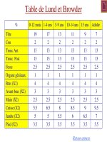
Practical Pediatric Gastrointestinal Endoscopy - part 7 ppt
Bạn đang xem bản rút gọn của tài liệu. Xem và tải ngay bản đầy đủ của tài liệu tại đây (367.22 KB, 22 trang )
124 CHAPTER 6
and recognition by the physicians performing the procedure
that things are not going well, with a decision to abort the pro-
cedure and precede with an open gastrostomy. Sometimes as
with percutaneous liver biopsy, complications are unavoidable
due to patient anatomy or underlying disease and the possi-
bility of these complications should be discussed with parents
prior to the endoscopic procedure. Reported minor complica-
tions that can become major complications include cellulitis, un-
complicated pneumoperitoneum, tube defects/disconnection,
GER, granulation tissue at insertion site, and pain at the in-
sertion site. Reported major complications include gastrocolic
fistula, gastroileal fistula, gastrocoloileal cutaneous fistula, in-
trahepatic placement, duodenal hematoma, complicated pneu-
moperitoneum, aspiration, peritonitis, catheter complications
including migration, buried bumper syndrome (Figs. 6.21–
6.23), partial gastric separation, catheter/bumper impaction if
not retrieved, intussusception secondary to catheter migration,
VP shunt infection, gastric or bowel perforation, and death.
Late complications include gastrocolic fistula, gastroileal fis-
tula, catheter migration/buried bumper syndrome/partial gas-
tric separation, gastric ulceration, cellulitis, fasciitis, gastric or
bowel perforation, catheter migration or other catheter-related
complications, bronchoesophageal fistula (following removal),
and aortic perforation (following cut and pass technique). PEG
tubes in children are not associated with a higher rate of sub-
sequent revision when compared to surgically placed open gas-
trostomy tubes if tube revisions due to unrecognized bowel per-
foration at initial PEG placement are excluded.
Fig. 6.21 Buried bumper
syndrome. The bumper of the
gastrostomy tube is no longer in
the stomach. However, it remains
in the abdominal wall close to the
stomach. The shadow of the
bumper is still visible.
Fig. 6.22 The gastrostomy tube
is buried in the abdominal wall,
although the stoma remains open.
This was confirmed by injection
of small amount of saline.
New uses of the PEG technique
Innovative pediatric and adult gastroenterologists and sur-
geons have further modified the techniques of PEG. Utilizing
modifications of the PEG technique, tubes can be placed directly
Fig. 6.23 The extramural type of buried bumper syndrome was
confirmed by CT scan.
THERAPEUTIC UPPER GI ENDOSCOPY 125
1 This is a procedure that is best done quickly. Once the endoscopic portion of the procedure starts, it is
usually accomplished by an experienced team within approximately 10 minutes. Longer procedures are
associated with excessive air insufflation, which makes identifying the gastric impression more difficult
and may increase the risk of distending the small bowel or colon with air, and therefore interposing a loop
of bowel between the stomach and the anterior gastric wall with its resultant complications.
2 If things are not going well in terms of positioning, the PEG tube should not be placed. There may be
something – liver, bowel, mesentery, etc. – between the trocar and the anterior gastric wall. Unless the
liver has been punctured, these complications are usually self-limited if the angiocatheter/trocar is
removed and the PEG is not placed.
3 If significant bleeding occurs or stool is visualized at any point, surgical consultation is appropriate.
4 When faced with a patient with atypical anatomy (cardiac surgery patients, patients with a scoliosis,
etc.,) the PEG may require placement in a nonstandard position (i.e., right side of the abdomen in a patient
with situs inversus). The endoscopic technique should be similar to the standard procedure. Avoid
location selection by formulas (i.e., one-third the distance between the xiphoid and the umbilicus). Pick
the location that is best, based on the individual patient’s anatomy.
5 The buried bumper syndrome. The gastrostomy bumper is no longer in the stomach. The complication
did occur in teenagers who suffered form severe botulism toxicity. Muscle paralysis was a contributing
factor to the rare complication.
6 The existing fistular was confirmed by injection of small amount of saline.
7 A CT scan showed extragastric location of the buried bumper.
Table 6.1 Tricks of the trade.
in the jejunum (PEJ) for feeding and in the cecum (PEC) for an-
tegrade colonic enemas. The PEJ technique currently has limited
applicability in young children due to equipment and size limita-
tions. If larger series confirm earlier reported success with PECs,
this is likely to become an increasingly reported technique in
children with neurologic abnormalities and developmental ab-
normalities resulting in chronic constipation.
Conclusions
PEGs are being increasingly utilized in pediatric patients. Place-
ment of a PEG tube does not increase the incidence of postopera-
tive GER and does not interfere with subsequent gastric surgery.
PEG placement is an advanced endoscopic procedure associ-
ated with a higher rate of complications than standard esopha-
gogastroduodenoscopy. Placement of PEGs in children requires
modification of the technique required in adults due to size and
anatomic considerations and also due to different anticipated
duration of use. The key points of the safe technique of the PEG
placement are summarized in Table 6.1.
NASOJEJUNAL TUBE PLACEMENT
A nasoduodenal or a nasojejunal tube feeding is commonly used
in children with severe GER as a bridge nutritional therapy be-
fore surgery or nutritional support for critically ill children with
various conditions in intensive care units.
126 CHAPTER 6
An enteral tube may be placed endoscopically if other options
such as spontaneous passage or installation under fluoroscopy
with the use of a radiopaque guidewire have failed.
After the appropriate tube is chosen, it should be prepared by
placement of one silk suture at the tip. The patient is sedated
and put in the left lateral decubitous position. The tube should
be inserted into the stomach via the nose, first, followed by the
endoscope. The tube may be found as either conveniently po-
sitioned along the greater curvature of the stomach pointing to
the antrum or coiled in the gastric body. In the second scenario,
it is pulled back until the tip is visible. The tube with an internal
guidewire can be advanced forward if it is not coiled. A smooth
surface of the antrum and lack of mucosal folds simplify grasp-
ing of the silk string. A regular biopsy forceps is preferable to use
for grasping because it usually eliminates sticking of the suture
to the grasper and accidental dislodgement of the tube from the
duodenum or jejunum back to the stomach during withdrawal
of the forceps. A significant friction between the scope and the
feeding tube creates a passive engagement of the nasoduodenal
or nasojejunal tube when the shaft is advanced toward pylorus.
Therefore, the external part of the tube should be secured to pre-
vent an excessive insertion and coiling of the tube in the stomach.
Once a regular forceps grasps the silk suture, it is dragged in
the biopsy channel to align the feeding tube with the tip of a
scope. The shaft of the endoscope is maneuvered through py-
lorus into the distal duodenum or proximal jejunum in a stan-
dard fashion. Then the forceps is pushed forward for a few cen-
timeters while the shaft is pulled back for the same distance
simultaneously. These “exchange’’ sequences are repeated until
the tip of the scope is drawn back to the antrum. A view of the
forceps and the tube engaging through the pylorus is reassuring
that the exchange procedure was performed successfully. After
that the biopsy forceps is opened to release the string attached
to the tube and pulled back into the stomach and closed before
complete removal. Finally, the shaft is pulled out using side-to-
side gentle rolling technique to decrease friction and accidental
dragging of a feeding tube back into the stomach. The position
of the tube along the lesser curvature is ideal (Fig. 6.24).
Simple postprocedure flat abdominal film or fluoroscopy con-
firms the appropriate position of the feeding tube.
A similar technique can be used for placement of the gastro-
duodenal or gastrojejunal feeding tube in children with an es-
tablished gastrostomy. The only difference is the introduction of
the feeding tube into the stomach through the gastrostomy.
Alternatively, nasojejunal intubation can be performed with
the so-called over-the-wire method. First, a pediatric gastroduo-
denoscope or colonoscope is inserted into the distal duodenum
or the proximal jejunum. Then, a Teflon-coated guidewire is
THERAPEUTIC UPPER GI ENDOSCOPY 127
Pylorus
Fig. 6.24 Nasojejunal tube. The adequate position of the tube is
achieved: the distal part of the tube is in the duodenum while the rest
of the tube is properly positioned in the stomach.
placed in the biopsy channel and advanced a few centimeters
beyond the tip of the scope. The next step involves synchronous
withdrawal of the shaft and insertion of the guidewire until the
endoscope is withdrawn completely. A soft lubricated tube is
advanced into the oro pharynx through the nose and removed
from the mouth by the index finger blindly or with the help of a
plastic grasper. After that, a guidewire is inserted into the tube
and rerouted through the nose.
The protective tube is removed. The final stage of the proce-
dure is performed under fluoroscopy. A lubricated nasojejunal
tube is advanced along the guidewire into the distal duodenum
or proximal jejunum. The position of the guidewire and the en-
teral tube is adjusted under fluoroscopy.
FURTHER READING
Benign esophageal stricture
Almendinger N, Hallisey MJ, Markowitz SK, et al. Balloon dilatation of
the esophageal strictures in children. J Pediatr Surg 1996;31:334–6.
Lan LCL, Wong KKY, Lin SCL, et al. Endoscopic balloon dilatation of
esophageal strictures in infants and children: 17 years’ experience and
a literature review. J Pediatr Surg 2003;38:1712–15.
Lang T, Hummer HP, Behrens R. Balloon dilatation is preferable
to bougienage in children with esophageal atresia. Endoscopy
2001;33:329–35.
Sandgren K, Malmfors G. Balloon dilatation of esophageal strictures in
children. Eur J Pediatr Surg 1998;8:9–11.
Pneumatic dilation in achalasia
Berquist WE,ByrneWJ, AmentME, Fonkalsrud EW,EulerAR. Achalasia:
diagnosis, management, and clinical course in 16 children. Pediatrics
1983;71:798–805.
128 CHAPTER 6
Boyle JT, Cohen S, Watkins JB. Successful treatment of achalasia in child-
hood by pneumatic dilatation. J Pediatrics 1981;99:35–40.
Gershman G, Ament ME, Vargas J. Frequency and medical management
of esophageal perforation after pneumatic dilatation in achalasia. J Pe-
diatr Gastroenterol Nutr 1997;25:548–53.
Hammond PD, Moore DJ, Davidson GP, Davis RP. Tandem balloon di-
latation for childhood achalasia. Pediatr Radiol 1997;27:609–13.
Mayberry JF, Mayell MJ. Epidemiological study of achalasia in children.
Gut 1988;29:90–3.
Myers NA, Jolley SG, Taylor R. Achalasia of the cardia in children: a
world survey. J Pediatr Surg 1994;29:1375–9.
Pineiro-Carrero VM, Sullivan CA, Rogers PL. Etiology and treatment of
achalasia in the pediatric age group. Gastrointest Endosc Clin N Am
2001;11(2):387–408.
Podas T, Eaden J, Mayberry M, Mayberry J. Achalasia: a critical review
of epidemiological studies. Am J Gastroenterol 1998;93:2345–7.
Foreign bodies
Arana A, Hauser B, Hachimi-Idrissi, Vandenplas Y. Management of in-
gested foreign bodies in childhood and review of the literature. Eur
J Pediatr 2001;160:468–72.
Gun F, Salman T, Abbasoglu L, Celik R, Celik A. Saftey-pin ingestion in
children: a cultural fact. Pediatr Surg Int 2003;19:482–4.
Kay M, Wyllie R. Pediatric foreign bodies and their management. Curr
Gastroenterol Rep 2005;7:212–8.
Khan S, Orenstein SR, Di Lorenzo C, et al. Eosinophilic esophagitis: stric-
tures, impections, dysphagia. Dig Dis Sci 2003;48:22–9.
Livovitz T, Schmitz BF. Ingestion of cylindrical and button batteries: an
analysis of 2382 cases. Pediatrics 1992;89:747–57.
Maves MD, Carithers JS, Birck HG. Esophageal burns secondary to disk
battery ingestion. Ann Otol Rhinol Laryngol 1984;93:364–9.
Olives JP. Ingested foreign bodies. J Pediatr Gastroenterol Nutr 2000;
31(suppl):S188.
Raval MV, Campbell BT, Phollips JD. Case of missing penny: tho-
racoscopic removal of a mediastinal coin. J Pediatr Surg 2004;39:
1758–60.
Sharieff GQ, Brousseau TJ, Bradshaw JA, Shad JA. Acute esophageal
coin ingestions: is immediate removal necessary? Pediatr Radiol
2003;33:859–63.
Tanaka J, Yamashita M, Yamashita M, Kajigaya H. Esophageal electro-
chemical burns due to button type lithium batteries in dogs. Vet Hum
Toxicol 1998;40:193–6.
Yardeny D, Yardeny H, Coran AG, Golladay ES. Severe esophageal dam-
age due to button battery ingestion: can it be prevented? Pediatr Surg
Int 2004;20:496–501.
Endoscopic hemostasis
American Society for Gastrointestinal Endoscopy. ASGE guide-line: the
role of endoscopy in acute non-variceal upper-GI hemorrhage. Gas-
trointest Endosc 2004;60:497–504.
Beppu K, Inokuchi K, Koyanagi N, et al. Prediction of variceal hemor-
rhage by esophageal endoscopy. Gastrointest Endosc 1981;27:213–8.
THERAPEUTIC UPPER GI ENDOSCOPY 129
Cano I, Urruzuno P, Medina E, et al. Treatment of esophageal varices by
endoscopic ligation in children. Eur J Pediatr Surg 1995;6:299–302.
Fox VL, Carr-Locke DL, Karrer FM, et al. Endoscopic ligation of
esophageal varices in children. J Pediatr Gastroenterol Nutr 1995; 20:
202–8.
Hassall E, Berquist WE, Ament ME, Vargas J, Dorney S. Sclerother-
apy for extrahepatic portal hypertension in childhood. J Pediatr
1989;115(1):69–74.
Howard ER, Stringer MD, Mowat AP. Assessment of injection slcerother-
apy in management of 152 children with oesophageal varices. Br J Surg
1988;75:404–8.
Hyams JS, Treem WR. Portal hypertensive gastropathy in children. J Pe-
diatr Gastroenterol Nutr 1993;17:13–18.
Khan K, Schwarzemberg SJ, Sharp H, et al. Argon plasma coagula-
tion: clinical experience in pediatric patients. Gastrointest Endosc
2003;57:110–12.
Laine L, Cook D. Endoscopic ligation compared with sclerotherapy for
treatment of esophageal variceal bleeding: a meta-analysis. Ann Intern
Med 1995;123:280–7.
Lee JG, Turnipseed S, Romano PS, et al. Endoscopy-based triage sig-
nificantly reduces hospitalization rates and cost of treating up-
per GI bleeding: a randomized controlled trial. Gastrointest Endosc
1999;50:755–61.
Lee YL, Oh JM, Park SE, et al. Successful treatment of a gastric Dieu-
lafoy’s lesion with a hemoclip in anewborn infant. Gastrointest Endosc
2003;57:435–6.
Lokesh TG, Jacobson K, Phang M, et al. Endoscopic hemostasis in a
neonate with a bleeding duodenal ulcer. Case report. J Pediatr Gas-
troenterol Nutr 2005;41:244–6.
Mumtaz R, Shaukat M, Ramirez FC. Outcomes of endoscopic treatment
of gastroduodenal Dieulafoy’s lesion with rubber band ligation and
thermal/injection therapy. J Clin Gastroenterol 2003;36:310–14.
Paquet KJ, Lazar A. Current therapeutic strategy in bleeding esophageal
varices in babies and children and long-term results of endoscopic
paravariceal sclerotherapy over twenty years. Eur J Pediatr Surg
1994;4:165–72.
Poddar U, Thapa BR, Singh K. Endoscopic sclerotherapy in children:
experience with 257 cases of extra hepatic portal venous obstruction.
Gastrointest Endosc 2003;57:683–6.
Price HR, Sartorelli KH, Karrer FM, et al. Management of esophageal
varices in children by endoscopic variceal ligation. J Pediatr Surg
1996;31:1056–9.
Raju GS, Gajula L. Endoclips for GI Endoscopy. Gastrointest Endosc
2004;59:267–79.
Reinoso MA, Sharp HL, Rank J. Endoscopic variceal ligation in pedi-
atric patients with portal hypertension secondary to liver cirrhosis.
Gastrointest Endosc 1997;46:244–6.
Saeed ZA, Michaletz PA, Winchester CB, et al. Endoscopic variceal liga-
tion in patientswhohave failed endoscopic sclerotherapy. Gastrointest
Endosc 1990;36(6):572–4.
Snady H, Feinman L. Prediction of variceal hemorrhage: a prospective
study. Am J Gastroenterol 1988;83(5):519–25.
Spolidoro JV, Kay M, Ament ME, et al. New endoscopic and diagnostic
techniques: working group report of the first world congress of pedi-
atric gastroenterology, hepatology and nutrition: management of GI
130 CHAPTER 6
bleeding, dysplasia screening and endoscopic training – issues for the
new millennium. J Pediatr Gastroenterol Nutr 2002;35(suppl 2):S196–
S204.
Stiegmann GV, Goff JS, Sun JH, Wilborn S. Endoscopic elastic band liga-
tion for active variceal hemorrhage. Am Surg 1989;55:124–8.
Stringer MD, Howard ER. Long-term outcome after injection sclerother-
apy for esophageal varices in children with extrahepatic portal hyper-
tension. Gut 1994;35:257–9.
Stringer MD, Howard ER, Mowat A. Endoscopic sclerotherapy in man-
agement of esophageal varices in 61 children with biliary atresia. J Pe-
diatr Surg 1989;24(5):438–42.
Thapa BR, Mehta S. Endoscopic sclerotherapy of esophageal varices in
infants and children. J Pediatr Gastroenterol Nutr 1990;10(4):430–4.
Zargar SA, Lavid G, Khan BA, et al. Endoscopic ligation compared with
sclerotherapy for bleeding esophageal varices in children with extra-
hepatic portal venous obstruction. Hepatology 2002;36:666–72.
Percutaneous endoscopic gastrostomy
Chaer RA, Rekkas D, Trevino J, et al. Intrahepatic placement of a PEG
tube. Gastrointest Endosc 2003;57(6):763–5.
Conlon SJ, Janik TA, Janik JS, et al. Gastrostomy revision: incidence and
indications. J Pediatr Surg 2004;39(9):1390–5.
Fox VL, Abel SD, Malas S, et al. Complications following percutaneous
endoscopic gastrostomy and subsequent catheter replacement in chil-
dren and young adults. Gastrointest Endosc 1997;45(1):64–71.
George DE, DoklerM. Percutaneous endoscopicgastrostomyin children.
Tech Gastrointest Endosc 2002;4(4):201–6.
Mathus-Vliegen EM, Koning H, Taminiau JA, et al. Percutaneous endo-
scopic gastrostomy and gastrojejunostomy in psychomotor retarded
subjects: a follow-up covering 106 patient years. J Pediatr Gatsroen-
terol Nutr 2001;33:488–94.
McCarter TL, Condon SC, Aguilar RC, et al. Randomized prospective
trial of early versus delayed feeding after percutaneous endoscopic
gastrostomy placement. Am J Gastroenterol 1998;93(3):419–21.
Panigrahi H, Shreeve DR, Tan WC, et al. Role of antibiotic prophylaxis
for wound infection in percutaneous endoscopic gastrostomy (PEG):
result of a prospective double blind randomized trial. J Hosp Infec
2002;50:312–15.
Segal D, Michaud L, Guimber D, et al. Late onset complications of percu-
taneous endoscopic gastrostomy in children. J Pediatr Gastroenterol
Nutr 2001;33:495–500.
Srinivasan R, Fisher RS. Early initiation of post PEG feeding: do
published recommendations affect clinical practice. Dig Dis Sci
2000;45(10);2065–8.
Taylor AL, Carroll TA, Jakubowski J, et al. Percutaneous endoscopic
gastrostomy in patients with ventriculoperitoneal shunts. Br J Surg
2001;88:724–7.
Van der Merwe WG, Brown RA, Ireland JD, et al. Percutaneous endo-
scopic gastrostomy in children – a 5-year experience. S Afr Med J
2003;93:781–5.
Wyllie R. Changing the tube: a pediatrician’s guide. Curr Opin Pediatr
2004;16(5):542–4.
THERAPEUTIC UPPER GI ENDOSCOPY 131
Nasojejunal tube placement
Gharpure V, MeertKL,Sarnaik AP, et al.Indicatorsof postpyloric feeding
tube placement in children. Crit Care Med 2000;28:2962–6.
Kirby DF, Delegge MH, Fleming C. American Gastroenterological As-
sociation technical review on tube feeding for enteral nutrition. Gas-
troenterology 1995;108:1282–1301.
Levy H. Nasogastric and nasoenteric feeding tubes. Gastrointest Endosc
Clin N Am 1998;8:529–49.
Lyons KA, Brilli RJ, Wieman RA, et al. Continuation of transpyloric feed-
ing during feeding of mechanical ventilation and tracheal extubation
in children: a randomized controlled trial. JPEN 2002;26:209–13.
Meert KL, Daphtary KM, Metheny NA. Gastric vs small-bowel feed-
ing in critically ill children receiving mechnical ventilation. Chest
2004;126:872–8.
Moore FA, Feliciano DV, Andrassy RJ, et al. Early enteral feeding, com-
pared with parenteral, reduces postoperative septic complications: the
results of meta-analysis. Ann Surg 1992;216:172–83.
Patrick PG, Marulendra S, Kirby DF, DeLegge MH. Endoscopic
nasogastric-jejunal feeding tube placement in critically ill patients.
Gastorintest Endosc 2997;45:72–6.
Pobiel RS, Bisset GS III,Pobiel MS. Nasojejunal feeding tube placement in
children: four years cumulative experience. Radiology 1994;190:127–9.
Stark SP, Sharpe JN,LarsonGM. Endoscopically placed nasoenteral feed-
ing tubes: indications and technique. Am Surg 1991;4:203–5.
Strong RM, Condon SC, Solinger MR, et al. Equal aspiration rates from
postpylorus and intragastric-placed small-bore nasoenteric feeding
tubes: a randomized, prospective study. JPEN 1992;16:59–63.
132
7
Pediatric Colonoscopy
INTRODUCTION
Colonoscopy is a challenging procedure not only for the begin-
ners but also for experts.The biggest obstacle is a relatively high
prevalence of abnormal fixation of the descending colon, and to
a lesser extent the ascending colon, which makes a colonoscopy
much more difficult and occasionally impossible to complete
even for experts.
However, an experienced colonoscopist is capable of manag-
ing the majority of cases successfully by using precise technique
and “intuitive’’ sense of“upstream’’ colon acquired during the
years of practice.On the contrary, b
eginners often create prob-
lems for themselves by resorting to inappropriate maneuvers,
transforming a “standard,’’easy to navigate colon into a twisted,
distended, and rigid tube.To avoid these “painful’’ mistakes, a
trainee should become familiar with the following:
r
Embryology and gross and endoscopic anatomyof the large
intestine
r
Main principles of colonoscopy technique
r
Specific maneuvers and approaches to the “difficult’’ colon
r
Endoscopic characteristics of common pathology
Another important aspect of training is achievement of acom-
petence level by the trainee to perform pediatric colonoscopy
safely and effectively. Although debatable, 100 diagnostic and
55 therapeutic procedures were chosen arbitrarily as a minimum
requirement. An additional source of training is colonoscopy
simulators, which may catalyze a learning process.
INDICATIONS FOR COLONOSCOPY
Traditionally, indications for colonoscopy are classified based
upon the goal of procedure: diagnostic or therapeutic.Over the
last decade, a new concept of high-volume low-yield indications
has been introduced in adult practice, as colonoscopy has been
used as a part of a large-scale screening program for the early
diagnosis of colon cancer. A low incidence of this disease in a
pediatric population virtually eliminates the needs for screening
colonoscopy except forasmall group of children with suspected
familial polyposis coli or other rare formsof polyposis.
The indications for diagnostic pediatric colonoscopy are fo-
cused primarily on clinical symptoms:“red flags’’ and additional
Practical Pediatric Gastrointestinal Endoscopy
George Gershman, Marvin Ament
Copyright © 2007 by Blackwell Publishing Ltd
PEDIATRIC COLONOSCOPY 133
Lower gastrointestinal bleeding
r
Hematochezia
r
Fecal occult blood
Inflammatory bowel disease
r
Diagnosis
r
Management
r
Extent and severity
r
Unclear response to treatment
r
Surveillance for colorectal cancer in chronic inflammatory bowel
disease
Unexplained chronic diarrhea
Evaluation of anatomic abnormalities seen on barium enema
Family history of a familial polyposis syndrome
Cancer surveillance
r
Ulcerative colitis
r
Polyposis syndrome
r
Adenomatous or mixed polyp
Abdominal pain and chronic diarrhea in patients with HIV and other
types of immunodeficiency disorders
Clinical signs of posttransplantation lymphoproliferative disorder
Intraoperatively
r
Detection of lesions that cannot be detected on palpation and/or
inspection
Therapeutic colonoscopy
r
Polypectomy
r
Treatment of bleeding, angiodysplasia
r
Removal of foreign body
r
Decompression of megacolon or colonic volvulus
r
Balloon dilation of stenotic lesions
Table 7.1 Indications for colonoscopy.
clues of serious pathology of the large intestine and the termi-
nal ileum obtained from radiological and other diagnostic proce-
dures or laboratory tests (Table7.1).Inaddition, colonoscopy and
biopsy are indicated for surveillance for detection of malignancy
in patients with long-standing inflammatory bowel disease.
Patients who have undergone small intestinal transplantation
may need to undergo ileoscopy and/or colonoscopy to obtain
specimens from transplanted bowel to look for rejection, viral
infection, and evidence of lymphoproliferative disease.
Diagnostic colonoscopy is not indicated in patients with
1 Acute self-limited diarrhea
2 Gastrointestinal (GI) bleeding with a demonstrated upper GI
source
3 Irritable bowel syndrome
134 CHAPTER 7
Peritonitis
Conditions with a high risk of preparation
r
Fulminant colitis
r
Toxic megacolon
r
Recent surgical anastomoses
Inability to visualize mucosa
r
Poor bowel preparation
r
Massive GI bleeding
Associated medical problems
r
Sepsis
r
Absolute neutropenia
r
Respiratory and cardiovascular distress
Table 7.2 Contraindications to colonoscopy.
4 Chronic non-specificabdominal pain
5 Constipation with or without impaction
6 Inflammatory bowel disease which is responsive to treatment
Diagnostic colonoscopy is absolutely contraindicated in any-
one with fulminant colitis or toxic megacolon, suspected per-
forated viscous, and recent intestinal resection (Table 7.2).
However, patients with acute severe colitis in which cultures are
negative for bacterial pathogens and parasites, such as Entamoeba
histolytica and Trichurus trichura, should have an examination of
the rectum and distal sigmoid colon to help establish whether
they have a specific type of colitis. In such cases, limiting the
area viewed, as indicated, does not pose an undue risk.There
are times when direct visualization of the mucosa gives a spe-
cific diagnosis such as when pseudomembranes or punched out
ulcers are seen.
Physicians should not consider performing colonoscopy in pa-
tients who have chronic or recurrent abdominal pain without
other signs and symptoms, such as weight loss,failure to grow,
loss of appetite, perianal disease, or positive indicators for in-
flammatory bowel disease, such as an elevated sedimentation
rate, increased C-reactive protein, and positive screening panel
for inflammatory bowel disease.
PREPARATION OF THE PATIENT
FOR COLONOSCOPY
Preparing infants and children for colonoscopy can bedifficult.
In children who are less than school-age, it is often very diffi-
cult to explain to them why they are asked to have a restrictive
diet, and a simple explanation of why the test is being done is all
PEDIATRIC COLONOSCOPY 135
that should be provided.The physician and family should try to
use words that the child will understand in order to clarify why
they are going to be tested. Children simply need to be told that
they are going to have a test to look at where their “poop’’ comes
from, and it has to be clean inside to take a good look.
In school-age children and adolescents more detailed explana-
tions may be provided depending on the level of sophistication
of the child. Itisuseful to show the children and parents dia-
gramsof the rectum and colon and distal small bowel to make
them aware of what is going to beexamined. Providing such
knowledge ahead of time may mak
e the child or adolescent more
amenable to the procedure and more cooperative in preparing
for the examination.They should be shown pictures of the in-
struments used and simple diagramsof what may be normally
seen.
Children at any age should be toldthat they will be given an in-
travenous infusion through which they will receive medications
to make them sleep and to minimize any pain or discomfort.
Because most colonoscopists use medication to alter memory,
such as Valium
R
or Versed
R
, the individuals and their families
should be told that they will have little memory of the proce-
dure other than going to sleep.They should be told that they
would have little or no pain during the procedure because of the
medications used to decrease their ability to sense pain.
They should be told, in preparation for the procedure, that
they will have devices attached to their fingers and arms, which
measure their blood pressure or how hard their heart pumps,
how fast their heart is beating, and the rate at which they are
breathing.They should also be told that devices would be used
to tell how much oxygen is in their blood.They should be told
that when they awake from sleep their parents would be nearby.
This type of explanation bef
ore the procedure in most children
will alleviate much of their anxiety.However, some children will
not becomforted by such explanations.
During preparation the most difficult thing to do is to prepare
the bowel so that it can be adequately visualized. A number of
different regimes are available that are based either on wash out
of the bowel (lavage) or on cathartics. Both methods are subject
to failure because they rely upon the cooperation of the child and
family.
Although it is debatable, we do not use any preparation of
the colon in infants less than 4 months old.T
hey have almost
liquid stool, which is easy to irrigate and aspirate during the
procedure.The best technique of colon preparation for infants
4–12 months of age is a combination of clear liquids and milk
of magnesia.Milk of magnesia 1 cc/kgof body weight should
be given two nights before the procedure and midday the day
136 CHAPTER 7
prior to the procedure.Magnesium citrate may also be used in
children above 1 year of age.This may be divided in two doses
12 hours before the colonoscopy.Some individuals become nau-
seated with this and other cathartics. Itisoften necessary to
give the dose of magnesium citrate in four fractions. Itisbest
given cold and over ice, or mixed with lemon-lime type soft
drinks.
The night before the colonoscopy, a glycerin suppository can
be used to enhance evacuation of the colon.This technique is
probably the most benign of the
methods available and is one in
which the infant or child is most likely to cooperate.
If a large-volume lavage method is chosen, the patient is al-
lowed to eat and drink up until the afternoon the day before
the procedure.The patient is then asked to fast for 4 hours. A
lavage solution contains nonabsorbable agents such as polyethy-
lene glycol and electrolytes.The solutions are available flavored.
The patient is given 5–10 ml/kgupto250 ml by mouth every
10 minutes.The patient continues taking this solution until the
rectal effluent is clear.
There are some adolescents and teenagers who will accom-
plish this preparation readily. In the younger age children, suc-
cess is less assured.Hospitali
zation for 24–48 hours may be nec-
essary before the procedure to cleanse the colon in uncooperative
patients. If one of these solutions is used in a younger child or an
uncooperative teenager, the placement of a nasogastric tube into
the stomach may be the only way that one can guarantee giving
the full volumeof the solution.
The patient can be given metoclopramide 0.1 mg/kgtoamax-
imum of10mg/20 min before the lavage is begun, to enhance
or speedup gastric emptying.The patient may develop vomiting
in response to the lavage. In these instances, the rate of
infusion
may have to be curtailed.One way that we have found that is ef-
fective in this regimeistoinfuse the solution continuously over
a period of12hours.This is very effective in individuals who
vomit the solution when it is given rapidly.The patient in this
instance may be given metoclopramide every 4 hours to enhance
gastric emptying.
If one uses the lavage technique, there should besome concern
if stool is not passed within the first 4 hours.The rate of infusion
is usually in the order of 100–200 ml/huptoafull volumeof
4L.We typically have an infusion going into a peripheral vein
to provide maintenance fluids and electrolytes.
In recent years, low-volume nonabsor
bable polyethylene gly-
col preparations and oral phosphosoda solution have been
proven safe and effective for colon preparation in children
over 2 years. Clinically significant hypernatremia or hyperphos-
phatemia have not been reported in pediatric patients before and
PEDIATRIC COLONOSCOPY 137
after colonoscopy.We use oral phosphosoda for children 3 years
and older.The regimen consists of two doses of oral phospho-
soda 7–8 hours apart the day prior to the procedure. Each dose
can be divided in two or three smaller portions, as a cold drink,
to prevent nausea or vomiting.
Enemas are not useful preparation for children with suspected
inflammatory bowel disease since they usually cause erythema,
edema, and petechiae of rectal and distalsigmoid mucosa, giving
a false-positive macroscopic image.
EQUIPMENT
Different types of pediatric colonoscopes less than 12 mm are
commercially available (Table 7.3). They have 3.2-mm biopsy
channels, which allow the use of many accessories, such as stan-
dard biopsy forceps, snares, needles, and laser probes.Someof
these colonoscopes have adjustable stiffeners.These instruments
are more suitable for children 2 years and older.
Colonoscopes specifically designed for infants and toddlers
do not exist. Instead, pediatric upper GI videoendoscopes can
be used. Itismore difficult to telescope the sigmoid colon with
these instru
ments, but their smaller diameter prevents excessive
stretching of the bowel, especially in infants.
Recently, the prototype of an ultrathin colonoscope with di-
ameter of only 9.8 mm has been developed by Pentax Corpo-
ration (Tokyo,Japan). The preliminary results in adults showed
comparable rate (96%) of cecal intubation between the standard,
pediatric, and ultrathin models.The application of this type of
colonoscope may be advantageous for pediatric practice espe-
cially for infants and toddlers.
Working Insertion tube Biopsy channel
length (mm) diameter (mm) diameter (mm)
Fujinon Corp
EC-250 MP5 1330 11.1 3.2
EC-250 LP5 1390 11.1 3.2
EC-450 MP5 1330 11.1 3.2
EC-450 LP5 1690 11.1 3.2
Olympus Corp
PCF-140 L 1680 11.5 3.2
PCF-160 L 1680 11.5 3.2
PCF-Q180 AL 1655 11.5 3.2
Pentax Corp
EC-3430 L 1700 11.7 3.5
Table 7.3 Some technical parameters of new models of pediatric videocolonoscopes.
138 CHAPTER 7
MAGNETIC IMAGING SYSTEM
A relatively high percentage of difficult colonoscopies in adults
defined as failure of advancement of the tip of a colonoscope for
at least 5 minutes stimulated development of a nonradiographic
imaging method for reconstruction of the position, shape of the
shaft within the colon, and optimal placement of manual hands
supporting pressure in real time.The prototype of the system
was developed in 1993 based on the principle of magnetic field
position screening.The modern version of the system is com-
mercially available as a Scopeguide (Olympus Optical Corpo-
ration). It is a porta
ble and mobile unit, which is easy to set
up and position at the site of the patient’s gurney.The device
produces a radiation similar to a modern TV set.The calibration
process is quite simple and may be performed in less than 2 min-
utes. Itisequipped with a three-dimensional image reconstruc-
tion processor, which imitates a spatial configuration of a special
colonoscope or inserted probe during colonoscopy. A pediatric
colonoscope with built-in coils for magnetic image receptive sys-
tem is not currently available.The existing probe is designed for
colonoscopes with 3.2-mm biopsy channel.This limits an appli-
cation of the technology
for infants and small toddlers. Gentle
insertion of the probeisrequired before procedure for calibra-
tion.The optimal position of the probe just above the tip of the
colonoscope is secured byasimple plastic–rubber anchoring de-
vice.The presence of the probe inside a biopsy channel dimin-
ishes the effectiveness of suction, which requires even more re-
striction of air insufflation compared with a standard technique.
Serial images help to verify a configuration of the probe and cor-
responding shape of the inserted shaft (Figs. 7.1–7.
3) and, more
importantly, simplify the straightening of the shaft.These are
also useful for trainees for faster understanding and learning of
Fig. 7.1 Alfa loop.The tip of
the scope is in the splenic
flexure.
Fig. 7.2 Configuration of the
scope after the Alfa loop was
reduced.
Fig. 7.3 The tip of the scope is
in the cecum.There are no visible
loops.The length of the inserted
scope is close to the real length of
the colon.
PEDIATRIC COLONOSCOPY 139
a torque-steering technique and building up skill in colonoscopy.
Development of a pediatric version of the colonoscope for a
Scopeguide system in the future will increase the application
of this technique for pediatric patients.
INFORMED CONSENT AND
PREPROCEDURE PREPARATION
The risks and benefits of the colonoscopy should be reviewed
with the family at the time that the procedure is scheduled.Ques-
tions and answers about the procedure may be discussed at that
time.
On the day of the procedure, informed consent is again ob-
tained.The child and parents or guardian may be brought to
the preprocedure area. In this area an intravenous infusion is
started.
In order to minimize the discomfort of the intravenous nee-
dle, EMLA
R
creammay be applied to three or four potential
intravenous sites 60 minutes before an appropriate angiocath is
placed.Once the angiocath is in position and functioning well, it
is secured and intravenous infusion is started.The patient is then
transferred to the procedure area, where all necessary prepara-
tions for sedations are taken care of.
SEDATION FOR COLONOSCOPY
These are three options to performing a colonoscopy in pedi-
atrics: without sedation, with sedation, or general anesthesia.
A colonoscopy without sedation is rather hypothetical but
practical option. Although it is feasible in the hands of an expe-
rienced gastroenterologist in the rare case of a very cooperative
patient and parents, it is not a common practice in the United
States and Europe.
Pediatric colonoscopy is routinely performed under sedation
or general anesthesia. Usually, an anxious and scared child does
not allow even digital rectal exam or proper positioning on the
gurney until deeply sedated.The definition of deep sedation
includes the following:
r
Patient is responsive only to painful stimuli
r
Spontaneous breathing
r
Presence of deep tendon reflexes
General anesthesia with commonly used medications such
as Ketamine
R
or Propofol
R
is not principally different from
deep sedation but requires a skillful anesthesiologist in case of
complications or need for endotracheal intubation.On the con-
trary, a pediatric gastroenterologist providing a deep sedation
should be capable of endotracheal intubation.The logistics of
the choice usually depends on the specific policy of an individual
140 CHAPTER 7
institution, availability of an anesthesiologist, and economics of
a particular medical practice.
The advantages of general anesthesia with Propofol are quick
induction time, minimal side effects, and short stay in recovery
rooms, which are attractive for pediatric gastroenterologists es-
pecially in private practice. It also may decrease the turnover
timeof each procedure and increase potential revenue.On the
other hand, a higher cost of routine colonoscopy under general
anesthesia may not be covered by all insurances.
The goal of any sedation for colonoscopy in children is maxi-
mal elimination of anxiety and pain during the procedure with
minimal risk of complication. An
xiety is relatively easy to over-
comeinmajority of children by appropriate dose of tranquilizers.
Pain control is a more complicated and controversial part of the
sedation. Itisimportant to accept that pain during colonoscopy
is always related to a loop formation and stretching of the colon.
A general rule is that the more skillful the endoscopist, the less
analgesics are required for sedation.
There is a real concern that deep sedation, and especially gen-
eral anesthesia, masks patient discomfort and stimulates exces-
sive activity by the less experienced endoscopist, which may
lead to overstretching of the sigmoid colon and increase the risk
of complications. Again it is important to accept the concept
that a sedated patient with slight discom
fort is comparable to a
screaming nonsedated child. It is wrong to give an extra dose of
anesthesia and/or tranquilizer to overcome this warning sign in
order to makesome progress with bowel intubation. It is a good
practice to stop and reassess the position of the colonoscope, and
to makesomeadjustments to reduce the loop before further ad-
vancement. Itisimportant to remember that it is better to abort a
colonoscopy rather than increase the risk of complications.Once
again, arefined technique of colonoscopy should be considered
as an important part of pain control
.
Following sedation the patient is placed in the left lateral de-
cubitus position.The parents are asked to leave the room once
the patient is sedated.
EMBRYOLOGY OF THE COLON
Abnormal rotation and fixation of the embryonic colon is prob-
ably the major reason foradifficult colon and incomplete
colonoscopy.The rotation of the primitive large intestine begins
when the embryo is only 10 mm long. It occurs as a result of
elongation of the intestinal tube, separation of the yolk stalk,
and stepwise herniation of the duodenojejunal loop into the um-
bilical cord.
A counterclockwise rotation around the superior mesenteric
artery is the main mechanism of“packaging’’ the growing
PEDIATRIC COLONOSCOPY 141
intestine in preparation for its return back to the abdomen. At
a stage of a 25-mm embryo, almost the entire intestine is within
the umbilical cord.When the embryo grows to 40 mm in length,
there is enough space in the abdomen to accommodate the small
and large intestine.
Additional counterclockwise rotation is again crucial for
proper relocation of the intestine into the peritoneal cavity. As
a result the cecum swings to the right hypochondric area above
the superior mesenteric artery. At the end of rotation, the cecum
migrates down to the right iliac fossa.Finally, the mesentery
of the descending and ascending colon fuses with the posterior
peritoneum and disappears being pushed backby heavy loops
of the small bowel.
In normal circumstances, the cecum also does not have a
mesentery because it is an outpouching of the antimesenteric
aspect of the ascending colon. Its incomplete posterior fixation al-
lows some mobilityofthececum, which does not create anyprob-
lems for colonoscopists unless the patient has a mobile cecum.
The rectum is derived from the cloacae and fuses with the
sigmoid colon by the eighth week of gestation and has some but
limited mobility.
Thus as a result of a normal rotation, the colon acq
uires two
zones offull fixation – the descending and ascending colon – as
well as two areas of partial fixation – the cecum and rectum. In
addition, the mobility of the splenic and hepatic flexure is some-
what limited by a phrenocolic ligament and the extension of the
hepatorenal ligament, respectively.Only the sigmoid and trans-
verse colons possess their own mesentery and are fully mobile.
It is not surprising that they became a target of various endo-
scopic maneuvers preventing or minimizing stretching of these
vulnerable segments of the intestine.
It is easy to imagine that ab
normal rotation or fixation of the
embryonic colon can multiply difficulties in telescoping of an
unusually mobile bowel. As a rule, this is a total surprise for
the endoscopist.Someof the anomalies can be suspected during
a procedure, e.g., fixation of the cecum in the right hypochon-
drium.
The intrinsic property of the embryonic colon to move from the
left iliac fossa to the right one as the result of a counterclockwise
rotation gives an important clue to understand the concept of a
torque-steering technique of a colonoscopy.
In general, counterclockwise rotation of an endoscope creates
some deviations of
the sigmoid colon to the right flank of the
abdomen.The degree of sigmoid stretching is proportional to
the length and plasticity of the attached mesentery and amount
offorce applied to the colonoscope to push it forward or ro-
tate it counterclockwise.Thus a stretching and looping of the
sigmoid colon should be anticipated during counterclockwise
142 CHAPTER 7
Dentate line
Fig. 7.5 Squamocolumnar junction or dentate line.
rotation of the endoscope.To the contrary, clockwise rotation
of the endoscope moves the colon to the left and helps to
telescope the sigmoid colon and minimize stretching and loop
formation.
ENDOSCOPIC ANATOMY
The anal canal is less than 2 cm in a newborn, reaching an adult
length of 3cmby 4 years of age. It is normally closed due to a
tonic contraction of the anal sphincter. If it is constantly open or
if sphincter tone is substantially decreased, spina bifida, trauma,
or sexual abuse should be ruled out (Fig. 7.4). Itisimportant
to remember that an axis of the anal canal is pointed anteriorly.
Proper insertion of the colonoscope will prevent the discomfort
due to excessive pressure and disorientation in the distal rectum
due to imbedding of the tip into the rectal mucosa.
Fig. 7.4 Unusually wide-open
anus.This finding is suspicious
for spina bifida, trauma, or sexual
abuse.
The proximal edge of the anal canal is demarcated bya
squamocolumnar junction or pectinate (dentate) line (Fig. 7.5).
Few longitudinal folds (the columns ofMorgani) run within the
anal canal and terminate at anal papillae (Fig.7.6). Occasionally,
Columns of Morgani
Columns of Morgani
Fig. 7.6 The longitudinal folds in the distal rectum (the columns ofMorgani) and enlarged anal papilla.The
u-turn maneuver in the rectum is useful for detail observation of the distal rectum close to the anal canal.
PEDIATRIC COLONOSCOPY 143
anal papillae may be quite prominent, cone like grayish struc-
tures.The rectumbecomes enlarged and fusiformbetween the
upper edge ofthecolumns ofMorgani andtherectosigmoid junc-
tion.This part of the rectum is called an ampulla. Itismarked by
three semilunar folds referred to as valves ofHouston (Fig. 7.7).
There are two such folds on the left and one on the right lateral
wall.The ampulla narrows at the level of rectosigmoid junction,
which is distanced from the anal verge by 9 cm in neonates and
15 cm in children 10
years and older.The rectal mucosa is smooth
and transparent and allows a good visualization of submucosal
veins (Fig. 7.8).
Multiple small lymphoid follicles on the rectal mucosa are
normally present in infants and toddlers.Scattered follicles less
than 3 mm can be seen in older children.
The sigmoid colon is the most “unpredictable’’ part of the
colon due to its long,V-shaped mesocolon.Stretching during
colonoscopy could double the lengthofthesigmoid colon.There-
fore, an absolute length of the sigmoid colon is not so i mportant
unless it is tremendously elongated.
The mobility and displacement of the sigmoid colon could
be
limited due to previous surgery, adhesions, or shortening of the
mesentery.
A relatively small sigmoid colon in infants and toddlers has
some disadvantages for the endoscopist:
First, it decreases a threshold for pain during stretching and
limits an application of standard pediatric colonoscopes sec-
ondary to the relatively large radius of curvature.
Second, it makes it impossible to perform the alpha loop ma-
neuver, leaving no choice but precise telescoping of the
sigmoid colon without any room for even small technical
mistakes.
Fig. 7.7
Semilunar folds of
Houston in the rectum.
Fig. 7.8 Typical vascular pattern
of the normal rectum.
Fig. 7.9 The sigmoid colon.The
endoscopic markers of normal
sigmoid colon are (i) rounded
lumen,(ii) circular folds, and (iii)
subtle vascular pattern.
The normal sigmoid colon appears tubular due to the promi-
nence of a circular muscle layer.The mucosa is less transparent
than in the rectum.There are multiple circular folds throughout
the sigmoid colon (Fig. 7.9).
The teniae coli are usually not visible along the sigmoid colon
except on the area adjacent to the sigmoid–descending junction.
The appearance of teniae coli in this area indicates significant
stretching of the sigmoid colon.
During colonoscopy, the sigmoid colon is “shaped up’’ in
somewhat predictable fashion. It becomes more spiral and
twisted clockwise between the posteriorly located rectum and
descending colon.The concave sacrum and a f
orward-projecting
sacral promontory determine the initial anterior deviation of
sigmoid loop. At this stage of the procedure, a colonoscope
can be palpated easily unless the sigmoid colon is extremely
stretched.
144 CHAPTER 7
In children, palpable loop can be reduced or modified byan
assistant in coordination with withdrawal maneuver performed
by the endoscopist.
The transition zone between the sigmoid and ascending colon
is usually located at the level of pelvic brim. It is rather an endo-
scopic as opposetoanatomic entity.Sharp angulationoccursusu-
ally secondary to twisting and stretching of the sigmoid colon.
The angle is sharper when the descending colon extends down
below the pelvic brim due to unusually low fixation and/or
when the sigmoid colon was stretched out extensively (Fig. 7.10).
Fig. 7.10 The angle is sharper
when the descending colon
extends down below the pelvic
brim due to unusually low
fixation and/or when the sigmoid
colon was stretched out
extensively.
Once the endoscope is passed through the junction between
the sigmoid and descending colon, the “surprises’’ are usually
over unless the patient has lax phrenocolic ligament or persisted
mesocolon of the ascending colon.
Normally the descending colon is relatively short, about 10 cm
in infants and 20 cm in toddlers. It is slightly wider and more
oval than the sigmoid colon (Fig. 7.11). It runs straight up toward
the left hypochondrium to joint the splenic flexure.The mucosa
of the descending colon appears grayish.
Fig. 7.11 The descending colon.
The descending colon is oval in
shape.
The stemsof the vessels run along the folds, i.e., perpendicular
to the lumen.The small branches spread around and across the
folds (Fig. 7.12). It may help to verify the axis of the colon without
a panoramic view of the lumen, when pulling back is limited
byextensively twisted bowel, which could untwist during the
withdrawal maneuver.
The folds of the descending colon are spread more apart rela-
tive to the folds of the sigmoid colon.The teniae coli are usually
not visible.These minor endoscopic changes help to verify the
position of the shaft in the descending colon during the advanc-
ing phase of colonoscopy.
T
he splenic flexure is marked by the bluish color of the transil-
luminated spleen (Fig. 7.13). This area should occupy the right
part of the lumen if the colonoscope was positioned properly in-
side the sigmoid and descending colon.The same color spot can
be seen occasionally when the tip of a colonoscope is trapped
Small branch
Vessel stem
Fig. 7.12 The vascular pattern of the descending colon.The stemsof
the vessels run along folds, i.e., parallel to the lumen.The small
branches spread around and across the folds and along the lumen.
PEDIATRIC COLONOSCOPY 145
within a very large sigmoid loop.Thus this color mark does not
definitively prove that the splenic flexure has been reached.
The splenic flexure is firmly attached to the diaphragmby the
phrenocolic ligament at the level of tenth and eleventh ribs.That
could explain occasional hiccups and transient hypoxia during
exploration of the transverse colon due to excessive pressure and
irritation of the phrenic nerve especially in infants and young
children.
The junction with the transverse colon is located along the
upper aspect of the medial wall of the splenic flexure. Itis“natu-
rally’’ angled by the mobile transverse colon, which hangs down
from the elevated splenic flexure.The j
unction is more sharply
angled and even folded when the patient is in the left lateral
position (Fig. 7.14).
Fig. 7.13 The splenic flexure. It
is marked by bluish discoloration.
The transverse colon is relatively short in children. Itisabout
14 cm in newborns and 30 cm in 10-year-olds, which is a big help
during pediatric colonoscopy. Relatively thin circular ratherthan
longitudinal layers of the muscularis propria are responsible for
the triangular shape of the transverse colon (Fig. 7.15).
Fig. 7.15 The transverse colon.
The triangular shape is the
endoscopic hallmark of the
transverse colon.
The slope of the transverse colon is pointed toward the hep-
atic flexure. Itismore voluminous than the adjacent colonic seg-
ments and has a blue-gray color acquired from the neighboring
liver (Fig. 7.16). The folds are circular at both ends of the hepatic
flexure.They are less prominent at the apex.
The junction between the hepatic flexure and the ascending
colon is located higher than that between the hepatic flexure and
1 Descending colon
2 Splenic flexure
3 Transverse colon
3
2
1
Fig. 7.14 The relationship of the angle between the descending colon
and the splenic flexure and the position of the patient during
colonoscopy.The irregular configuration encountered at the splenic
flexure and adjacent descending colon is created by the transverse
colon, which is hanging down during colonoscopy when the patient is
in the left lateral position.