Practical Pediatric Gastrointestinal Endoscopy - part 8 ppt
Bạn đang xem bản rút gọn của tài liệu. Xem và tải ngay bản đầy đủ của tài liệu tại đây (604.61 KB, 22 trang )
146 CHAPTER 7
Direction to the
ascending colon
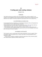
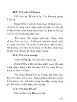
Fig. 7.17 The hepatic flexure. Itisdome-shaped.The junction
between the hepatic flexure and the ascending colon is always hidden
in the right upper corner of the screen behind the mucosal fold.
Steering of the shaft counterclockwise, pulling it back, and elevation of
the tip help to stretch the folded lumen.Subsequent clockwise rotation
and deviation of the tip to the right and decompression of the colon
facilitate exploration of the ascending colon.
the adjacent transverse colon. It points toward the right lobeof
the liver and is sharply angled posteriorly (Fig. 7.17).
The ascending colon is a short, retroperitoneal, and fixed seg-
ment of the right colon. It runs between the cecum anteriorly and
the lower pole of the right kidney posteriorly.The lumen of the
ascending colon is wide and constantly opened. It terminates as
a “blind’’ pouch cecum, which has two landmarks:
r
appendiceal orifice and
r
ileocecal valve
The appendiceal orifice is usually oval or rounded (Fig. 7.18)
and is located at the intersection of the teniae coli.The ileocecal
valve is situated at the posterior medical aspect of the cecum.
It usually stays aside from the forward-oriented optical system
of a colonoscope.That is why it is only partially seen as a focal
widening of the circular fold (Fig. 7.19).
Fig. 7.16 The hepatic flexure.
The mucosa of this area is paler
and has light bluish tinge
acquired from the adjacent liver.
Fig. 7.18 The appendiceal
orifice.
In the newborns, the cecum is cone-shaped, with the appendix
in the middle.Later on, the cecum expands sideways by unequal
enlargement of the haustra: a lateral sac becomes more spacious
Direction to the
ileocecal valve
Fig. 7.19 The ileocecal valve. A focal widening of the circular fold in
the cecum is the sign of the hiding ileocecal valve.
PEDIATRIC COLONOSCOPY 147
than the medial one.Thusthececum assumes an eccentric shape.
The thickness of the cecal wall is the smallest along the colon,
which should be kept in mind during polypectomy.
TORQUE-STEERING TECHNIQUE
A special colonoscopy technique has been developed to over-
come high flexibility, elasticity, and multiple angulations of the
large intestine (the sigmoid colon in particular). The main prin-
ciple of this technique, often called torque-steering technique,
isasubstitution of a corkscrew maneuvering around an angled
segment of the colon for pushing forward approach, which leads
to a loop formation.
Following are the elements of the technique:
r
Rotation around bended colon segments instead of pushing
up against them
r
Slow rather than rapid start of each maneuver with a colono-
scope
r
Frequent pulling back for shortening the sigmoid and trans-
verse colon and straightening of twisted segments of the large
intestine
r
Prediction rather searching foralumen
r
Pulling back when orientation is lost
r
Ascertainment of a correct axis of the colon before manipu-
lations with a colonoscope (this is much more important for
progress than search for a fully opened lumen)
r
Substitution of clock- or counterclockwise torque and up and
down angulations for manipulations with the R/L knob
r
Utilization of the R/L control knob as little as possible (knob-
induced tip deflection gets less and less effective with advance-
ment of the shaft)
r
Avoidance offull angulations of the tip. It will not slide along
the colon
r
Anticipation of a spring effect of twisted colon and preven-
tion of spontaneous untwisting of coiled segment by repeated
clock- and counterclockwise rotations
r
Programmed rotation of the lumen: the colon usually moves
in an opposite direction to the rotation of a shaft
r
Minimize insufflations: excessive air in the colon makes it
ridged and elongated
r
Frequent air suction and infrequent suction offluid
Sharing “inherited’’ similarity, pediatric colonoscopy is not
a traditional colonoscopy forasmall patient.The most impor-
tant difference in technique of colonoscopy between adult and
children is a low efficacy of an “Alfa’’ maneuver and more detri-
mental effect of a loop formation for children, especially the
younger ones.The rule of thumb is that the younger the child,
the more difficult to bypass the sigmoid–descending junction if
a big loop occurred.
148 CHAPTER 7
Handling a colonoscope:There are two ways to perform a
colonoscopy:
r
By the endoscopist managing all manipulation with a control
panel and the shaft with the left and right hand, respectively
(one person – single-handed approach).
r
By the endoscopist working with the control panel and the
assistant handling the shaft according to the endoscopist’sor-
ders.(two persons – two-handed approach).
It is generally accepted that one-person single-handed tech-
nique is the most effective way to conduct a colonoscopy.The
benefits of this approach are:
r
Precise control of an entire colonoscope
r
Coordinated activity of the left-hand-operated up/down con-
trol knob and the right-hand-rotated shaft
r
Almost immediate response to a changing position of the colon
r
Constant assessment and control of the bowel resistance
r
Anability to prevent unwinding of the telescoped bowel
A colonoscope is held similar to an upper GI videoendoscope
(see Chapter 5). Attention should be paid to a constant grip of
the shaft by the right thumb and by index and middle fingers.
The intensity of grip varies from light to firm with continuous
rotation. A common mistakeof the beginner is to lose hold of
a shaft with an attempt to use an R/L control knob. A released
shaft untwists immediately, allowing the bowel to escape from
telescoped and straighten condition.
A three-finger rotation technique is the most eff
ective way to
torque a colonoscope for a full 360
◦
. An additional 180
◦
rotation
can be achieved by moving a wrist in clock- or counterclockwise
direction.
If continuous rotation is needed, an assistant can hold the shaft
while the endoscopist adjusts a grip. Alternatively the endo-
scopist moves a leftarm with the control panel within the forth
and fifth fingers under the right arm, squeezes the shaft tight be-
tween the index and middle fingers and the control panel, and
then adjusts the grip of the right hand without “loosing’’ a tele-
scoped bowel. A colonoscope should be maximally straightened
to optimize transmission of the rotating force from the control
panel to the shaf
t. It can be achieved by keeping an appropri-
ate distance between the child and the endoscopist and repeat
pulling backmaneuvers.One of the common mistakes of the
beginner is holding the shaft too close to the anus. Grasping a
colonoscopy to the level of20–25 cm from the tip decreases the
need for frequent changes of handgrip and facilitates an appli-
cation of torque and control of rotation.
Position of the patient and insertion technique:Traditionally,
colonoscopy is performed with the patient in the left decubitus
position.The child’s head is resting on a small firm pillow.The
arms are relaxed along the torso;
left leg is stretched while the
PEDIATRIC COLONOSCOPY 149
right bended leg is positioned across the left one. It protects
the patient from accidentally rolling back or turning prone.
The insertion of the colonoscope into the rectum and control
of the shaft is easier when the patient is in the left decubitus than
in the supine position. In addition, if the child is placed close to
the endoscopist’s side of the gurney, the shaft hangs down and
can be kept in the desired position, by trapping it between the en-
doscopist’s right thigh and the edge of the gurney without being
held.There are threedisadvantagesoftheleft decubitus position:
r
Less precise control of the sigmoid colon, which is easier to pal-
pate and support by hand pressure when the patient is supine
r
The sigmoid colon tends to crumple down toward the left
flank, making the transition into the descending colon more
angled and difficult to bypass
r
The transverse colon flops down and narrows the connection
with the splenic flexure
Thus, a procedure could be started with the child in the left de-
cubitus position, and then the patient can be turned supine when
the sigmoid–descending junction is approached. Alternatively, a
supine position can be used from the beginning of colonoscopy
in infants, toddlers, and preschool children.
Insertion technique: Before insertion, the entire equipment and
suction system should be checked for proper function. A gur-
ney is lifted to the height comfortable for the endoscopist.The
distal 20 cm o
f the shaftislubricated. A rectal exam prior to the
procedure serves two purposes:
r
Lubrication of the anal channel
r
Reassurance that the patient has been adequately prepared
and sedated
If there are any doubts about the quality of bowel prepara-
tion, a rectal exam should be performed before sedation to avoid
unnecessary exposure to medication.
The assistant gently lifts up the right buttock to expose the
anus.The endoscopist grips the shaftat20–30-cmmarks, posi-
tions the tip into a gentle contact with the anus, and aligns the
bending portion of the shaft with the axis of the anal channel,
which runs toward anterior abdominal wall. Insufflation of the
anal canal and slight clockwise torque of the shaf
t facilitate slid-
ing of the tip into a distal rectum with minimal pressure.This
technique virtually eliminates any pain or accidental traumaof
the distal rectum. Right after initial exploration of the rectum,
a colonoscope is pulled back slightly and angled upward to
establish a panoramic view of the rectal ampulla. Any liquid
stool can be easily aspirated to simplify the approach to the dis-
tal rectum. Do not aspirate semiformed stool at the beginning
of colonoscopy to avoid problems with the suction channel. It
will lead to overinflation of the colon with air and difficulty in
completing a total colonoscopy. After that the colonoscope is
150 CHAPTER 7
advanced toward the rectosigmoid area. It is distant from den-
tate line for about 10–15 cm.This is the first but not the last time
when the lumen may disappear.
Endoscopic clues of a hidden lumen: In order to reach the splenic
flexure reasonably quickly, it is important to accept the concept
that a constant search for a full lumen is not a productive way to
conduct colonoscopy. It creates more problems than benefits for
the endoscopist and the patient.First of all, it is not possible be-
cause many segments of the colon, especially the sigmoid colon,
are sharply angulated during exploration.Second, a long opened
upstream segment of the sigmoid colon indicates a big loop for-
mation and should be avoided.Third, an extensive search for
a fully open lumen leads to overinflation of the colon, which
makes it ridged and elongated. Distention of the colon induces
discomfort and pain, leading to oversedation and increased risk
of complications. Instead, the endoscopist should not waste time
searching for a full lumen but concentrate on an effort to recreate
the axis of the upstream colon and the way to approach it.
In general, intubation of the colon and the sigmoid colon in
particular creates clusters of
sharply angled and bent segments,
which have a saw-tooth pattern. It means that the axis between
two adjacent colonic segments runs in opposite directions; e.g.,
if the visible segment climbs up diagonally from right to left
to 11 o’clock, the following segment falls down in the opposite
direction toward 5 o’clock.
This rule helps to accept the concept that initial position of
the twisted lumen gives a clue to a pattern of colonic “behavior’’
and direction for steering until a sharply angulated segment sets
the endoscopist off track. Disappearance of the lumen can be
explained by unequal shortening of the m
esenteric and antime-
senteric edges of the sigmoid colon during rotation and pulling
backmaneuvers and positioning of the tip close to the mucosa
with sudden loss of orientation.
Two strategies are useful in these circumstances:
r
Search for a hidden lumen and colonic axis using endoscopic
clues
r
Simply pull back slowly
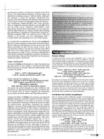
A narrowed slot-likeordimpled lumen of a twisted colon is
usually located in three areas: between 10 and 12 o’clock,1and
3o’clock, or 4 and6o’clock (Fig. 7.20). Another clue to an obscure
lumen is converging folds pointed to the slightly depressed,
grove-like area (Fig. 7.21). It is useful to remember that main
submucosal vessels are parallel to circular folds.However, their
small branches are usually spreading around between the folds
and can highlight the axis of the lumen (Fig. 7.22).
When the tip is close to the sig
moid–descending junction, a
prominent tenia coli or a center of the convexfolds indicates a
direction of the colonic axis and the location of the next segment
(Fig. 7.23).
Fig. 7.20 Common locations of the lumen.The leftimage: the lumen is located at 9 o’clock; the middle
image: the lumen is between 1 and 2 o’clock; the right image: the lumen is located at 5 o’clock.
Merging folds point
toward the lumen
Fig. 7.21 Slightly depressed groove-like area and merging folds are
the signs of the hidden lumen.
Small branches are pointed
toward the lumen
Fig. 7.22 The main submucosal veins and their branches.The main
vessels are parallel to the circular folds.The small branches are pointed
toward the lumen.This endoscopic clue may be useful when the tip of
the scope is distant from the mucosa for at least 1 or 2 cm.
Tenia coli
Fig. 7.23 Prominent tenia coli. An appearance of the tenia while
approaching the sigmoid–descending junction indicates the presence of
the significant loop in the sigmoid colon.
152 CHAPTER 7
The following is a description of the corkscrew technique,
which is particularly useful for sliding through the sharply an-
gled segments of the sigmoid colon and sigmoid–descending
junction:
r
Orient the tip toward a narrowed lumen and advance the shaft
forward slowly. If the lumen is located at 11 o’clock, rotate the
shaft counterclockwise and angle the tip up. As soon as the
edge of the lumen is approached, rotate the shaft clockwise
and pull it back. If the lumen is located between 4 and 6 o’clock,
rotate the shaft clockwise and pull it back. It will untwist thelu-
men and facilitate sliding of the tip into the proximal segment
of the colon. If the next segment is straight, advance the shaft
a few centimeters
forward. Rotate it clockwise and pull it back
to telescope (shortening) the colon. Repeat this maneuver sev-
eral times until the sigmoid–descending junction is reached.
This technique is equally applicable to the rectosigmoid area
and the junction between the splenic flexure and the transverse
colon.
EXPLORATION OF THE SIGMOID COLON AND
SIGMOID–DESCENDING JUNCTION
The sigmoid colon is the most vulnerable part of the large intes-
tine. It is not as long in children as in adults.However, children
especially infants and toddlers are less tolerant to stretching of
the sigmoid colon. A relatively short mesentery is less elastic,
which decreases the threshold for pain.
Nevertheless, in deeply sedated infants and toddlers, a less ex-
perienced endosocopist can create a huge loop which is not pal-
pable through the abdominal wall because it occupies both lat-
eral gutters and pushes up against the liver and left diaphragm.
It may create a false impression of a properly performed pro-
cedure without significant loop.The clinical clues to this dan-
gerous condition are sudden changes in oxygen saturation, hic-
cups, shallow breathing, and irritability of the patient,followed
by signs o
f respiratory distress. Immediate reduction of the loop
and interruption of the procedure is mandatory until the child
becomes stable.
During exploration of the sigmoid colon small loops are un-
avoidable, but easily reducible and are considered a routine part
of the procedure.However,formation of the larger loops should
be prevented.
There are several clues to recognition of clinically significant
loops:
r
Discomfort and pain
r
Long tubular segment of the bowel ahead
r
Loss of“one-to-one’’ relationship between pushing of the
colonoscope and advancement in the colon
PEDIATRIC COLONOSCOPY 153
r
Paradoxical movement of the lumen away from the tip with
attempts to advance the shaft
r
Increased stiffness of the angulations control and increased
resistance to the shaft
The elements of the most effective technique for preventing a
big loop from forming are:
r
Corkscrew sliding around sharply angled colonic segments
r
Establishing an appropriate angle for corkscrew sliding ma-
neuvers
r
Avoidance offorceful advancement (push through a signifi-
cant resistance)
r
Frequent pulling back with simultaneous clockwise rotation
of the shaft
r
Minimal insufflations
r
Transabdominal hand pressure support of the sigmoid colon
r
Changing the patient’s position
The presence of a big loop is a sign of two possible scenarios:
r
Formation of a large “N’’ loop
r
Existence of a large Alfa loop or atypical loops
The second variant is less likely in children. In any case, it is
reasonable to assume that the tip is in close proximity to the
sigmoid–descending junction. A supporting endoscopic sign of
this location is a prominent tenia coli pointed toward the right
upper corner of the screen. It is worse trying to turn this unde-
sirable situation into your favor.For successful reduction of a
sigmoid loop and advancement of the tip into descending colon,
proceed with the following:
First, turn the patient to the back to decrease the sharpness of
the sigmoid–descending junction.
Second, try to palpate the domeof the loop and show your as-
sistant how to support it. If the dom
eof the loop is in the right
part of the abdomen, an Alfa loop is most likely formed. If a
loop is palpated in the left part of the abdomen, an N loop has
most likely been created.
Third, in case of an Alfa loop scenario pull the shaft back slowly
and rotate it clockwise.The assistant should feel the loop con-
stantly and push it gently toward the left hypochondrium syn-
chronously with the endoscopist’s maneuvers. Atypical loop
should be suspected if the lumen slips away from the tip.Stop
withdrawing; move the sha
ft to the initial position and then
pull it back slowly with simultaneous vigorous counterclock-
wise rotation.Significant reduction of resistance and effective
withdrawal of at least 20–30 cm of the shaft with a stable po-
sition of the tip is a sign of successful loop reduction. If the N
loop is suspected, locate and support the loop with hand pres-
sure, rotate the shaft clockwise until the lumen opens up and
the slightly grayish mucosa of the descending colon appears
on the screen. Pull the shaft back slightly until the ridge of the
154 CHAPTER 7
next bent segment is reached; rotate the sha ft clockwise and
advance it forward when a reasonably long segment of the
descending colon appears. At this point the shaft is advanced
deep into the descending colon and is stable enough to com-
plete the reduction of the N loop by pulling the shaft back.
In the majority of cases the sigmoid colon is explored with-
out a big loop. During shortening and rotation maneuvers the
bowel becomes twisted and creates enough force to untwist
spontaneously and slip away from the shaft.The likelihood
of this undesirable effect increases when the tip is very close
to or inside the junction between the sigmoid and descending
colon. A
ll manipulation with the shaft should be very careful,
slow, and sequential. As mentioned above, the supine posi-
tion reduces a sharp angle of the sigmoid–descending colon
junction.Hand-pressure stabilization of the sigmoid colon is
very appropriate for the moment.The key for success is a
vigorous clockwise rotation, which facilitates sliding of the
tip into the descending colon. If an additional segment is lo-
cated ahead at 11 o’clock, pull the shaft back slowly, elevate the
tip up above the edge of the fold, and rotate the shaft clock-
wise until a wide-open oval lumen of the descending colon
appears.Then advance the shaft and align the tip with the
axis of the upstream segment.The lumen of the descending
colon is more oval, compared to the sigmoid colon.The folds
are less frequent, the color is more grayish, and the vascu-
lar pattern is more prominent.Once the descending colon is
reached, advance the shaft quickly to the level of the splenic
flexure. It is one of the easiest steps of colonoscopy if the shaft
is fully straight and the descending colon is normally fixed in
retroperitoneum.
SPLENIC FLEXURE AND
TRANSVERSE COLON
In order to straighten the sigmoid colon, and untwist the ex-
ternal portion of the colonoscope, the shaft should be rotated
counterclockwise. Attention should be given to the lumen of the
bowel in odder to avoid laceration of the mucosa by the tip of
the colonoscope.This maneuver facilitates an exploration of the
splenic flexure.
Tosimplify the entrance into the transverse colon, pull the
shaft back gently, rotate it counterclockwise, and angle it toward
11 o’clock. Initially, the lumen of the transverse colon appears
as a slot along the line between 7 and 11 o’clock.
An additional
deflection in the same direction and counterclockwise rotation
make the lumen wider. At this point, rotate the shaft clockwise
to a quarter turn and bring the tip down slowly. It is necessary
to turn the shaft counterclockwise again and elevate the tip up
PEDIATRIC COLONOSCOPY 155
before pushing the shaft into the transverse colon. Exploration
of the transverse colon does not require forceful advancement of
the colonoscope. In the absence of visible progress or in case of
increasing resistance, pull the shaftafew centimeters back while
keeping the lumen opened, and then elevate the tip and push it
forward, applying clockwise torque simultaneously. Repeat this
maneuver two or three times. If no significant progress has been
made, rotate the patient into right lateral position, straighten the
colonoscope by pulling it back, apply pressure to stabilize the
sigmoid colon, and advance the shaft
forward. Decreased re-
sistance and progression of the tip forward indicate successful
exploration of the transverse colon, which has a distinctive tri-
angular lumen. At this point, the hepatic flexure can by reached
almost momentarily by either pulling the shaft back with simul-
taneous counterclockwise rotation or pushing it gently forward.
Itisextremely unlikely to create a so-called “gamma’’ loop
in pediatric patients.The formation of this loop manifests by
increasing resistance and paradoxical movement of the proxi-
mal transverse colon away from the tip
, with attempts to push
the shaft forward.Successful reduction of agamma loop can be
challenging.First, rotate the patient to the back, and then pull
the shaft back and rotate it counterclockwise intensively. If the
tip remains stable during the withdrawal phase of the maneu-
ver, continue pulling back until the shaft is straightened. Itis
possible that a fter initial counterclockwise rotation a clockwise
torque should be tried.
HEPATIC FLEXURE, ASCENDING COLON,
AND CECUM
Exploration of the hepatic flexure may be challenging for be-
ginners. Itisimportant to remember that the axis of the hepatic
flexure has a reverse gamma configuration.The entrance to the
area is always located at an 11 o’clock position. A vigorous search
in the wrong direction may induce pain secondary to pressure
and distention of the bowel, small mucosal trauma, or retroflex-
ion of the bent portion of the colonoscope.The correct approach
to the hepatic flexure consists offew steps:(i) Orientation:The
transitional area between the transverse colon and the hepatic
flex
ure often appears as a blind pouch.The right part of the
pouch is convex with few circular folds creating an illusion of
the lumen.The left wall of the pouch is short due to rotation
and spiral configuration of the bowel. Attention should be fo-
cused on the upper portion of this area.(ii) Withdrawal: Pull the
shaft back slowly and orient the tip to the 11 o’clock direction.
Continue withdrawing and deflection of the tip in the same di-
rection until the lumen starts to open up with an initial slot-like
appearance.(iii) Decompression: Decompress the bowel until the
156 CHAPTER 7
lumen begins to collapse.(iv) Switching direction: Rotate the shaft
clockwise and move the tip to the right and slightly down using
the R/L knob.(v) Advancement: Advance the shaft forward and
adjust the position by counterclockwise rotation and elevation
of the tip, enough to keep it in the center of the lumen.
TERMINAL ILEUM
The ileocecal valve is tucked behind the folds. It is usually lo-
cated between the 9 and 11 o’clock position (Fig. 7.24). How-
ever, occasionally it could be found in the lower aspect of the
cecumbetween 5 and 7 o’clock position (Fig. 7.25). The ileoce-
cal valve appears as a lip-shaped thickening of the mucosal fold.
Anexploration of the terminal ileumbegins with detection of the
ileocecal valve by pulling the shaft away from the appendiceal
orifice.Once the valve is located, the tip is moved forward closer
to the appendix. The following steps should beadjusted to the
actual position of the ileocecal valve. If it is located at 11 o’clock,
the endoscopist should (i) decompress the cecum,(ii) orient the
tip to 11 o’clock, and (iii) slowly pull the shaft back until the tip
slips into the terminal ileum.The position of the ileocecal valve
between 5 or 7 o’clock dictates bending the tip down and to the
right toward the target, clockwise rotation, and pulling the shaft
back.Successful exploration of the terminal ileum is manifested
by the change incolorand texture of the mucosa;whilethececu
m
appears gray and smooth with prominent vessels, the terminal
ileum is pink with a slight yellow tinge and velvet mucosa with
multiple small (less than 3 mm) lymphoid follicles (Fig. 7.26).
The mucosal pattern of the colon is best evaluated as the in-
strument is slowly withdrawn.However, some stretching of the
bowel during advancement of a colonoscope makes the circular
Fig. 7.24 The ileocecal valve. It
is usually located between the 9
and 11 o’clock position of the
cecum.
Fig. 7.25 The less common
position of the ileocecal valve.
The ileocecal valve is at 5 o’clock
position.
Fig. 7.26 The terminal ileum.
Velvet texture, yellowish tinge,
and lymphoid follicles are the
main endoscopic characteristics
of the mucosa of the terminal
ileum in children.
PEDIATRIC COLONOSCOPY 157
folds more flat and easy to explore. Itisuseful for detection of
small lesions such a sessile polyp.
COMPLICATIONS
Routine use of colonoscopy in children would beimpossible
without solid proof that the procedure is safe. It does not mean,
however, that it is free from complications.This issue should be
fully disclosed and explained to the parents or caretaker as a part
of informed consent.
Complications associated with colonoscopy in children can be
classified according to
1 a necessity for hospitalization and
2 an absence or presence of structural damage of the intestine
and or adjacent organs (Table 7.4).
The incidence of minor complications is difficult to estimate.
Most likely it is underreported.First, it is unlikely that all mi-
nor complications were and are going to be counted.Second,
somecomplications are clinically silent: serosal tears and small
mesenteric hematomas have been accidentally discovered dur-
ing unrelated surgery soon after colonoscopy in adults.
The reported frequency of serious complications related to
pediatric colonoscopy is about 0.2%, which is similar to the data
from large-scale multicenter studies in adults. Perforation is a
ma
jor complication associated with colonoscopy and it can occur
due to four reasons:
r
Excessive pressure created by advancing forward or forcefully
withdrawing the shaftof a colonoscope
r
A tip imbedded into the bowel wall
r
Excessive air pressure
r
Inappropriate technique of polypectomy, hemostasis, or bal-
loon dilation of a benign stricture
Minor complications: Major complications:
no need for requirement for
hospitalization hospitalization
Structural damage of the
intestine or adjacent
organs
Small, nonobstructing mucosal or
submucosal hematomas, small
mucosal lacerations, petechiae
Perforation, bleeding requiring
blood transfusion and endoscopic or
surgical hemostasis,
postpolypectomy syndrome
Absence of structural
damage
Transient abdominal pain, bloating,
abdominal distention resolving after
passing gas, mild dehydration
secondary to bowel preparation
Cardiovascular and respiratory
distress, prolonged episode of
hypoxia requiring resuscitation
and/or endotracheal intubation
Table 7.4 Complications associated with pediatric colonoscopy.
158 CHAPTER 7
Three types of perforations related to diagnostic colonoscopy
have been described.Shaft-induced perforations are the result of
big loop formation. It is usually larger than expected and located
on the antimesenteric wall.
Tip perforations are smaller and typically occur when the
“sliding by’’ technique is used inappropriately or a tip is trapped
in wide diverticula or imbedded into mucosa when orientation
is lost.
Excessive air pressure perforation has been documented pri-
marily with strictures of the left colon. Attempts to bypass the
narrowed area create intermittent obstruction of the colon, accu-
mulation of air in the upstream colon, and increased hydrostatic
pressure, which could reach a critical level of81mm Hg f
or the
cecum.Hydrostatic pressure of169 mm Hgisrequired to perfo-
rate of the sigmoid colon in adults.This could explain the fact
that majority of air pressure related perforation has occurred in
the cecum and even in the ileum after the so-called uneventful
colonoscopy.Hydrostatic perforations have not been described
in children.
Most large traumatic perforations are immediately obvious.
One of the presenting symptoms could be sudden onset of irre-
ducible abdominal distention, decreased resistance to insertion
of a colonoscope,failure to insufflate the collapsed colon, visible
organs of a peritoneal cavity
, and severe and progressively in-
creasing abdominal pain. Immediate discontinuation of the pro-
cedure and request for plain abdominal films are mandatory.
Closed perforations are less dramatic. Almost 10% of patients
with a perforated colon can be initially symptoms free. In addi-
tion, another 10–15% of patients may develop mild to moderate
abdominal pain or discomfort. Absence offree are in the peri-
toneal cavity does not rule out perforation.High level of suspi-
cion and careful postprocedure observation are clues for early
recognition o
f complications. Persistent abdominal pain and/or
low-grade fever should be considered a sign of perforation un-
til proven otherwise. Early diagnosis in these circumstances is
absolutely crucial to prevent or decrease morbidity and mortal-
ity associated with perforation of the colon.Treatment of colonic
perforation can be nonoperative or surgical. Patients with a well-
prepared colon and therefore decreased risk of significant con-
tamination of the peritoneal cavity, absence of peritonitis, and
otherwise stable can be treated medically with bowel rest, broad-
spectrum antibiotics, and parenteral nutrition. Deterioration of
a patient’s condition,
signs of peritoneal irritation, and suspicion
of a large spillage of intestinal contents into the peritoneal cavity
mandate a surgical exploration. According to large-scale studies
in adults, the frequency of colonic perforation after polypectomy
is usually higher by two or three fold. It results from excessive
thermal coagulation of the tissue either due to inappropriate
PEDIATRIC COLONOSCOPY 159
setting of power and mode of current (more often when a
“blended’’ mode is used), cutting the large sessile polyp more
than 2 cm without a piece-meal technique or due to accidental
contact of the adjacent mucosa with the head of a cut polyp.
These perforations are often small and subtle and cause late on-
set of abdominal pain a few hours after the procedure.Severity of
pain usually increases with time.Fever is another common sign
of deep tissue necrosis.The treatment of these complications
(polypectomy syndrome) is similar to uncomplicated divertic-
ulitis, i.e.,
aggressive treatment with broad-spectrum antibiotics,
bowel rest, and good hydration.
Bleeding after a diagnostic colonoscopy is quite rare and can
be prevented by a thorough history and physical exam.His-
tory should be focused on a family history of bleeding diathesis,
frequent nasal bleeding, oozing from gumsafter the brushing of
teeth, and easy bruising without obvious trauma. A simple ques-
tion about recent treatments with aspirin and/or nonsteroidal
anti-inflammatory drugs is an effective way to prevent bleeding
secondary to platelets dysfunction.
B
leeding disorders are not a contraindication to pediatric
colonoscopy. Even patients with moderate to severe hemophilia
could be undergoing successful colonoscopy with biopsy or
polypectomyafter special preparations conducted by pediatric
hematologists. According to American Society for Gastrointesti-
nal Endoscopy, colonoscopy and colonoscopic polypectomy are
classified as a low risk for bacteremia. In recent publications, a
transient bacteremia has been reported in less than4% of patients
after an uneventful colonoscopy.The patients usually remain
asymptomatic without requiring any medical treatment. If pa-
tient becomes febrile
,flat abdominal and cross-table films, blood
culture, and empirical treatment with broad-spectrum antibi-
otics are mandatory.Malnourished, immunodeficient patients
and children with congenital or acquired valve defects are at
risk of infectious complications and endocarditis due to tran-
sient bacteremia.These children should receive antibiotics prior
to colonoscopy. Careful observation in a recovery room (until
the child is fully awake and ambulatory) and next day telephone
follow-up should be a routinepartof the postprocedure protocol.
COMMON PATHOLOGY
Rectal bleeding
Every child with hematochezia does not require colonoscopy.
Careful history and physical examination are essential for diag-
noses of bacterial, protozoal, or antibiotic-associated colitis, or
an anal fissure. In the pediatric patients with persistent or re-
current hematochezia, and no identifiable cause, colonoscopy is
160 CHAPTER 7
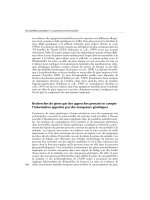
Fig. 7.27 Ulcerative colitis. Diffuse inflammation is typical for ulcerative colitis: erythema, exudates, loss of
vascular pattern.
the procedure of choice to search for mucosal changes or other
lesions associated with bleeding.
Ulcerative colitis is characterized by continuous and cir-
cumferential inflammation, diffuse erythema, edema, increased
mucosal friability, disappearance of vascular pattern, granular
appearance, grayish exudates, and microulcerations or shallow
ulcer (Fig. 7.27). Ulcerative colitis typically begins in the rec-
tum and spreads proximally. It may be mild or intense and may
involve the rectum and the left or entire colon.“Cecal patch’’
of local inflammation surrounding the appendiceal orifice
may
coexist with left-sided colitis (Fig. 7.28). Signs of the so-called
“back-washed’’ ileitis can be found in the terminal ileum: dif-
fuse mild to moderate erythema, edema, and petechiae within
5–10 cm of the ileum adjacent to the ileocecal valve.Severe form
of ulcerative colitis presents endoscopically with some degree of
narrowing and tubular appearance of the bowel due to severe
edema and loss of circular folds, striking erythema, large amount
of pus, and shallow ulcerations (Fig. 7.29).
Deep ulcers are not typical
for ulcerative colitis even with se-
vere form of the disease. Chronic and relapsing course of ul-
cerative colitis leads to unequal distribution of inflammation,
formation of pseudopolyps, and attenuation of vascular pattern
(Fig.7.30).
Appendiceal orifice
Microabscess
Fig. 7.28 Rare case of“cecal patch’’ in a child with left-sided ulcerative colitis.Left picture: multiple
microabscess aroud the appendiceal orifice (close-up view); Right picture: appendiceal orifice.
PEDIATRIC COLONOSCOPY 161
Fig. 7.29 Severe form of ulcerative colitis.Large amount of pus,
severe edema, loss of vascular pattern, and small ulcerations are seen.
Colitis in patients with Crohn’s disease is rather patchy than
uniform. It could be mild or intense, and may involve all or just
a part of the colon.Fifty percent of patients with Crohn’s colitis
have rectal sparing. At least half of children with Crohn’s disease
have ileocecal involvement. A so-called skip lesion is common.
Aphthous ulcer is a common manifestation of Crohn’s disease.
Itisasmall 4–5-mm ulcer surrounded by a thin rim of erythema
(Fig.7.31).
Aphthous ulcers can be clustered in few colonic segments or
spread throughout the colon.The other characteristic features o
f
Crohn’s disease are the signs of deep inflammation: narrowing
of the lumen, strictures, mucosal bridging, and different kinds
of ulcers such as stellate, longitudinal, tortuous, and serpiginous
(Figs.7.32–7.35).
The importance of the colonoscopy in patients with inflam-
matory bowel disease is to define the extent of inflammation,
to obtain sample tissues to look at histologically to establish the
specific diagnosis, and as an aid to planning therapy.
Allergic colitis is typically seen in infants.The endoscopic
markers of this condition are hem
orrhages, edema, and focal
erythemaof the rectum and the sigmoid colon (Fig.7.36).
Pseudopolyp
Fig. 7.30 Pseudopolyp in a patient with long-standing ulcerative
colitis.
162 CHAPTER 7
Aphthoid ulcer
of the ileum
Multiple aphthoid ulcers
in the colon
(a)
(b)
(c)(d)
Fig. 7.31 Aphthoid ulcer. Itissmall, shallow lesion with the rim of
erythema.(a) Aphthoid ulcer of the ileum;(b) multiple aphthoid ulcers
in the colon;(c) multiple aphthoid ulcers in the colon;(d) a close-up
view of the aphthoid ulcer.
Fig. 7.32 Deep longitudinal
ulcers in a patient with Crohn’s
disease.
Fig. 7.33 Mucosal bridging in
the cecum in 14-year-old patient
with Crohn’s disease.
Fig. 7.34 Tight stricture and
severe inflammation of the
ileocecal area in a patient with
Crohn’s disease.
PEDIATRIC COLONOSCOPY 163
Fig. 7.35 Severe inflammation
and large pseudopolyp in the
ileocecal region in a patient with
Crohn’s disease.
Fig. 7.36 Allergic colitis.
Focal erythema, small
aphthoid-like lesions, and
edemaof the sigmoid colon.
Arteriovenous
malformation of
the right colon
Hemangioma of
the colon
Fig. 7.37 Vascular lesions in the colon.
Rare lesions such as arteriovenous malformations or heman-
giomas may be discovered (Fig.7.37).
Isolated petechiae or small ulcerations in the rectum and
the distal sigmoid colon could be results of bowel preparation
(Fig.7.38).
Fig. 7.38 Small aphthoid-like
lesions can be occasionally
induced by bowel preparation.
Polypoid lesions, polyps, and
polyposis syndromes
Nodular lymphoid hyperplasia of the colon is typically seen in
early infancy and is characterized by the umbilicated lesions in
the rectum, sigmoid, and/or more proximal colon (Fig.7.39).
Lymphoid follicles
Fig. 7.39 Numerous lymphoid follicles in the sigmoid colon.
164 CHAPTER 7
Fig. 7.40 Sessile juvenile polyp.
Juvenile or inflammatory polyps are not uncommon in chil-
dren.They are most prevalent in the 4–6-year age group but
may be present as early as in 1-year-olds.They are uncommon
after age 18. Although autoamputation may occur in these cases,
many will not spontaneously disappear.
Fig. 7.41 Pedunculated juvenile
polyp.
Fig. 7.42
Large juvenile polyp
in the descending colon.
The coexistence ofjuvenile polyps in the rectum, the sigmoid
colon, and more proximal colon has been documented in at least
third of children.For this reason a colonoscopy with polypec-
tomy is the procedure of choice for children with recurrent
painless rectal bleeding. A typical juvenile polyp is small (less
than 1 cm) sessile or pedunculated structure. It has raspberry
or smooth appeared “head’’ with or without a stalk (Figs. 7.40
and 7.41). A large juvenile polyp is usually located in the sig-
moid colon. In rare cases it could be found in the descending
and transverse colon (Fig. 7.42). Such a polyp may induce an in-
termittent pain due to colonic intussusception.The endoscopic
marker o
f a nearby large juvenile polyp is the so-called goose
skin sign (Fig. 7.43).
Different type of hereditary polyposis syndromes can be diag-
nosed during pediatric colonoscopy. Diagnostic criteria for juve-
nile polyposis are the presence of 3–5 or more juvenile polyps in
“Goose skin”
sign
The base of the
removed polyp
“Goose skin”
sign
Fig. 7.43 The “goose skin’’ sign.The mucosa around a large juvenile polyp has specific pattern induced by
lipid-loaded macrophages.
PEDIATRIC COLONOSCOPY 165
Fig. 7.44 Juvenile polyposis.Multiple juvenile polyps in the rectum
and the colon.
the colon (Fig. 7.44). Surveillance colonoscopy is indicated due
to increased risk of colon cancer.
Peutz-Jeghers’s syndrome rarely presents with isolated
colonic hamartomas.More often clusters of gastric, small bowel,
and colonic polyps can be seen.The optimal diagnostic and
therapeutic strategy consists of combined upper GI endoscopy,
push enteroscopy, colonoscopy with polypectomy, and capsule
endoscopy surveillance to prevent chronic intussusception and
malignant transformation.Laparoscopy-assisted enteroscopy is
the procedure of choice for treatment children with the small
bowel hamartomas. A new method of a double balloon en-
teroscopy has not been validated in pediatric patients yet.
Colonoscopy has aleadingrole in diagnosis offa
milial polypo-
sis coli, Gardner polyposis, and other formsof hereditary poly-
posis in children. It is also a tool for colorectal cancer surveillance
in these patients.The colon may contain dozens or hundreds of
usually small sessile polyps (Fig. 7.45).
Multiple biopsies and polypectomyof the largest polyps pro-
vide tissues for initial diagnosis of low- or high-grade dysplasia.
Upper GI endoscopy should be performed in these patients, es-
pecially in children with Gardner’s syndrome who care a high
166 CHAPTER 7
Fig. 7.45 Multiple colon polyps in 5-year-old boy with Gardner’s
syndrome.
risk of synchronous lesions in the gastric body and the second
portion of the duodenum.
Asymptomatic children of patients with inherited polyposis
syndromes should undergo a surveillance colonoscopy since
11 years of age.Once the patient is diagnosed with familial poly-
posis coli, a colectomy with ileal–anal pull-through procedure
should be planned.
Chronic diarrhea
Chronic nonbloody diarrhea is an uncommon indication for
colonoscopy; however, if the diarrhea has indeed been chronic
in nature and the stool cultures and ova/parasites have been
nondiagnostic, colonoscopy can help to establish a correct diag-
nosis.Microscopic colitis has been described in children present-
ing with chronic diarrhea, abdominal pain, loss of appetite, and
weight loss.
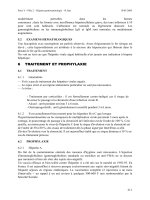
Fig. 7.46 Adenocarcinomaof the right colon in 11-year-old boy with significant weight loss, anemia, and
ascites. Colonoscopy revealed severe edemaof the distal part of the ascending colon.Further exploration of
the ascending colon showed ulcerated large tumor.The biopsy confirmed the diagnosis of mucinous
adenocarcinoma.
PEDIATRIC COLONOSCOPY 167
Fig. 7.47 Non-Hodgkin’slymphomaof the ileum.The indications for a colonoscopy were intermittent
severe right low quadrant pain, weight loss, and anemia.The intussusception was found in the descending
colon. It was gently reduced after the tissue samples were cautiously obtained.
Cancer surveillance
Development of adenocarcinomaof the colon in children is ex-
tremely rare but does occur even in children who never had
ulcerative colitis. It typically presents with a progressive weight
loss,fatigue, intermittent rectal bleeding, and anemia.Tumor is
equally located in the left or right colon. Itisquite difficult to
identify an ulcerated mass due to almost complete obstruction
and severe edemaof the surrounding tissue. Usually the edge of
the firm, easily fragmented during biopsy, discolored mass can
be seen (Fig. 7.46). Non-Hodgkin’slym
phomaof the ileum can
be discovered during colonoscopy in children with intermittent
abdominal pain and weight loss. Pain is a result of the ileocolonic
intussusception; red irregular mass occupying the intestinal lu-
men could be found in ascending colon (Fig. 7.47). Abiopsy
cares a risk of pealing of a quite large fragment of tissue. Proper
fixative solution is important for morphological and cytogenetic
diagnosis.
Adenocarcinoma of the colon
in ulcerative colitis
The determining factor in who develops cancer in ulcerative col-
itis seemstobe the severity of the original disease as well as the
extent of mucosal involvement and the duration of colitis.
The cancer risk for patients with pancolitis is 3% in the first
decade of disease and1–2% peryearthereafter.Patientswithuni-
versal colitis should begin biyearly colonoscopy,10years after
onset of disease.Multiple biopsies within few cm intervals are
recommended. Any flat or elevated lesions should be additional
targets. Chromoendoscopy has been found useful to increase the
yield offinding high-grade dysplasia in adults.