Textbook of Interventional Cardiovascular Pharmacology - part 6 pdf
Bạn đang xem bản rút gọn của tài liệu. Xem và tải ngay bản đầy đủ của tài liệu tại đây (2 MB, 68 trang )
308 Utilization of antiproliferative and antimigratory compounds
Figure 9
Inhibition of restenosis by paclitaxel in the rat carotid artery
injury model. Paclitaxel inhibits the accumulation of smooth
muscle cells 11 days after balloon catheter injury of rat carotid
artery. Animals were treated with 2 mg/kg body weigh paclitaxel
in vehicle (control animals were treated with vehicle alone) two
hours after injury and daily for the next four days.
Representative hematoxylin- and eosin-stained cross sections
from (
AA
) uninjured, (
BB
) vehicle-treated, and (
CC
) paclitaxel-treated,
injured rat carotid arteries. X240.
Source
: From Ref. 47.
Clinical trials investigating stent-
based delivery of paclitaxel
A number of randomized clinical trials (RCTs) have investi-
gated stent-based delivery of paclitaxel. These studies utilized
a number of different delivery methods, including polymeric
sleeves, nonpolymeric drug delivery and from drug-polymer
coatings on stents.
The Study to COmpare REstenosis rate between QueSt
and QuaDDS-QP2 trial was designed to control neointimal
proliferation through prolonged high-dose (800 µg) delivery
of the paclitaxel derivative 7-hexanoyltaxol (QP2) via acrylate
polymer membranes on the QuaDDS stent (Quanam
Medical, Santa Clara, California, U.S.A.) (64). Despite a
potential antirestenotic effect, enrollment in the trial was
terminated early, due to an unacceptable safety profile, as
seen by high rates of early stent thrombosis and MI. The very
high doses of paclitaxel used in this study and the unknown
vascular compatibility of the polymeric sleeve used for deliv-
ery could be a few of the many reasons responsible for failure
of the study.
Data from the European EvaLuation of pacliTaxel ElUting
Stent clinical trial, in which a Cook V-Flex Plus DES (Cook
Incorporated, Bloomington, Indiana, U.S.A.) was coated with
escalating doses of paclitaxel (0.2, 0.7, 1.4, and 2.7 µg/mm
2
)
applied directly to the abluminal surface of the stent, showed
a binary restenosis rate of 3.1% in the paclitaxel-eluting stent
group compared with 20.6% in the BMS group (65). In the
Asian Paclitaxel-Eluting Stent Clinical Trial, patients were
randomized to placebo (BMS) or one of two doses of pacli-
taxel (1.3 or 3.1 µg/mm
2
) on a Supra G
™
stent (Cook
Incorporated, Bloomington, Indiana, U.S.A.) (66). These
studies demonstrated a positive result using angiographic
endpoints and were used as the basis for the larger Drug
ELuting coronary stent systems in the treatment of patients
with de noVo nativE coronaRy lesions (DELIVER I) study.
However, no significant reduction in angiographic restenosis
rate or target vessel failure (TVF) was seen in the DELIVER-I
trial (67). Therefore, despite the improvement seen in angio-
graphic parameters in the earlier clinical trials, delivery of
paclitaxel via a nonpolymeric approach did not demonstrate a
positive clinical benefit. This failure may have several causes,
such as the loss of the drug to the systemic circulation before
its deployment at the target site, as well as variability of
the drug-release kinetics and dose delivered. The use of
polymers to control the release of a drug is discussed in
Chapter 22, “The Application of Controlled Drug Delivery
Principles to the Development of Drug-Eluting Stents.”
The TAXUS DES, which utilizes a polymeric delivery
approach for paclitaxel, has been examined across multiple
patient and lesion types in various clinical trials with successful
results demonstrating its antirestenotic potential. These
clinical data are described next.
Clinical studies using
the TAXUS Express
®
paclitaxel-eluting stent
The first study of the TAXUS paclitaxel-eluting stent in
humans, TAXUS I, reported major adverse cardiac events at
one-year follow-up at 3.2% for the TAXUS DES group
versus 10.0% for the BMS control group (p = NS) (68).
TAXUS I, now has data through four years and these bene-
fits were maintained for the TAXUS group (Fig. 12).
These data formed the basis of the most comprehensive
RCT program of a DES to date, evolving to encompass
higher patient numbers and higher-risk lesions and patients.
Over 6200 patients have been enrolled in the clinical trial
1180 Chap25 3/14/07 11:34 AM Page 308
program and a number of peri- and post-approval registries
have also been completed.
The TAXUS II study compared slow-release (SR) and
moderate-release (MR) formulations of the PES with BMS in
patients with relatively noncomplex lesions (69,75). At three
years, the TLR rate was 5.4% for the SR group and 3.7% for
the MR group, compared with 15.7% for the combined
control groups (p = 0.0001) (Fig. 12). TAXUS III was a single-
arm, pilot study assessing the feasibility of implanting up to two
PES for the treatment of ISR (70). The TAXUS IV pivotal study
in the United States is the largest ongoing PES RCT designed
to assess the safety and efficacy of the SR TAXUS Express™
DES for the treatment of de novo, coronary artery lesions (62,
63). In this study, TLR rates at three years were significantly
lower with the TAXUS DES group than the BMS control
group [6.9% vs. 18.6%, respectively (P Յ 0.0001); Fig. 12].
The remaining trials, TAXUS V and VI, incorporated higher-
risk patients or patients with higher-risk lesions. TAXUS V
expanded on the TAXUS IV pivotal study by including a higher
proportion of diabetic patients (31%) as well as those with
Antirestenotic agents incorporated into drug-eluting stents 309
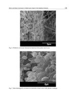
Figure 10
(
See color plate
.) Inhibition of restenosis
by paclitaxel inhibits in a porcine coronary
model. Photomicrographs demonstrating
neointimal thickness in arteries 28 days
after stent deployment. (
AA
) Uncoated
(bare) stent without paclitaxel;
(
BB
) chondroitin sulphate and
gelatin-coated stent with paclitaxel;
(
CC
) chondroitin-sulphate and gelatin stent
containing 1.5 µg of paclitaxel;
(
DD
) chondroitin-sulphate and gelatin
stent containing 8.6 µg of paclitaxel;
(
EE
) chondroitin-sulphate and gelatin
stent containing 20.2 µg of paclitaxel;
and (
FF
) chondroitin-sulphate and gelatin
stent containing 42.0 µg of paclitaxel.
Movat pentochrome stain; Scale
bar represents 0.12 mm.
Source
: From
Ref. 61.
1.5
1.0
0.5
0.0
010203040
Days after stenting
Uncoated stent
Poly(lactide-co-Σ-caprolactone)-coated stent
Intimal area (mm
2
)
50 60
*
Poly(lactide-co-Σ-caprolactone)-coated paclitaxel-releasing stent
**
Figure 11
(
See color plate
.) Sustained reduction in neointimal hyperplasia
in the rabbit iliac model.
Source
: From Ref. 107.
TAXUS VI
(MR)
n=
1 yr 2yr 3yr 4yr
100
70
100
70
100
70
100
70
219
227
PES
BMS
PES
BMS
SR MR PES
BMS
BMS
PES
662
652
131
135
270
31
30
TAXUS IV
(SR)
TAXUS II
(SR/MR)
TAXUS I
(SR)
Figure 12
(
See color plate
.)
Sustained freedom from target lesion
revascularization in TAXUS clinical trials.
Abbreviations
: BMS,
bare-metal stent; MR, moderate-release; PES, paclitaxel-eluting
stent; SR, slow-release.
Source
: From Ref. 73.
1180 Chap25 3/14/07 11:34 AM Page 309
small or large vessels, and patients with long lesions requiring
multiple overlapping stents (71). In this study, PES reduced the
nine-month TLR rate from 15.7% for BMS-treated patients to
8.6% for TAXUS DES-treated patients (p = 0.0003). The
TAXUS VI moderate release paclitaxel-eluting stent study
comprised the longest mean lesion lengths and highest-risk
patient population of any DES study to date, and currently has
data for three years of follow-up. A total of 28% of the patients
had long lesions with overlapping stents; the small vessel
subpopulation was also 28% of the total patient population.
Diabetic patients represented 20% of the study population.
Even in this more challenging study population, two-year TLR
rates were low in the PES group (9.7%) compared with the
BMS control group (21.0%) (p = 0.0013) (68).
Similar findings to those demonstrated in RCTs have been
seen in postapproval registries (72,73), corroborating the
findings of RCTs with “real-world” data. In addition, recent
studies have demonstrated significant benefit by DES when
used for the treatment of ISR, comparable with that seen
with intracoronary radiation (71,74). These findings point to
the potential utility of DES platforms in scenarios other than
de novo lesions, emphasizing the need to continue to under-
stand and assess this technology for unmet clinical needs.
Conclusions
Stent-based delivery of antirestenotic agents, now considered
a major technological advance in the interventional cardiology
area, was the first successful application of controlled drug
delivery technology in the management of occlusive coronary
artery disease. The success of DES in preventing coronary
restenosis has opened doors to other potential indications
suitable for local and regional drug delivery. Various pharma-
cotherapeutic options and delivery modalities are being
considered for a number of pathologies, such as vulnerable
plaque, stroke, valvular heart disease, and congestive heart fail-
ure (76). A thorough understanding of disease biology, drug
pharmacology, and a delivery technology appropriate for the
intended clinical application would be critical elements of a
successful therapeutic strategy.
Acknowledgments
The authors would like to thank Cecilia Schott, PharmD, and
Michael Eppihimer, PhD, for their assistance in the prepara-
tion of this chapter.
References
1 Investigators TBARIB. Comparison of coronary bypass surgery
with angioplasty in patients with multivessel disease. The
Bypass Angioplasty Revascularization Investigation (BARI)
Investigators. N Engl J Med 1996; 335(4):217–225.
2 Serruys PW, Unger F, Sousa JE, et al. Comparison of coronary-
artery bypass surgery and stenting for the treatment of
multivessel disease. N Engl J Med 2001; 344(15):1117–1124.
3 Wiskirchen J, Schober W, Schart N, et al. The effects of pacli-
taxel on the three phases of restenosis: smooth muscle cell
proliferation, migration, and matrix formation: an in vitro study.
Invest Radiol 2004; 39(9):565–571.
4 Hiatt BL, Ikeno F, Yeung AC,Carter AJ. Drug-eluting stents for
the prevention of restenosis: in quest for the Holy Grail.
Catheter Cardiovasc Interv 2002; 55(3):409–417.
5 Farb A, Sangiorgi G, Carter AJ, et al. Pathology of acute and
chronic coronary stenting in humans. Circulation 1999;
99(1):44–52.
6 Farb A, Weber DK, Kolodgie FD, Burke AP, Virmani R.
Morphological predictors of restenosis after coronary stenting
in humans. Circulation 2002; 105(25):2974–2980.
7 Hoffmann R, Mintz GS, Dussaillant GR, et al. Patterns and
mechanisms of in-stent restenosis. A serial intravascular ultra-
sound study. Circulation 1996; 94(6):1247–1254.
8 Virmani R, Farb A. Pathology of in-stent restenosis. Curr Opin
Lipidol 1999; 10(6):499–506.
9 Wahlgren CM, Frebelius S, Swedenborg J. Inhibition of neo-
intimal hyperplasia by a specific thrombin inhibitor. Scand
Cardiovasc J 2004; 38(1):16–21.
10 Costa MA, Simon DI. Molecular basis of restenosis and drug-
eluting stents. Circulation 2005; 111(17):2257–2273.
11 Welt FG, Rogers C. Inflammation and restenosis in the stent
era. Arterioscler Thromb Vasc Biol 2002; 22(11):1769–1776.
12 Chesebro JH, Lam JY, Badimon L, Fuster V. Restenosis after
arterial angioplasty: a hemorrheologic response to injury. Am J
Cardiol 1987; 60(3):10B–16B.
13 Ferns GA, Raines EW, Sprugel KH, Motani AS, Reidy MA,
Ross R. Inhibition of neointimal smooth muscle accumulation
after angioplasty by an antibody to PDGF. Science 1991;
253(5024):1129–1132.
14 Ip JH, Fuster V, Israel D, Badimon L, Badimon J, Chesebro JH.
The role of platelets, thrombin and hyperplasia in restenosis
after coronary angioplasty. J Am Coll Cardiol 1991; 17(6 suppl
B):77B–88B.
15 Willerson JT, Yao SK, McNatt J, et al. Frequency and severity of
cyclic flow alternations and platelet aggregation predict the
severity of neointimal proliferation following experimental
coronary stenosis and endothelial injury. Proc Natl Acad Sci
USA 1991; 88(23):10624–10628.
16 Kamath KR, Barry JJ, Miller KM. The Taxus drug-eluting stent:
a new paradigm in controlled drug delivery. Adv Drug Deliv
Rev 2006; 58(3):412–436.
17 Lau K-W, Sigwart U. Restenosis—an accelerated arteriopathy:
pathophysiology, preventive strategies and research horizons.
In: Edelman ER, Levy RJ, eds. Molecular Interventions and
Local Drug Delivery. London: W. B. Saunders Co. Ltd,
1995:1–28.
18 Chorny M, Fishbein I, Golomb G. Drug delivery systems for
the treatment of restenosis. Crit Rev Ther Drug Carrier Syst
2000; 17(3):249–284.
19 Lincoff AM, Topol EJ, Ellis SG. Local drug delivery for the
prevention of restenosis. Fact, fancy, and future. Circulation
1994; 90(4):2070–2084.
310 Utilization of antiproliferative and antimigratory compounds
1180 Chap25 3/14/07 11:34 AM Page 310
20 Kavanagh CA, Rochev YA, Gallagher WM, Dawson KA,
Keenan AK. Local drug delivery in restenosis injury: thermo-
responsive co-polymers as potential drug delivery systems.
Pharmacol Ther 2004; 102(1):1–15.
21 Bailey SR. Local drug delivery: current applications. Prog
Cardiovasc Dis 1997; 40(2):183–204.
22 Camenzind E, Kint PP, Di Mario C, et al. Intracoronary heparin
delivery in humans. Acute feasibility and long-term results.
Circulation 1995; 92(9):2463–2472.
23 Mitchel JF, McKay RG. Treatment of acute stent thrombosis
with local urokinase therapy using catheter-based, drug deliv-
ery systems: a case report. Cathet Cardiovasc Diagn 1995;
34(2):149–54.
24 Tahlil O, Brami M, Feldman LJ, Branellec D, Steg PG. The
Dispatch catheter as a delivery tool for arterial gene transfer.
Cardiovasc Res 1997; 33(1):181–187.
25 Bailey SR. Coronary restenosis: a review of current insights
and therapies. Catheter Cardiovasc Interv 2002;
55(2):265–271.
26 Lambert CR, Leone JE, Rowland SM. Local drug delivery
catheters: functional comparison of porous and microporous
designs. Coron Artery Dis 1993; 4(5):469–475.
27 Hanke H, Strohschneider T, Oberhoff M, Betz E, Karsch KR.
Time course of smooth muscle cell proliferation in the intima
and media of arteries following experimental angioplasty. Circ
Res 1990; 67(3):651–659.
28 Holmes DR Jr, Simpson JB, Berdan LG, et al. Abrupt closure:
the CAVEAT I experience. Coronary angioplasty versus exci-
sional atherectomy trial. J Am Coll Cardiol 1995; 26(6):
1494–1500.
29 Linde J, Strauss BH. Pharmacological treatment for preven-
tion of restenosis. Expert Opin Emerg Drugs 2001; 6(2):
281–302.
30 Rogers C, Edelman ER, Simon DI. A mAb to the beta2-leuko-
cyte integrin Mac-1 (CD11b/CD18) reduces intimal thickening
after angioplasty or stent implantation in rabbits. Proc Natl
Acad Sci U S A 1998; 95(17):10134–10139.
31 Simon DI, Dhen Z, Seifert P, Edelman ER, Ballantyne CM,
Rogers C. Decreased neointimal formation in Mac-1(Ϫ/ Ϫ)
mice reveals a role for inflammation in vascular repair after
angioplasty. J Clin Invest 2000; 105(3):293–300.
32 Fidler J. Clinical Oncology. New York: Churchill Livingstone,
2000.
33 Li JJ, Gao RL. Should atherosclerosis be considered a cancerof
the vascular wall? Med Hypotheses 2005; 64(4):694–698.
34 Serruys PW, Ormiston JA, Sianos G, et al. Actinomycin-eluting
stent for coronary revascularization: a randomized feasibility
and safety study: the ACTION trial. J Am Coll Cardiol 2004;
44(7):1363–1367.
35 Marx SO, Jayaraman T, Go LO, Marks AR. Rapamycin-FKBP
inhibits cell cycle regulators of proliferation in vascular smooth
muscle cells. Circ Res 1995; 76(3):412–417.
36 Bruns CJ, Koehl GE, Guba M, et al. Rapamycin-induced
endothelial cell death and tumor vessel thrombosis potentiate
cytotoxic therapy against pancreatic cancer. Clin Cancer Res
2004; 10(6):2109–2119.
37 Adams JD, Flora KP, Goldspiel BR, Wilson JW, Arbuck SG,
Finley R. Taxol: a history of pharmaceutical development and
current pharmaceutical concerns. J Natl Cancer Inst Monogr
1993; 15:141–147.
38 Ramaswamy B, Puhalla S. Docetaxel: a tubulin-stabilizing agent
approved for the management of several solid tumors. Drugs
Today (Barc) 2006; 42(4):265–279.
39 Mullins DW, Koci MD, Burger CJ, Elgert KD. Interleukin-12
overcomes paclitaxel-mediated suppression of T-cell prolifera-
tion. Immunopharmacol Immunotoxicol 1998; 20(4):473–492.
40 Tange S, Scherer MN, Graeb C, et al. The antineoplastic drug
paclitaxel has immunosuppressive properties that can effec-
tively promote allograft survival in a rat heart transplant model.
Transplantation 2002; 73(2):216–223.
41 Hui A, Min WX, Tang J, Cruz TF. Inhibition of activator protein
1 activity by paclitaxel suppresses interleukin-1-induced colla-
genase and stromelysin expression by bovine chondrocytes.
Arthritis Rheum 1998; 41(5):869–876.
42 Rowinsky EK, Donehower RC. Paclitaxel (taxol). N Engl J Med
1995; 332(15):1004–1014.
43 Schiff PB, Fant J, Horwitz SB. Promotion of microtubule
assembly in vitro by taxol. Nature 1979; 277(5698):665–667.
44 Jordan MA, Thrower D, Wilson L. Mechanism of inhibition of
cell proliferation by Vinca alkaloids. Cancer Res 1991;
51(8):2212–2222.
45 Schiff PB, Horwitz SB. Taxol stabilizes microtubules in
mouse fibroblast cells. Proc Natl Acad Sci U S A 1980;
77(3):1561–1565.
46 Jordan MA, Toso RJ, Thrower D, Wilson L. Mechanism of
mitotic block and inhibition of cell proliferation by taxol at low
concentrations. Proc Natl Acad Sci U S A 1993;
90(20):9552–9556.
47 Sollott SJ, Cheng L, Pauly RR, et al. Taxol inhibits neointimal
smooth muscle cell accumulation after angioplasty in the rat. J
Clin Invest 1995; 95(4):1869–1876.
48 Ganansia-Leymarie V, Bischoff P, Bergerat JP, Holl V. Signal
transduction pathways of taxanes-induced apoptosis. Curr
Med Chem Anti-canc Agents 2003; 3(4):291–306.
49 Blagosklonny MV, Darzynkiewicz Z, Halicka HD, et al.
Paclitaxel induces primary and postmitotic G1 arrest in human
arterial smooth muscle cells. Cell Cycle 2004;
3(8):1050–1056.
50 Giannakakou P, Robey R, Fojo T, Blagosklonny MV. Low
concentrations of paclitaxel induce cell type-dependent p53,
p21 and G1/G2 arrest instead of mitotic arrest: molecular
determinants of paclitaxel-induced cytotoxicity. Oncogene
2001; 20(29):3806–3813.
51 Axel DI, Kunert W, Goggelmann C, et al. Paclitaxel inhibits
arterial smooth muscle cell proliferation and migration in vitro
and in vivo using local drug delivery. Circulation 1997;
96(2):636–645.
52 Blagosklonny MV, Demidenko ZN, Giovino M, et al.
Cytostatic activity of paclitaxel in coronary artery smooth
muscle cells is mediated through transient mitotic arrest
followed by permanent post-mitotic arrest: comparison with
cancer cells. Cell Cycle 2006; 5(14):1574–1579.
53 Oberhoff M, Herdeg C, Al Ghobainy R, et al. Local delivery of
paclitaxel using the double-balloon perfusion catheter before
stenting in the porcine coronary artery. Catheter Cardiovasc
Interv 2001; 53(4):562–568.
54 Celletti FL, Waugh JM, Amabile PG, Kao EY, Boroumand S,
Dake MD. Inhibition of vascular endothelial growth factor-
mediated neointima progression with angiostatin or paclitaxel.
J Vasc Interv Radiol 2002; 13(7):703–707.
References 311
1180 Chap25 3/14/07 11:34 AM Page 311
55 Patterson C, Mapera S, Li HH, et al. Comparative effects of
paclitaxel and rapamycin on smooth muscle migration and
survival. Role of Akt-dependent signaling. Arterioscler Thromb
Vasc Biol 2006; 26(7):1479–1480.
56 Drachman DE, Edelman ER, Seifert P, et al. Neointimal thick-
ening after stent delivery of paclitaxel: change in composition
and arrest of growth over six months. J Am Coll Cardiol 2000;
36(7):2325–2332.
57 Heldman AW, Cheng L, Jenkins GM, et al. Paclitaxel stent coat-
ing inhibits neointimal hyperplasia at 4 weeks in a porcine model
of coronary restenosis. Circulation 2001; 103(18):2289–2295.
58 Hou D, Rogers PI, Toleikis PM, Hunter W, March KL.
Intrapericardial paclitaxel delivery inhibits neointimal prolifera-
tion and promotes arterial enlargement after porcine coronary
overstretch. Circulation 2000; 102(13):1575–1581.
59 Kolodgie FD, John M, Khurana C, et al. Sustained reduction of
in-stent neointimal growth with the use of a novel systemic
nanoparticle paclitaxel. Circulation 2002; 106(10):1195–1198.
60 Signore PE, Machan LS, Jackson JK, et al. Complete inhibition
of intimal hyperplasia by perivascular delivery of paclitaxel in
balloon-injured rat carotid arteries. J Vasc Interv Radiol 2001;
12(1):79–88.
61 Farb A, Heller PF, Shroff S, et al. Pathological analysis of local
delivery of paclitaxel via a polymer-coated stent. Circulation
2001; 104(4):473–479.
62 Stone GW, Ellis SG, Cox DA, et al. A polymer-based,
paclitaxel-eluting stent in patients with coronary artery disease.
N Engl J Med 2004a; 350(3):221–231.
63 Stone G, Ellis S, Cox D, et al. One-year clinical results
with the slow-release, polymer-based, paclitaxel-eluting
TAXUS stent: the TAXUS-IV trial. Circulation 2004b;
109(16):1942–1947.
64 Grube E, Lansky A, Hauptmann KE, et al. High-dose 7-
hexanoyltaxol-eluting stent with polymer sleeves for coronary
revascularization: one-year results from the SCORE random-
ized trial. J Am Coll Cardiol 2004; 44(7):1368–1372.
65 Gershlick A, De Scheerder I, Chevalier B, et al. Inhibition of
restenosis with a paclitaxel-eluting, polymer-free coronary
stent: the European evaLUation of pacliTaxel Eluting Stent
(ELUTES) trial. Circulation 2004; 109(4):487–493.
66 Hong MK, Mintz GS, Lee CW, et al. Paclitaxel coating reduces
in-stent intimal hyperplasia in human coronary arteries: a serial
volumetric intravascular ultrasound analysis from the Asian
Paclitaxel-Eluting Stent Clinical Trial (ASPECT). Circulation
2003; 107(4):517–520.
67 Silber S. Paclitaxel-eluting stents: are they all equal? An analysis
of six randomized controlled trials in de novo lesions of 3,319
patients. J Interv Cardiol 2003; 16(6):485–490.
68 Grube E, Silber S, Hauptmann KE, et al. TAXUS I: six- and
twelve-month results from a randomized, double-blind trial on
a slow-release paclitaxel-eluting stent for de novo coronary
lesions. Circulation 2003; 107(1):38–42.
69 Colombo A, Drzewiecki J, Banning A, et al. Randomized study
to assess the effectiveness of slow- and moderate-release
polymer-based paclitaxel-eluting stents for coronary artery
lesions. Circulation 2003; 108(7):788–794.
70 Tanabe K, Serruys PW, Grube E, et al. TAXUS III Trial: in-stent
restenosis treated with stent-based delivery of paclitaxel incor-
porated in a slow-release polymer formulation. Circulation
2003; 107(4):559–564.
71 Stone GW, Ellis SG, Cannon L, et al. Comparison of a
polymer-based paclitaxel-eluting stent with a bare metal stent
in patients with complex coronary artery disease: a random-
ized controlled trial. JAMA 2005; 294(10):1215–1223.
72 Abizaid A, Chan C, Lim Y-T, et al. Twelve-month outcomes
with a paclitaxel-eluting stent transitioning from controlled trials
to clinical practice: the WISDOM registry. Am J Cardiol 2006;
98:1028–1032.
73 Lasala JM, Stone GW, Dawkins KD, et al. An overview of the
TAXUS
®
EXPRESS
®
paclitaxel-eluting stent clinical trial
program. J Interv Cardiol 2006; 19:422–431.
74 Saia F, Lemos PA, Hoye A, et al. Clinical outcomes for
sirolimus-eluting stent implantation and vascular brachytherapy
for the treatment of in-stent restenosis. Catheter Cardiovasc
Interv 2004; 62(3):283–288.
75 Tanabe K, Serruys PW, Degertekin M, et al. Chronic arterial
responses to polymer-controlled paclitaxel-eluting stents:
comparison with bare metal stents by serial intravascular ultra-
sound analyses: data from the randomized TAXUS-II trial.
Circulation 2004; 109(2):196–200.
76 Sousa JE, Serruys PW, Costa MA. New frontiers in cardiology:
drug-eluting stents: Part II. Circulation 2003; 107(18):
2383–2389.
77 Whitworth HB, Roubin GS, Hollman J, et al. Effect of nifedip-
ine on recurrent stenosis after percutaneous transluminal
coronary angioplasty. J Am Coll Cardiol 1986; 8(6):
1271–1276.
78 Corcos T, David PR, Val PG, et al. Failure of diltiazem to
prevent restenosis after percutaneous transluminal coronary
angioplasty. Am Heart J 1985; 109(5 Pt 1):926–931.
79 Hoberg E, Dietz R, Frees U, et al. Verapamil treatment after
coronary angioplasty in patients at high risk of recurrent steno-
sis. Br Heart J 1994; 71(3):254–260.
80 Schwartz L, Bourassa MG, Lesperance J, et al. Aspirin and
dipyridamole in the prevention of restenosis after percuta-
neous transluminal coronary angioplasty. N Engl J Med 1988;
318(26):1714–1719.
81 Bertrand ME, Allain H, Lablanche JM. Results of a randomized
trial of ticlopidine vs placebo for prevention of acute closure
and restenosis after coronary angioplasty. The TACT study.
Circulation 1990; 82(suppl 3):190.
82 Okamoto S, Inden M, Setsuda M, Konishi T, Nakano T. Effects
of trapidil (triazolopyrimidine), a platelet-derived growth factor
antagonist, in preventing restenosis after percutaneous translu-
minal coronary angioplasty. Am Heart J 1992; 123(6):
1439–1444.
83 Serruys PW, Rutsch W, Heyndrickx GR, et al. Prevention of
restenosis after percutaneous transluminal coronary angioplasty
with thromboxane A2-receptor blockade. A randomized,
double-blind, placebo-controlled trial. Coronary Artery
Restenosis Prevention on Repeated Thromboxane-Antagonism
Study (CARPORT). Circulation 1991; 84(4):1568–1580.
84 Knudtson ML, Flintoft VF, Roth DL, Hansen JL, Duff HJ. Effect
of short-term prostacyclin administration on restenosis after
percutaneous transluminal coronary angioplasty. J Am Coll
Cardiol 1990; 15(3):691–697.
85 Urban P, Buller N, Fox K, Shapiro L, Bayliss J, Rickards A. Lack
of effect of warfarin on the restenosis rate or on clinical
outcome after balloon coronary angioplasty. Br Heart J 1988;
60(6):485–488.
312 Utilization of antiproliferative and antimigratory compounds
1180 Chap25 3/14/07 11:34 AM Page 312
86 Ellis SG, Roubin GS, Wilentz J, Douglas JS Jr, King SB III. Effect
of 18- to 24-hour heparin administration for prevention of
restenosis after uncomplicated coronary angioplasty. Am Heart
J 1989; 117(4):777–782.
87 Stone GW, Rutherford BD, McConahay DR, et al. A random-
ized trial of corticosteroids for the prevention of restenosis in
102 patients undergoing repeat coronary angioplasty. Cathet
Cardiovasc Diagn 1989; 18(4):227–231.
88 Pepine CJ, Hirshfeld JW, Macdonald RG, et al. A controlled trial
of corticosteroids to prevent restenosis after coronary angio-
plasty. M-HEART Group. Circulation 1990; 81(6): 1753–1761.
89 Multicenter European Research Trial with Cilazapril after
Angioplasty to Prevent Transluminal Coronary Obstruction
and Restenosis (MERCATOR) Study Group. Does the new
angiotensin converting enzyme inhibitor cilazapril prevent
restenosis after percutaneous transluminal coronary angio-
plasty? Results of the MERCATOR study: a multicenter,
randomized, double-blind placebo-controlled trial. Circulation
1992; 86(1):100–110.
90 Desmet W, Vrolix M, De Scheerder I, Van Lierde J, Willems
JL, Piessens J. Angiotensin-converting enzyme inhibition with
fosinopril sodium in the prevention of restenosis after
coronary angioplasty. Circulation 1994; 89(1):385–392.
91 O’Keefe JH Jr, McCallister BD, Bateman TM, Kuhnlein DL,
Ligon RW, Hartzler GO. Ineffectiveness of colchicine for the
prevention of restenosis after coronary angioplasty. J Am Coll
Cardiol 1992; 19(7):1597–1600.
92 Kent KN, Williams DO, Cassagneau B, et al. Double-blind,
controlled trial of the effect of angiopeptin on coronary
restenosis following coronary angioplasty. Circulation 1993;
88(suppl 1):5063.
93 Eriksen UH, Amtorp O, Bagger JP, et al. Continuous
angiopeptin infusion reduces coronary restenosis following
balloon angioplasty. Circulation 1993; 88(suppl 1):594.
94 Serruys PW, Klein W, Tijssen JP, et al. Evaluation of ketanserin
in the prevention of restenosis after percutaneous transluminal
coronary angioplasty. A multicenter randomized double-blind
placebo-controlled trial. Circulation 1993; 88(4 Pt 1):
1588–1601.
95 Sahni R, Maniet AR, Voci G, Banka VS. Prevention of resteno-
sis by lovastatin after successful coronary angioplasty. Am
Heart J 1991; 121(6 Pt 1):1600–1608.
96 Bairati I, Roy L, Meyer F. Double-blind, randomized,
controlled trial of fish oil supplements in prevention of recur-
rence of stenosis after coronary angioplasty. Circulation 1992;
85(3):950–956.
97 Dehmer GJ, Popma JJ, van den Berg EK, et al. Reduction in
the rate of early restenosis after coronary angioplasty by a
diet supplemented with n-3 fatty acids. N Engl J Med 1988;
319(12):733–740.
98 Grigg LE, Kay TW, Valentine PA, et al. Determinants of
restenosis and lack of effect of dietary supplementation with
eicosapentaenoic acid on the incidence of coronary artery
restenosis after angioplasty. J Am Coll Cardiol 1989;
13(3):665–672.
99 Nye ER, Ablett MB, Robertson MC, Ilsley CD, Sutherland WH.
Effect of eicosapentaenoic acid on restenosis rate, clinical course
and blood lipids in patients after percutaneous transluminal
coronary angioplasty. Aust N Z J Med 1990; 20(4):549–552.
100 Reis GJ, Boucher TM, Sipperly ME, et al. Randomised trial of
fish oil for prevention of restenosis after coronary angio-
plasty. Lancet 1989; 2(8656):177–181.
101 Muller DW, Topol EJ, Abrams GD, Gallagher KP, Ellis SG.
Intramural methotrexate therapy for the prevention of
neointimal thickening after balloon angioplasty. J Am Coll
Cardiol 1992; 20(2):460–466.
102 Gradus-Pizlo I, Wilensky RL, March KL, et al. Local delivery
of biodegradable microparticles containing colchicine or a
colchicine analogue: effects on restenosis and implications for
catheter-based drug delivery. J Am Coll Cardiol 1995; 26(6):
1549–1557.
103 Margolin L, Fishbein I, Banai S, et al. Metalloproteinase
inhibitor attenuates neointima formation and constrictive
remodeling after angioplasty in rats: augmentative effect of
alpha(v)beta(3) receptor blockade. Atherosclerosis 2002;
163(2):269–277.
104 van Beusekom HM, Post MJ, Whelan DM, de Smet BJ,
Duncker DJ, van der Giessen WJ. Metalloproteinase inhibi-
tion by batimastat does not reduce neointimal thickening in
stented atherosclerotic porcine femoral arteries. Cardiovasc
Radiat Med 2003; 4(4):186–191.
105 Wu CH, Pan JS, Chang WC, Hung JS, Mao SJ. The molecu-
lar mechanism of actinomycin D in preventing neointimal
formation in rat carotid arteries after balloon injury. J Biomed
Sci 2005; 12(3):503–512.
106 Suzuki T, Kopia G, Hayashi S, et al. Stent-based delivery of
sirolimus reduces neointimal formation in a porcine coronary
model. Circulation 2001; 104(10):1188–1193.
References 313
1180 Chap25 3/14/07 11:34 AM Page 313
1180 Chap25 3/14/07 11:34 AM Page 314
Introduction
The role of immune cells and inflammatory mediators in
cardiovascular disease has been well documented.
Atherosclerosis has been described as a chronic inflammatory
syndrome, a systemic disorder characterized by focal lesions
throughout the vasculature (1,2). Immune cells such as T-cells
and macrophages are recruited to the vascular wall where
they and their signaling molecules play important roles at all
stages of lesion development including plaque initiation,
progression, and rupture leading to thrombotic events (3,4).
Compositionally, varying sections of the plaque may be
engorged with soft, pliable lipid (cholesterol ester) and
immune components such as foam-cell-like macrophages,
typical of either newly formed or shoulder regions of mature
lesions versus regions with more stable transformations
comprised of proliferated smooth muscle cells (SMCs), fibrob-
lasts, and matrix (5–7). With growth and maturation,
remodeling occurs with thickening and breakdown of the
architecture and function of the vascular wall, ultimately
impinging on the size of the lumen and reducing blood flow. It
is these larger lesions, those more easily identified by angiog-
raphy, that are typically treated with interventional procedures.
Attempts at treating stenotic vessels due to vascular plaque
have included surgical interventions such as bypass and, since
the late 1970s, angioplasty. Unfortunately, in nearly 30% to
40% of patients, these procedures failed leading to re-occlusion
of the vessel within 6 to 12 months (8). Pathologically, this fail-
ure has been ascribed to either an acute closure from
stretching and recoil of the vessel or a more chronic biologi-
cally mediated lumen loss. This longer-term failure, or
restenosis, is due to a response to the mechanical disruption
and endothelial denudation from the procedure and results
from a cellular response to repair the injury. The major
component of restenotic plaque is neointima, primarily
misaligned, proliferated/migrated SMCs and fibroblasts, and
matrix material appearing somewhat in disarray. Early
attempts to treat restenosis focused on the local proliferative
process, primarily SMC expansion, with numerous therapeu-
tic agents and approaches investigated over more than two
decades (9).
Recently, a breakthrough has been achieved leading to a
significant shift in therapeutic paradigm, initially by use of the
Cypher sirolimus drug-eluting stent (DES). Sirolimus, an
immune suppressant approved for use in patients undergoing
kidney transplant, has pleotropic effects on cellular metabolism.
Specifically, the compound appears to act as an inhibitor of cell
cycle progression, and based on this, may combine the activi-
ties required on the numerous mechanisms and cell types
purported to participate in the restenotic process. Utilizing this
approach, a clear improvement has occurred in outcomes,
despite the reality that we really still do not completely under-
stand the restenotic participants or mechanisms.
This chapter focuses on percutaneous transluminal coro-
nary angioplasty (PTCA), provides a summary of the
underlying immune activities of the diseased vasculature, and
focuses in part on the role of immune and inflammatory
mediators in the restenotic process. In addition, the mecha-
nism of action of sirolimus, the drug used in the first successful
DES for reduction of restenosis will be highlighted. Finally, the
potential role for immune mediators on the overall processes
of atherosclerosis will be explored.
Percutaneous transluminal
coronary angioplasty
Today, standard therapy for myocardial infarction or luminal
narrowing includes thrombolytics, anticoagulants, and often
interventional procedures such as PTCA. With its introduction
26
Anti-inflammatory drugs, sirolimus, and
inhibition of target of rapamycin and its
effect on vascular diseases
Steven J. Adelman
1180 Chap26 3/14/07 11:35 AM Page 315
in the late 1970s, improvement was seen in the treatment of
luminal narrowing from obstructive coronary artery disease or
blockage due to myocardial infarction. The procedure involves
placing a balloon-tipped catheter at the site of occlusion and
disrupting and expanding the occluded vessel by inflating the
balloon. Although initially successful at removal of the blockage
and achieving luminal enlargement, the process also damages
the blood vessel wall extensively including the loss of the
endothelial lining. The ensuing response to this severe injury is
often enhanced expression of cytokines and growth factors
and, subsequently, a rapid reclosure or recoil, and/or a slow
progressive re-occlusion or restenosis of the vessel. With the
introduction of stents, metal-based cage/tube-like structures
placed into the vessel lumen, a step toward improving
outcomes was achieved. Coronary stents provide luminal
scaffolding, eliminating elastic recoil which can occur rapidly
following an interventional procedure. Unfortunately, although
acute reclosure was reduced, neointimal hyperplasia was not,
and in fact, the procedure lead to an increase in the prolifera-
tive comportment of restenosis (10).
As a consequence of PTCA, a neointima is formed within
the vascular wall, typically including myointimal hyperplasia,
proliferation and migration of SMCs and fibroblasts, connec-
tive tissue matrix remodeling, and formation of thrombus.
Restenosis, referring to the renarrowing of the vascular
lumen following an intervention such as balloon angioplasty, is
defined clinically as Ͼ50% loss of the initial luminal diameter
gain following the interventional procedure and has affected
anywhere from 30% to 40% of treated vessels.
Restenosis: role of
inflammation
Initial attempts at treating or preventing restenosis focused
primarily on inhibition of the proliferation of vascular SMCs
(VSMCs). A series of agents successful at inhibition of SMC
proliferation in vitro as well as in vivo in animal models such
as carotid injury models in the rat failed to demonstrate bene-
fit in the clinic. More recently, it has been shown in addition
to effects on SMCs, that mechanical intervention also activates
the recruitment and activation of immune cells. Cell signaling
through cytokines, chemokines, and adhesion molecule
expression results in the recruitment to the vascular wall of
cells of many types, as well as their proliferation, migration,
and/or maturation.
As with atherosclerosis itself, recruitment of inflammatory
cells is now recognized as an essential step in the pathogene-
sis of neointima formation in humans (11,12). In various
animal models, reduction of leukocyte recruitment by selec-
tive blockade of adhesion molecules significantly reduced
neointima formation and restenosis (13–16). Recent studies
also concluded a role of pre-existing inflammation within the
treated lesion itself and also, a correlation with systemic
markers of inflammation. Interestingly and in addition, there
are also current data suggesting a mobilization of
hematopoeitic progenitor cells (HPC) contributing to
restenosis, both from studies in mice and in humans (17).
Activation of inflammation
Following PTCA, responses within the vascular wall are typi-
cal of a response to injury. Numerous studies in animals
demonstrate that the inflammatory response is strongly
related to degree of arterial injury, with balloon dilation
damaging the endothelial lining and stimulating cytokine and
adhesion molecule expression (12,18). A layer of platelets and
fibrin forms at the injured site and circulating cells are
recruited. P-selectin mediates the adhesion of activated
platelets with monocytes and neutrophils and the rolling of
leukocytes on the endothelium (14,15). This is the main
pathophysiological process linking inflammation with throm-
bosis after arterial wall injury.
Leukocytes are recruited to the site of injury and NFkB is
activated. Recent findings support a role for nuclear factor-
kappa B (NFkB) as a key player in restenosis. NFkB, a central
mediator of expression of inflammatory genes including
cytokines and interleukins (ILs), is activated by degradation of
its inhibitor IkB through the ubiquitin–proteasome system.
This system regulates mediators of proliferation, inflamma-
tion, and apoptosis that are fundamental mechanisms for the
development of restenosis. In animal studies, blocking the
proteasome system reduced intimal hyperplasia (19,20)
showing that inflammation contributes significantly. Activation
of cytokines enhances the migration of leukocytes across the
platelet–fibrin layer into the tissue. Growth factors are
released from platelets and leukocytes, and SMCs and fibrob-
lasts proliferate and undergo a transformation to
myofibroblasts 3 to 14 days after the intervention (11). With
the release of growth factors, the initiation of the first phase
(G1) of the cell cycle is activated, regulated by the assembly
and phosphorylation of cyclin/cyclin-dependent kinase (CDK)
complexes. Growth factors trigger signaling pathways that
activate these CDK complexes.
Studies using human arterial segments strongly support a
role for inflammation in restenosis. Immediately following
stent implantation, studies by Grewe et al. (21) demonstrate
that a mural thrombus is formed, followed by invasion of
SMCs, T-lymphocytes, and macrophages. Additional studies
in atherectomy specimens following PTCA demonstrate an
increase of monocyte chemoattractant protein-1 and speci-
mens from restenotic lesions show an increased number of
macrophages (22). These results indicate that local expres-
sion of macrophage activity may be associated with the
mechanisms of intimal hyperplasia. A correlation was found
between stent strut penetration with inflammatory cell
316 Inhibition of target of rapamycin and its effect on vascular diseases
1180 Chap26 3/14/07 11:35 AM Page 316
density and neointimal thickness (23). Neointimal inflamma-
tory cell content was 2.4-fold greater in segments with
restenosis, and inflammation was associated with neoangio-
genesis. Coronary stenting that is accompanied by medial
damage or penetration of the stent into the lipid core induces
increased arterial inflammation, which is associated with
increased neointimal growth.
Circulating markers of
inflammation
Similar to a growing body of evidence in studies of athero-
sclerosis and cardiovascular disease, assessment of markers
from blood samples has provided information regarding the
role of inflammation after PTCA. Included among markers for
atherosclerosis are C-reactive protein (CRP), IL-6, serum
amyloid A (SAA), and even white blood cell (WBC) count.
With respect to PTCA, many of these same markers provide
insight. In studies by Serrano et al. (24) coronary sinus blood
samples taken 15 minutes after angioplasty showed evidence
of leukocyte and platelet activation with increased adhesion
molecule expression on the surface of neutrophils and mono-
cytes. Late lumen loss was correlated with the changes in IL-6
concentrations post-PTCA and MAC-1 activation in coronary
sinus blood (25,26). Recent studies demonstrated that stent
deployment is associated with an increase in CRP (27).
Interestingly, CRP plasma levels were significantly higher and
more prolonged in patients with restenosis compared with
patients without restenosis. Similar findings were reported in
a series of patients with stable angina who underwent PTCA
(28). The association between the extent of vascular inflam-
matory response with long-term outcome was even
observed in patients with stable angina undergoing stent
implantation (29). Finally, a recent study showed that the
inflammatory response after stent implantation can be
assessed by measuring the circulating monocytes in the
peripheral blood. The maximum monocyte count after stent
implantation showed a significant positive correlation with in-
stent neointimal volume at six-month follow-up. In contrast,
other fractions of WBCs were not correlated with in-stent
neointima volume (30). These findings demonstrate that
there is an inflammatory stimulus following PTCA, which
needs to be assessed for the risk stratification for restenosis.
Pre-existing inflammation
The studies discussed earlier demonstrate that vascular injury
caused by PTCA triggers inflammation. Importantly, however,
at the time of stent implantation, the overall inflammatory status
is not equivalent in all patients and, critically, in all atherosclerotic
plaques. Therefore, PTCA in an already inflamed plaque may
have significant impact on clinical and angiographic outcome.
Studies in patients with unstable angina and elevated baseline
CRP, SAA, and IL-6 values showed an enhanced inflammatory
response to angioplasty. Pretreatment CRP level is an indepen-
dent predictor for one-year major adverse cardiac events
(MACE), including the need for re-intervention in patients not
receiving statins. CRP levels were significantly higher in patients
with recurrent angina compared with asymptomatic patients
(31,32). Walter et al. (33) found that tertiles of CRP levels were
independently associated with a higher risk of MACE and
angiographic restenosis after stenting, and Buffon et al. (34)
found that baseline CRP and SAA levels were independent
predictors of clinical restenosis. Additionally, Patti et al. (35)
found that preprocedural IL-1 receptor antagonist (IL-1Ra)
plasma levels were an independent predictor of MACE during
the follow-up period. Furthermore, the overall activation status
of the immune system, estimated by the amount of IL-1
β
produced by monocytes, had positive correlation with late
lumen loss, while the expression of CD66 by granulocytes has
shown to prevent luminal renarrowing (36). Finally, the
concentration of macrophages was also reported to be an
independent predictor for restenosis (23).
The role of pre-existing inflammation in clinical outcome
after stenting was also studied by measuring the temperature
of the culprit lesion (37), a marker of inflammation. Patients
with MACE had increased plaque temperature before the
intervention. During a clinical follow-up of 18 months, the
incidence of MACE in patients with increased temperature
was higher compared with those without increased thermal
heterogeneity. The adverse cardiac events were mainly due
to restenosis at the culprit lesions.
It appears that the overall and local inflammatory status at
the time of PTCA plays a significant role in the development
of restenosis. The current evidence arises from studies
combining data from the clinical syndrome and peripheral
markers of inflammation. For patients with unstable clinical
syndromes and with increased levels of monocytes and CRP,
there is strong evidence for increased risk of restenosis. The
measurement of other inflammatory indices, such as SAA,
IL-6, IL-1
β
, IL-1Ra plasma levels, Lp(a), and fibrinogen,
seems to provide additional information.
Thus, overall, there is considerable evidence for an impor-
tant role for inflammation contributing to the restenotic
process.
Sirolimus: molecular
mechanism of action
Sirolimus (rapamycin, Rapamune) is a naturally occurring
macrocyclic lactone produced by Streptomyces hygroscopicus,
a streptomycete isolated from a soil sample collected from
Sirolimus: molecular mechanism of action 317
1180 Chap26 3/14/07 11:35 AM Page 317
Easter Island (Rapa Nui) first discovered and characterized by
Sehgal in 1975 (38). Initially identified as an antifungal agent,
the compound was subsequently found to posses potent
immunosuppressive activities, initially demonstrated through
its ability to prevent adjuvant-induced arthritis and experi-
mental allergic encephalomyelitis in rodent models. As a
potent immunosuppressive agent, sirolimus has been devel-
oped and marketed by Wyeth Pharmaceuticals for the
prevention of renal transplant rejection (Rapamune
®
) (39).
Sirolimus has pleotropic effects on a wide variety of cell
types with relevance to restenosis. The underlying mecha-
nism of action of the compound is as an inhibitor of the cell
cycle, with its principal effect on the G1 to S transition (40).
Importantly, sirolimus affects the numerous cell types thought
to be involved in the restenotic process including cells typi-
cally resident to the vascular wall, such as SMCs, as well as
those recruited from the circulation at times of injury such as
immune constituents. As the complete delineation of the
steps and mechanisms of restenosis remain to be deter-
mined, the benefit of sirolimus may be due to its ability to
affect the multiple cell types involved.
Although the mechanism of action of sirolimus is unique, it
belongs to a class of immunosuppressive agents whose cellu-
lar activity depends on their complexing to specific cytosolic
binding proteins called immunophilins. Cyclosporin A and
tacrolimus (FK506) are also members of this class. Specific to
cyclosporin A and tacrolimus, when complexed to their
respective immunophilins, the phosphatase calcineurin is
inhibited, thus blocking its ability to dephosphorylate the cyto-
plasmic subunit of NF-AT, a transcription factor contributing to
cytokine production (41–43). Without dephosphorylatin,
translocation to the nucleus is blocked, resulting in reduced
transcription of cytokines (44,45). In contrast, although
sirolimus binds to the same immunophilin, FKBP12, as does
tacrolimus (46), but rather than affecting calcineurin, puts the
complex into a conformation that interacts with and blocks
activation of target of rapamycin (TOR), a kinase critical to cell
cycle progression from G1 to S (47). Consequently, rather
than proliferative, cells generally are driven to a more quies-
cent or differentiated state.
This critical nuclear protein TOR [also known as FKBP12
rapamycin-associated protein (FRAP), rapamycin and FKBP12
target 1 (RAFT 1), sirolimus effector protein (SEP), and regu-
latory associated protein of mTOR (RAPT)] is a 289 kDa
protein highly conserved across species with similarities to
several PI kinases and is thought to be an important mediator
of cellular proliferation/differentiation processes (48–50).
Through its complex formation, sirolimus inhibits the activa-
tion of the kinase, p70S6 k, an enzyme involved in the
phosphorylation of the S6 ribosomal protein, regulating the
translocation of critical cell-cycle regulating proteins (51–53).
In addition, through its effects on TOR, sirolimus diminishes
the kinase activity of the CDK-4/cyclin D and CDK2/cyclin E
complexes that peak in mid-to-late G1 in the cell cycle
(54,55). Normally, this activation involves a change in
stoichiometry with the CDK inhibitors p21 and p27kip1 (56).
Sirolimus blocks the elimination of kip1 and the activation of
CDK/cyclin complexes. Consequently, downstream events
including hyperphospohorylation of retinoblastoma proteins
and dissociation of Rb:E2F complexes are inhibited resulting
in decreased synthesis of cell cycle proteins cdc2, cyclin A,
and TTK, a serine threonine tyrosine kinase. Sirolimus does
not affect early response genes c-fos/c-jun and c-myc, but
inhibits transcription of bcl-2, a proto-oncogene induced by
IL-2 critical for cell cycle progression (57,58).
Based on the activities described earlier, sirolimus had been
found to have effects on several cells of the immune
response. Similar to other immunosuppressive drugs,
sirolimus inhibits T-cell proliferation (59). In contrast to
cyclosporin A and tacrolimus which inhibit calcineurin and
subsequent IL-2 production, however, the antiproliferative
effect of sirolimus results from the inhibition of the kinase
TOR and regulation of the CDK inhibitor p27kip1 (60–62).
The T-cell proliferative effects of sirolimus are not limited to
inhibition of IL-2 or IL-4 mediated growth as it has also been
found to inhibit intermediate or late-acting IL-12, IL-7, and
IL-15, driven proliferation of activated T-cells, demonstrated
by the findings that it blocks lymphocyte proliferation even
when added up to 12 hours after stimulation. In addition to
effects on T-cell activity, sirolimus has been found to inhibit IL-
2-dependent and -independent proliferation of B-cells in the
mid-G1-phase of the cell cycle and to prevent cytokine-
induced B-cell differentiation into antibody-producing cells,
thereby decreasing IgM, IgG, and IgA production.
The role and benefit of sirolimus on the restenotic process
may be due to its ability to affect the many cell types and
many mechanisms involved. As well summarized by Marks
(63), in addition to immune cells, sirolimus also has inhibitory
effects on SMC proliferation and migration through pathways
that are similar or identical to those observed in the immune
cells. Inhibition of TOR by sirolimus results in the upregula-
tion of p27kip1 and p21cip, leading to growth arrest of
cultured VSMCs. In addition, recent evidence by Martin et al.
(64) also suggests an effect of sirolimus on SMC differentia-
tion. Upon injury of the arterial wall, VSMC de-differentiate
into a synthetic, proliferative phenotype and these studies
suggest that sirolimus may play a new role as differentiator of
vascular smooth muscle (SM) phenotype, with a focus on the
TOR/p70 S6K1 pathway regulating differentiation. TOR inhi-
bition promotes the coordinated regulation of not only cell
cycle progression but also the expression of contractile
proteins to induce the differentiated phenotype. Sirolimus
treatment of primary human, porcine, or rat VSMC caused a
marked increase in expression of SM-myosin heavy chain,
SM-actin, and calponin. Interestingly, overexpression of the
TOR target p70 S6 kinase (S6K1) reversed the effects on
contractile protein and p21cip expression. Although regula-
tion of PI3-K/Akt (upstream activators of TOR) signaling has
been shown to change platelet-derived growth factor-
induced proliferative response of VSMC toward enhanced
318 Inhibition of target of rapamycin and its effect on vascular diseases
1180 Chap26 3/14/07 11:35 AM Page 318
contractile protein expression (65), the study by Martin et al.
provides the first evidence that S6K1 actively opposes VSMC
differentiation. Moreover, because VSMC dedifferentiation
(characterized by decreased contractile protein expression) is
a prerequisite for the transformation of VSMC into a migra-
tory, proliferative phenotype, these novel results add new
mechanistic insight for the prevention of restenosis. It is possi-
ble that the drug may promote the maintenance of functional,
quiescent VSMC at the site of injury. Finally, Nuhrenberg et al.
(17) has demonstrated both the recruitment of HPCs to the
vascular wall with restenosis and the inhibition of their recruit-
ment in the presence of sirolimus.
Effects of sirolimus on
percutaneous transluminal
coronary angioplasty: animal
models
In vivo, studies have demonstrated efficacy of sirolimus on
vascular disease from a diverse array of animal models
thought to mimic aspects of human vascular disorders.
Initially, Gregory et al. (66) and Morris et al. (67) demon-
strated that sirolimus was a potent inhibitor of the intimal
thickening that occurs following balloon injury of the carotid
artery in the rat. In these studies, short-term (–3–13 days)
treatment with sirolimus combined with mycophenolic acid
reduced arterial intimal thickening when studied out to 44
days following mechanical injury. Endothelial replacement was
also observed. Subsequent studies by Gallo et al. (68)
reported that sirolimus significantly reduced the arterial prolif-
erative response after PTCA in the pig. Administration was
associated with a significant inhibition in coronary stenosis in
treated (36% stenosis) versus control (63%; p Ͻ 0.001)
animals, resulting in a concomitant increase in luminal area
(3.3 vs. 1.7 mm
2
; p Ͻ 0.001) after PTCA. Drug administra-
tion significantly reduced the arterial proliferative response
after PTCA in the pig by increasing the level of the CDKI
p27kip1 and inhibition of pRb phosphorylation within the
vessel wall. These studies demonstrating efficacy on induced
vascular injury in the pig ultimately led to the investigation and
development of the Cypher
®
stent, the first drug (sirolimus)-
eluting coronary stent as discussed further below.
Clinical observations
With the recent development of angioplasty combined with
DES such as the Cypher-Coronary Stent marketed by
Cordis/J&J Pharmaceuticals, treatment of the culprit vessel in
myocardial infarction has had a significant and meaningful
advance (69,70). By engineering the device to elute sirolimus
over ~14 days (71), the intimal thickening and restenosis
formally associated with angioplasty is now reduced to near
zero over the long-term, and utilization of these DES has
brought about a new era in the practice of interventional
cardiology. Importantly, its use has greatly reduced the
burden of follow-up procedures. Sirolimus, the agent utilized
in this first successful DES is an immune mediator shown to
quiet the local immune activation and also to reduce or elim-
inate cellular proliferation. Locally, the DESs have been
shown to be of substantial benefit to the culprit lesion, effec-
tively reducing the restenotic process and maintaining the
patency of the treated vessel over the long term. Their use
has changed the practice of interventional cardiology.
As shown in a human organ culture model (17), sirolimus
combines antiproliferative and anti-inflammatory properties
and reduces neointima formation after angioplasty in patients.
Vascular wall inflammation is attenuated as are progenitor cell
promoters as assessed by gene expression during neointima
formation.
In the RAVEL trial (69), as studied by intravascular ultra-
sound (IVUS), the difference in neointimal hyperplasia (2 vs.
37 mm
3
) and percent of volume obstruction (1% vs. 29%) at
six months between the two groups were highly significant
( p Ͻ 0.001), emphasizing the nearly complete abolition of
the proliferative process inside the DES. In an update by
Kipshidze et al. (72), it is quoted that the introduction of DES
to interventional cardiology practice has resulted in a signifi-
cant improvement in the long-term efficacy of percutaneous
coronary interventions. DES successfully combines mechani-
cal benefits of bare-metal stents in stabilizing the lumen, with
direct delivery and the controlled elution of a pharmacologi-
cal agent to the injured vessel wall to suppress further
neointimal proliferation. The dramatic reduction in restenosis
has resulted in the implementation of DES in clinical practice
and has rapidly expanded the spectrum of successfully treat-
able coronary conditions, particularly in high-risk patients and
complex lesions.
In long-term follow-up of the RAVEL trial (73), clinical
benefit with sirolimus-eluting coronary stents has been main-
tained. Using cumulative one to three-year event-free
survival rates, treatment with sirolimus-eluting stents was
associated with a sustained clinical benefit and very low rates
of target lesion revascularization up to three years after device
implantation. As recently shown by both Kastrati and cowork-
ers (74) and Windecker et al. (75), the Cypher stent eluting
sirolimus is highly effective and may have clinical benefit
beyond alternative DES products.
Cardiovascular disease and
immune mechanisms
Despite the success of the DESs, the incidence of atheroscle-
rosis and accompanying acute coronary syndromes remain
Cardiovascular disease and immune mechanisms 319
1180 Chap26 3/14/07 11:35 AM Page 319
significant issues. With an estimated 180 million individuals
affected at various stages of the disease process, clinically
symptomatic disease accounts for ~34 million patients world-
wide (76). It has recently been recognized that myocardial
infarctions often occur in patients with plaques with only mild
to moderate obstruction, more often than not, in vessels with
Ͻ50% stenosis (77–79). These most dangerous lesions are
typically not detected with routine imaging techniques such as
angiography and, thus, are not treated. Recently, the concept
of a vulnerable plaque has emerged, characterized by a lipid
core, an excessive inflammatory cell component, and a thin
fibrous cap (80–82). The presence of increased macrophage
and activated T-cell infiltration may be critical, as these appear
to be the lesions that are more likely to rupture and are
responsible for many of the acute coronary thrombosis lead-
ing to myocardial infarction (83). Mortality here remains high
and, short of death, rupture of plaques is associated with signif-
icant morbidities including stable and unstable angina as well as
non-ST elevation myocardial infarction and ST elevation
myocardial infarction (84,85). Consequently, vulnerable
plaques and vulnerable patients, those having a high systemic
total plaque burden, remain of substantial concern.
Although treatment of the culprit lesion is now possible
with DES and the overall event rate including the need for re-
intervention is reduced, the more serious events such as a
second myocardial infarction have not changed significantly. In
addressing this issue, it has been found in patients undergoing
angioplasty due to an event with plaque rupture, that there
was clear evidence of additional ruptures at sites distal to the
culprit or treated lesion. By utilizing IVUS in patients under-
going angioplasty for an infarcted artery, Rioufol et al. (86)
observed distal ruptures in at least 80% of patients examined.
These ruptures occurred in plaques that were Ͻ50%
stenosed and thus their detection likely would have been
missed by angiography. This finding suggests that treating the
culprit lesion alone as is accomplished with stent therapy is
not sufficient and that intervention at multiple active lesion
sites will be required to reduce secondary events and
mortality.
Finally, in addition to the issues of costs and secondary
events, treatment is also lacking for many more at-risk
patients who cannot undergo successful angioplasty. These
patients, who may have either diffuse, nonstentable, bifur-
cated lesions, or multivessel disease (i.e., diabetics), are not
benefiting as much from DES, and improved treatments here
also remain a clear clinical need. Often there is a systemic and
local activation of the immune response, followed by a
consequent local vascular incident. The role of the systemic
immune response in these individuals, as well as in cardiovas-
cular patients in general, is evidenced by the numerous
reports of correlation of disease with increases in plasma
markers such as CRP, tumor necrosis factor, and even circu-
lating white cell counts (87–89).
The understanding of atherosclerosis as a chronic inflam-
matory process represents an interesting paradigmatic shift.
Plasma concentrations of immune markers such as CRP, SAA,
IL-6, and WBC count may reflect the intensity of occult
plaque inflammation and the vulnerability to rupture.
Monocyte chemoattractant protein-1 and IL-8 play a
crucial role in initiating atherosclerosis by recruiting mono-
cytes/macrophages to the vessel wall (90), which promotes
atherosclerotic lesions and plaque vulnerability. In addition,
circulating levels of these proinflammatory cytokines increase
in patients with acute myocardial infarction and unstable
angina, but not in those with stable angina. Based on the
above information, there is clearly a need for new therapies
to quiet the inflammation within areas of disease of the vascu-
lar wall. Such therapy would be of importance for secondary
intervention following an initial event as described above,
where there is documentation of multiple sites of rupture, for
patients with nonstentable diffuse or multivessel disease and
potentially for use as primary prevention in those patients
with documented atherosclerotic disease and elevated
immune markers.
Potential for immune/
inflammation intervention in
atherosclerotic vascular disease
In addition to induced injury models, recent studies suggest
that drugs such as sirolimus may have benefit beyond PTCA
and may include atherosclerosis itself. In a series of studies in
the apoprotein E deficient mouse model of atherosclerosis
(91,92), it has been found that sirolimus can eliminate the
development of lesion formation. This was observed despite
an excessively high circulating lipid load, with total cholesterol
exceeding 1300 mg/dL in these animals. Based on morpho-
logical evidence, as well as on vascular cholesterol/cholesteryl
ester content, sirolimus-treated animals developed no lesions
at doses ranging from 2 to 8 mg/kg q.o.d. Spleen expression
of T-cell markers for TH-1 (IL-12 p40, interferon γ) and
TH-2 (IL-10) was reduced and TGFβ expression was
increased. Atherogenic lipids such as total cholesterol, triglyc-
erides, and LDL cholesterol were either not effected or, in
some instances, were increased from control. Waksman et al.
(93) and Naoum et al. (94) also demonstrated inhibitory
effects on lesion development in similar models with sirolimus
administration and also on vascular expression, at the tran-
scriptional level, of a variety of genes thought to be involved
in vascular disorders.
More recently, studies of sirolimus in a vascular allograft
rejection model in nonhuman primates by Ikonen et al. (95), a
severe immune-mediated vascular disorder, have shown lesion
inhibition and possibly regression. Finally, clinical studies by
Mancini et al. (96) and Eisen et al. (97) with a sirolimus analog
on vasculopathy and also by Keogh et al. (98) on coronary
320 Inhibition of target of rapamycin and its effect on vascular diseases
1180 Chap26 3/14/07 11:35 AM Page 320
artery disease in subjects who have undergone heart trans-
plantation have demonstrated that sirolimus (or analogs) has
the ability to maintain patency and potentially reverse stenosis
of coronary vessels in patients.
Thus, the TOR pathway and sirolimus in particular has
been shown to be a promising approach to the treatment of
a variety of vascular disorders, both mechanistically at the
preclinical level and verified in the clinic. Clearly, there are
serious liabilities and toxicities with this approach if it were to
be used in a chronic systemic fashion. Immune modulation
with such a powerful agent would not be an acceptable
approach for treatment of cardiovascular disease. However,
results here do point to pathways for study and opens possi-
ble further understanding of the potential for intervention in
this serious condition affecting millions of patients.
References
1 Ross R. Atherosclerosis—an inflammatory disease. N Engl
J Med 1999; 340:115–126.
2 Libby P. Inflammation in atherosclerosis. Nature 2002;
420:868–874.
3 Hansson GK. Inflammation, atherosclerosis, and coronary
artery disease. N Engl J Med 2005; 352:1685–1695.
4 Tousoulis D, Davies G, Stefanadis C, Toutouzas P, Ambrose JA.
Inflammatory and thrombotic mechanisms in coronary ather-
osclerosis. Heart 2003; 89:993–997.
5 Choudhury RP, Fuster V, Badimon JJ, Fisher EA, Fayad ZA. MRI
and characterization of atherosclerotic plaque: emerging appli-
cations and molecular imaging. Arterioscler Thromb Vasc Biol
2002;:22:1065–1074.
6 Schroeder AP, Falk E. Pathophysiology and inflammatory
aspects of plaque rupture. Cardiol Clin 1996; 14:211–220.
7 Fuster V, Stein B, Ambrose JA, Badimon L, Badimon JJ,
Chesebro JH. Atherosclerotic plaque rupture and thrombosis.
Evolving concepts. Circulation 1990; 82(suppl 3):II47–II59.
8 Mintz GS, Hoffmann R, Mehran R, et al. In-stent restenosis:
the Washington Hospital Center experience. Am J Cardiol
1998; 81(7A):7E–13E.
9 Kester M, Waybill P, Kozak M. New strategies to prevent
restenosis. Am J Cardiovasc Drugs 2001; 1(2):77–83.
10 Edelman ER, Rogers C. Hoop dreams. Stents without
restenosis. Circulation 1996; 94(6):1199–1202.
11 Toutouzas K, Colombo A, Stefanadis C. Inflammation and
restenosis after percutaneous coronary interventions. Eur
Heart J 2004; 25(19):1679–1687.
12 Kornowski R, Hong MK, Tio FO, et al. In-stent restenosis:
contributions of inflammatory responses and arterial injury to
neointimal hyperplasia. J Am Coll Cardiol 1998; 31:224–230.
13 Welt FG, Tso C, Edelman ER, et al. Leukocyte recruitment and
expression of chemokines following different forms of vascular
injury. Vasc Med 2003; 8(1):1–7.
14 Wang K, Zhou Z, Zhou X, et al. Prevention of intimal hyper-
plasia with recombinant soluble P-selectin glycoprotein
ligand-immunoglobulin in the porcine coronary artery balloon
injury model. J Am Coll Cardiol 2001; 38:577–582.
15 Hayashi S, Watanabe N, Nakazawa K, et al. Roles of P-selectin
in inflammation, neointimal formation, and vascular remodel-
ing in balloon-injured rat carotid arteries. Circulation 2000;
102:1710–1717.
16 Conde ID, Kleiman NS. Arterial thrombosis for the interven-
tional cardiologist: from adhesion molecules and coagulation
factors to clinical therapeutics. Catheter Cardiovasc Interv
2003; 60:236–246.
17 Nuhrenberg TG, Voisard R, Fahlisch F, et al. Rapamycin atten-
uates vascular wall inflammation and progenitor cell promoters
after angioplasty. FASEB J 2005;19(2):246–248.
18 Schwartz RS, Holmes DR Jr, Topol EJ. The restenosis para-
digm revisited: an alternative proposal for cellular mechanisms.
J Am Coll Cardiol 1992; 20:1284–1293.
19 Breuss JM, Cejna M, Bergmeister H, et al. Activation of nuclear
factor-kappa B significantly contributes to lumen loss in a rabbit
iliac artery balloon angioplasty model. Circulation 2002;
105:633–638.
20 Meiners S, Laule M, Rother W, et al. Ubiquitin–proteasome
pathway as a new target for the prevention of restenosis.
Circulation 2002; 105:483–489.
21 Grewe PH, Deneke T, Machraoui A, et al. Acute and chronic
tissue response to coronary stent implantation: pathologic
findings in human specimen. J Am Coll Cardiol 2000;
35:157–163.
22 Hokimoto S, Oike Y, Saito T, et al. Increased expression of
monocyte chemoattractant protein-1 in atherectomy speci-
mens from patients with restenosis after percutaneous
transluminal coronary angioplasty. Circ J 2002; 66:114–116.
23 Moreno PR, Bernardi VH, Lopez-Cuellar J, et al. Macrophage
infiltration predicts restenosis after coronary intervention in
patients with unstable angina. Circulation 1996;
94:3098–3102.
24 Serrano CV Jr, Ramires JA, Venturinellie M, et al. Coronary
angioplasty results in leukocyte and platelet activation with
adhesion molecule expression. Evidence of inflammatory
responses in coronary angioplasty. J Am Coll Cardiol 1997;
29:1276–1283.
25 Hojo Y, Ikeda U, Katsuki T, et al. Interleukin 6 expression in
coronary circulation after coronary angioplasty as a risk factor
for restenosis. Heart 2000; 84:83–87.
26 Inoue T, Uchida T, Yaguchi I, et al. Stent-induced expression
and activation of the leukocyte integrin Mac-1 is associated
with neointimal thickening and restenosis. Circulation 2003;
107:1757–1763.
27 Gottsauner-Wolf M, Zasmeta G, Hornykewycz S, et al. Plasma
levels of C-reactive protein after coronary stent implantation.
Eur Heart J 2000; 21:1152–1158.
28 Almagor M, Keren A, Banai S. Increased C-reactive protein
level after coronary stent implantation in patients with stable
coronary artery disease. Am Heart J 2003; 145:248–253.
29 Gaspardone A, Crea F, Versaci F, et al. Predictive value of
C-reactive protein after successful coronary-artery stenting in
patients with stable angina. Am J Cardiol 1998; 82:515–518.
30 Fukuda D, Shimada K, Tanaka A, et al. Circulating monocytes
and in-stent neointima after coronary stent implantation. J Am
Coll Cardiol 2004; 43:18–23.
31 Chan AW, Bhatt DL, Chew DP, et al. Relation of inflammation
and benefit of statins after percutaneous coronary interven-
tions. Circulation 2003; 107:1750–1756.
References 321
1180 Chap26 3/14/07 11:35 AM Page 321
32 Rahel BM, Visseren FL, Suttorp MJ, et al. Preprocedural serum
levels of acute-phase reactants and prognosis after percutaneous
coronary intervention. Cardiovasc Res 2003; 60:136–140.
33 Walter DH, Fichtlscherer S, Sellwig M, et al. Preprocedural
C-reactive protein levels and cardiovascular events after coro-
nary stent implantation. J Am Coll Cardiol 2001; 37:839–846.
34 Buffon A, Liuzzo G, Biasucci LM, et al. Preprocedural serum
levels of C-reactive protein predict early complications and
late restenosis after coronary angioplasty. J Am Coll Cardiol
1999; 34:1512–1521.
35 Patti G, Di Sciascio G, D’Ambrosio A, et al. Prognostic value of
interleukin-1 receptor antagonist in patients undergoing percu-
taneous coronary intervention. Am J Cardiol 2002;
89:372–376.
36 Pietersma A, Kofflard M, de Wit LE, et al. Late lumen loss after
coronary angioplasty is associated with the activation status of
circulating phagocytes before treatment. Circulation 1995;
91:1320–1325.
37 Stefanadis C, Toutouzas K, Tsiamis E, et al. Increased local
temperature in human coronary atherosclerotic plaques: an
independent predictor of clinical outcome in patients under-
going a percutaneous coronary intervention. J Am Coll Cardiol
2001; 37:1277–1283.
38 Vezina C, Kudelski A, Sehgal SN. Rapamycin (AY-22,989), a
new antifungal antibiotic. I. Taxonomy of the producing strep-
tomycete and isolation of the active principle. J Antibiot (Tokyo)
1975; 10:721–726.
39 Sehgal SN. Sirolimus: its discovery, biological properties, and
mechanism of action. Transplant Proc 2003; 35(suppl
3):7S–14S. Prog Cell Cycle Res 1995; 1:53–71.
40 Wiederrecht GJ, Sabers CJ, Brunn GJ, Martin MM, Dumont FJ,
Abraham RT. Mechanism of action of rapamycin: new insights
into the regulation of G1-phase progression in eukaryotic cells.
Prog Cell Cycle Res 1995; 1:53–71.
41 Liu J, Farmer JD Jr, Lane WS, Friedman J, Weissman I, Schreiber
SL. Calcineurin is a common target of cyclophilin–cyclosporin A
and FKBP-FK506 complexes. Cell 1991; 66(4):807–815.
42 Liu J, Albers MW, Wandless TJ, et al. Inhibition of T cell signaling by
immunophilin-ligand complexes correlates with loss of calcineurin
phosphatase activity. Biochemistry 1992; 31(16):3896–3901.
43 Parsons JN, Wiederrecht GJ, Salowe S, et al. Regulation of
calcineurin phosphatase activity and interaction with the FK-
506.FK-506 binding protein complex. J Biol Chem 1994;
269(30):19610–19616.
44 Flanagan WM, Corthesy B, Bram RJ, Crabtree GR. Nuclear
association of a T-cell transcription factor blocked by FK506
and cyclosporin A. Nature 1992; 352:803–807.
45 McCaffrey PG, Perrino BA, Soderling TR, Rao A. NF-ATp, a T
lymphocyte DNA-binding protein that is a target for
calcineurin and immunosuppressive drugs. J Biol Chem 1993;
268(5):3747–3752.
46 Fruman DA, Wood MA, Gjertson CK, Katz HR, Burakoff SJ,
Bierer BE. FK506 binding protein 12 mediates sensitivity to
both FK506 and rapamycin in murine mast cells. Eur J
Immunol 1995; 25(2):563–571.
47 Cardenas ME, Zhu D, Heitman J. Molecular mechanisms of
immunosuppression by cyclosporine, FK506, and rapamycin.
Curr Opin Nephrol Hypertens 1995; 4(6):472–477.
48 Sarbassov Dos D, Ali SM, Sabatini DM. Growing roles for the
mTOR pathway. Curr Opin Cell Biol 2005; 17(6):596–603.
49 Wullschleger S, Loewith R, Hall MN. TOR signaling in growth
and metabolism. Cell 200610; 124(3):471–484.
50 Dann SG, Thomas G. The amino acid sensitive TOR pathway
from yeast to mammals. FEBS Lett 200622; 580(12):2821—
2829.
51 Chung J, Kuo CJ, Crabtree GR, Blenis J. Rapamycin-FKBP
specifically blocks growth-dependent activation of and signaling
by the 70 kda S6 protein kinases. Cell 1992; 69(7):1227–1236.
52 Kuo CJ, Chung J, Fiorentino DF, Flanagan WM, Blenis J,
Crabtree GR. Rapamycin selectively inhibits interleukin-2 acti-
vation of p70 S6 kinase. Nature 1992; 358(6381):70–73.
53 Price DJ, Grove JR, Calvo V, Avruch J, Bierer BE. Rapamycin-
induced inhibition of the 70-kilodalton S6 protein kinase.
Science 199214; 257(5072):973–977.
54 Sehgal SN, Molnar-Kimber K, Ocain TD, Weichman BM.
Rapamycin: a novel immunosuppressive macrolide. Med Res
Rev 1994; 14(1):1–22.
55 Wood MA, Bierer BE. Rapamycin: biological and therapeutic
effects, binding by immunophilins and molecular targets of
action. Perspect Drug Discov Design 1994; 2:163–184.
56 Nourse J, Firpo E, Flanagan WM, et al. Interleukin-2-mediated
elimination of the p27Kip1 cyclin-dependent kinase inhibitor
prevented by rapamycin. Nature 1994; 372:570–573.
57 Flanagan WM, Crabtree GR. Rapamycin inhibits p34cdc2
expression and arrests T lymphocyte proliferation at the G1/S
transition. Ann N Y Acad Sci 1993; 696:31–37.
58 Miyazaki T, Liu ZJ, Kawahara A, et al. Three distinct IL-2 signal-
ing pathways mediated by bcl-2, c-myc, and lck cooperate in
hematopoietic cell proliferation. Cell 1995; 81:223–231.
59 Dumont FJ, Staruch MJ, Koprak SL, Melino MR, Sigal NH.
Distinct mechanisms of suppression of murine T cell activation
by the related macrolides FK-506 and rapamycin. J Immunol
1990;144:251–258.
60 Aagaard-Tillery KM, Jelinek DF. Inhibition of human B lympho-
cyte cell cycle progression and differentiation by rapamycin.
Cell Immunol 1994; 156(2):493–507.
61 Kim HS, Raskova J, Degiannis D, Raska K Jr. Effects of
cyclosporine and rapamycin on immunoglobulin production by
preactivated human B cells. Clin Exp Immunol 1994;
96:508–512.
62 Ferraresso M, Tian L, Ghobrial R, Stepkowski SM, Kahan BD.
Rapamycin inhibits production of cytotoxic but not noncyto-
toxic antibodies and preferentially activates T helper 2 cells that
mediate long-term survival of heart allografts in rats. J Immunol
19941; 153(7):3307–3318.
63 Marks AR. Rapamycin: signaling in vascular smooth muscle.
Transplant Proc 2003; 35(suppl 3):231S–233S.
64 Martin KA, Rzucidlo EM, Merenick BL, et al. The mTOR/p70
S6K1 pathway regulates vascular smooth muscle cell differen-
tiation. Am J Physiol Cell Physiol 2004; 286(3):C507—C517.
65 Hegner B, Weber M, Dragun D, Schulze-Lohoff E. Differential
regulation of smooth muscle markers in human bone marrow-
derived mesenchymal stem cells. J Hypertens 2005;
23(6):1191–202.
66 Gregory CR, Huang X, Pratt RE, et al. Treatment with
rapamycin and mycophenolic acid reduces arterial intimal
thickening produced by mechanical injury and allows endothe-
lial replacement. Transplantation 1995; 59:655–661.
67 Morris RE, Cao W, Huang X, et al. Rapamycin (Sirolimus)
inhibits vascular smooth muscle DNA synthesis in vitro and
322 Inhibition of target of rapamycin and its effect on vascular diseases
1180 Chap26 3/14/07 11:35 AM Page 322
suppresses narrowing in arterial allografts and in balloon-
injured carotid arteries: evidence that rapamycin antagonizes
growth factor action on immune and nonimmune cells.
Transplant Proc 1995; 27(1):430–431.
68 Gallo R, Padurean A, Jayaraman T, et al. Inhibition of intimal
thickening after balloon angioplasty in porcine coronary arter-
ies by targeting regulators of the cell cycle. Circulation 1999;
99:2164–2170.
69 Morice MC, Serruys PW, Sousa JE, et al. A randomized compar-
ison of a sirolimus-eluting stent with a standard stent for coronary
revascularization. N Engl J Med 2002; 346:1773–1780.
70 Moses JW, Leon MB, Popma JJ, et al. Sirolimus-eluting stents
versus standard stents in patients with stenosis in a native coro-
nary artery. N Engl J Med 2003; 349:1315–1323.
71 Klugherz BD, Llanos G, Lieuallen W, et al. Twenty-eight-day
efficacy and pharmacokinetics of the sirolimus-eluting stent.
Coron Artery Dis 2002; 13(3):183–188.
72 Kipshidze NN, Tsapenko MV, Leon MB, Stone GW, Moses JW.
Update on drug-eluting coronary stents. Expert Rev
Cardiovasc Ther 2005; 3:953–968.
73 Fajadet J, Morice MC, Bode C, et al. Maintenance of long-term
clinical benefit with sirolimus-eluting coronary stents: three-year
results of the RAVEL trial. Circulation 2005; 111(8):1040–1044.
74 Dibra A, Kastrati A, Mehilli J, et al. Paclitaxel-eluting or
sirolimus-eluting stents to prevent restenosis in diabetic
patients. N Engl J Med 2005; 353(7):663–670.
75 Windecker S, Remondino A, Eberli FR, et al. Sirolimus-eluting
and paclitaxel-eluting stents for coronary revascularization.
N Engl J Med 2005; 18:353(7):653–662.
76 Rosamond W, Flegal K, Friday G, et al. Heart disease and
stroke statistics—2007 update. A Report from the American
Heart Association Statistics Committee and Stroke Statistics
Subcommittee. Circulation 2006; 115:e69–171e.
77 Hausmann D, Johnson JA, Sudhir K, et al. Angiographically
silent atherosclerosis detected by intravascular ultrasound in
patients with familial hypercholesterolemia and familial
combined hyperlipidemia: correlation with high density
lipoproteins. J Am Coll Cardiol 1996; 27:1562–1570.
78 Schoenhagen P, Nissen SE. Assessing coronary plaque burden
and plaque vulnerability: atherosclerosis imaging with IVUS
and emerging noninvasive modalities. Am Heart Hosp J 2003;
1(2):164–169.
79 Schoenhagen P, McErlean ES, Nissen SE. The vulnerable coro-
nary plaque. J Cardiovasc Nurs 2000; 15:1–12.
80 Schroeder AP, Falk E. Vulnerable and dangerous coronary
plaques. Atherosclerosis 1995; 118 (suppl):S141—S149.
81 Naghavi M, Libby P, Falk E, et al. From vulnerable plaque to
vulnerable patient: a call for new definitions and risk assessment
strategies: Part I. Circulation 20037; 108(14):1664–1672.
82 Vink A, Schoneveld AH, Richard W, et al. Plaque burden,
arterial remodeling and plaque vulnerability: determined by
systemic factors? J Am Coll Cardiol 2001; 38(3):718–723.
83 Corti R, Hutter R, Badimon JJ, Fuster V. Evolving concepts in
the triad of atherosclerosis, inflammation and thrombosis. J
Thromb Thrombolysis 2004; 17(1):35–44.
84 Virmani R, Burke AP, Farb A, Kolodgie FD. Pathology of the
vulnerable plaque. J Am Coll Cardiol 2006; 47(suppl 8):
C13–C18.
85 Klein LW. Clinical implications and mechanisms of plaque
rupture in the acute coronary syndromes. Am Heart Hosp J
2005; 3(4):249–255.
86 Rioufol G, Finet G, Ginon I, et al. Multiple atherosclerotic
plaque rupture in acute coronary syndrome: a three-vessel
intravascular ultrasound study. Circulation 2002; 106:
804–808.
87 Jialal I, Devaraj S. Inflammation and atherosclerosis: the value
of the high-sensitivity C-reactive protein assay as a risk marker.
Am J Clin Pathol 2001; 116(suppl):S108–S115.
88 Ridker PM, Brown NJ, Vaughan DE, Harrison DG, Mehta JL.
Established and emerging plasma biomarkers in the prediction
of first atherothrombotic events. Circulation 2004; 109(25
suppl 1):IV6–IV19.
89 Ridker PM. C-reactive protein in 2005. J Am Coll Cardiol
2005; 46(1):CS2–CS5.
90 Gerszten RE, Garcia-Zepeda EA, Lim YC, et al. MCP-1 and IL-8
trigger firm adhesion of monocytes to vascular endothelium
under flow conditions. Nature 1999; 398:718–723.
91 Elloso MM, Azrolan N, Sehgal SN, et al. Protective effect of
the immunosuppressant sirolimus against aortic atherosclero-
sis in apo E-deficient mice. Am J Transplant 2003; 3:562–569.
92 Basso MD, Nambi P, Adelman SJ. Effect of sirolimus on the
cholesterol content of aortic arch in ApoE knockout mice.
Transplant Proc 2003; 35(8):3136–3138.
93 Waksman R, Pakala R, Burnett MS, et al. Oral rapamycin
inhibits growth of atherosclerotic plaque in apoE knock-out
mice. Cardiovasc Radiat Med 2003; 4(1):34–38.
94 Naoum JJ, Woodside KJ, Zhang S, Rychahou PG, Hunter GC.
Effects of rapamycin on the arterial inflammatory response in
atherosclerotic plaques in Apo-E knockout mice. Transplant
Proc 2005; 37(4):1880–1884.
95 Ikonen TS, Gummert JF, Hayase M, et al. Sirolimus
(rapamycin) halts and reverses progression of allograft vascular
disease in non-human primates. Transplantation 2000;
70(6):969–975.
96 Mancini D, Pinney S, Burkhoff D, et al. Use of rapamycin slows
progression of cardiac transplantation vasculopathy. Circulation
2003; 108(1):48–53.
97 Eisen HJ, Tuzcu EM, Dorent R, et al. Everolimus for the
prevention of allograft rejection and vasculopathy in cardiac-
transplant recipients. N Engl J Med 2003; 349:847–858.
98 Keogh A, Richardson M, Ruygrok P, et al. Sirolimus in de novo
heart transplant recipients reduces acute rejection and
prevents coronary artery disease at 2 years: a randomized
clinical trial. Circulation 2004; 110(17):2694–2700.
References 323
1180 Chap26 3/14/07 11:35 AM Page 323
1180 Chap26 3/14/07 11:35 AM Page 324
Cell migration: a target for
the control of restenosis
It has long been considered that restenosis following balloon
angioplasty is the result of the formation of excessive neoin-
tima. More recently, both animal and human studies have
shown that constrictive arterial remodeling is the major
determinant of restenosis after balloon angioplasty, and it is
responsible for up to 70% of late lumen loss. Arterial remod-
eling in this context means a structural change of the vessel
wall, where re-organization of cells and matrix at sites of
injury leads to decreased lumen diameter. At the heart of
this remodeling process is the degradation of the extra cellular
matrix by a group of enzymes known as matrix metallopro-
teinases (MMPs), secreted predominantly by vascular smooth
muscle cells (VSMCs) and also by macrophages and
monocytes.
The matrix
metalloproteinases
The MMPs are a family of zinc-dependent neutral endopep-
tidases that share structural domains but differ in substrate
specificity, cellular sources, and inductivity (Table 1). All the
MMPs are important for remodeling of the extra cellular
matrix and share the following functional features: (i) they
degrade extracellular matrix components, including
fibronectin, collagen, elastin, proteoglycans, and laminin, (ii)
they are secreted in a latent proform and require activation
for proteolytic activity, (iii) they contain zinc at their active site
and need calcium for stability, (iv) they function at neutral pH,
and (v) they are inhibited by specific tissue inhibitors of metal-
loproteinases (TIMPs).
The activity of the MMPs is controlled at the transcriptional
level by activation of the latent proenzymes and by their
endogenous inhibitors, the TIMPs. Although low-level
expression of most MMPs is generally found in normal adult
tissue, it is upregulated during certain physiological and patho-
logical remodeling processes. Induction or stimulation at
transcriptional level is mediated by a variety of inflammatory
cytokines, hormones, and growth factors, such as IL-1, IL-6,
tumor necrosis factor-
␣
, epidermal growth factor, platelet-
derived growth factor, basic fibroblast growth factor, and
CD40. Binding of these stimulatory ligands to their receptors
triggers a cascade of intracellular reactions that are mediated
through at least three different classes of mitogen-activated
protein (MAP) kinases: extracellular signal-regulated kinase,
stress activated protein kinase/Jun N-terminal kinases, and
p38. Activation of these kinases culminates in the activation of
a nuclear AP-1 transcription factor, which binds to the AP-1 cis
element and activates the transcription of corresponding
MMP gene. Other factors such as corticosteroids, retinoic
acid, heparin, and IL-4 have been demonstrated to inhibit
MMP gene expression (1).
The role of matrix
metalloproteinases in
restenosis
Although the precise role of MMPs in inducing VSMC migra-
tion is not fully understood, there are multiple proposed
mechanisms of action, which include the removal of physical
restraints by the severing of cell-matrix contacts via integrins
or cell–cell contacts via adherins. Additionally, contact with
interstitial matrix components may be facilitated and migration
may be stimulated through exposure of cryptic extracellular
27
Anti-migratory drugs and mechanisms
of action
Ivan De Scheerder, Xiaoshun Liu, and Yanming Huang
1180 Chap27 3/24/07 10:17 AM Page 325
matrix sites, production of extracellular matrix fragments, and
the release of matrix or cell-bound growth factors (2). Other
recent studies also demonstrate that MMP activity is required
for lymphocyte transmigration across endothelial venules into
lymph nodes, providing some evidence for the concept that
MMPs are important players in transendothelial migration (3).
Coronary angioplasty inevitably produces a mechanical
injury to the vessel. Damage to the endothelia is thought to
trigger phenotypic modulation of medial VSMCs, changing
them from a normal contractile (differentiated) phenotype to
a synthetic (proliferative) state. To enable VSMC migration,
remodeling of the basement membrane and the interstitial
collagenous matrix that maintains VSMCs in a quiescent state
must occur. Intimal thickening ensues because of the migration
of medial VSMCs to the intima, where they proliferate and
secrete extracellular matrix proteins. This is supported by
studies on aortic explants (4), in rat carotid arteries (5), and in
human saphenous vein (2) which have shown that mechanical
injury stimulates the production of MMPs. More specifically,
remodeling following injury in the rat carotid artery model
has been shown to be associated with increased expression
of the gelatinases, MMP-9 and MMP-2, and subsequently
with increased migration and proliferation of VSMCs (6).
Furthermore, the response to arterial balloon injury involves
MMP-dependent VSMC migration and can be attenuated by
TIMP-1 expression. In vivo arterial gene transfer of TIMP-1
attenuates neointimal hyperplasia after vascular injury, with a
marked reduction in VSMC migration but without altering
proliferation (7). These results confirm that the balance of
MMPs/TIMPs is important and support the supposition
326 Anti-migratory drugs and mechanisms of action
Enzyme MMP classification Substrate(s)
Collagenases
Interstitial collagenase MMP-1 Collagen types I, II, III, VII, and X, gelatin, entactin, aggrecan
Neutrophil collagenase MMP-8 Collagen types I–III, aggrecan
Collagenase-3 MMP-13 Collagen types I–III, gelatin, fibronectin, laminins, tenascin
Collagenase-4 MMP-18 Not known
Gelatinases
Gelatinase A MMP-2 Collagen types I, IV, V, and X, fibronectin, laminins,
aggrecan, tenascin-C, vitronectin
Gelatinase B MMP-9 Collagen types IV, V, XIV, aggrecan, elastin, entactin,
vitronectin
Stromelysins
Stromelysin 1 MMP-3 Collagen types III, IV, IX, and X, gelatin, fibronectin,
laminins, tenascin-C, vitronectin
Stromelysin 2 MMP-10 Collagen IV, fibronectin, aggrecan
Stromelysin 3 MMP-11 Collagen IV, fibronectin, aggrecan, laminins, gelatin
Membrane-type (MT-MMPs)
MT1-MMP MMP-14 Collagen types I–III, fibronectin, laminins, vitronectin,
proteoglycans; activates proMMP-2
MT2-MMP MMP-15 Activates proMMP-2
MT3-MMP MMP-16 Activates proMMP-2
MT4-MMP MMP-17 Not known
MT5-MMP MMP-24 Activates proMMP-2
MT6-MMP MMP-25 Not known
Nonclassified MMPs
Matrilysins MMP-7 Gelatin, fibronectin, laminins, elastin, collagen IV,
vitronectin, tenascin-C, aggrecan,
Metalloelastase MMP-12 Elastin
Unnamed MMP-19 Not known
Enamelysin MMP-20 Aggrecan
MMP-23 Not known
Endometase MMP-26 Not known
Abbreviations
: MMP, matrix metalloproteinase; MT-MMP, membrane-type matrix metalloproteinase.
Source
: From Ref. 16.
Table 1 Matrix metalloproteinase family
1180 Chap27 3/24/07 10:17 AM Page 326
that targeting can be a powerful approach to control the
migratory capabilities of the cells and, consequently, to control
restenosis following balloon angioplasty and stenting.
Batimastat: mode of action
Batimastat, (4-N-Hydroxyamino)-2R-isobutyl-3s-(thiopen-2-
ylthiomethyl)-succinyl-l-phenylalanin-n-methylamide, was
originally developed by British Biotech Pharmaceuticals
Limited as a broad-spectrum matrix metalloproteinase
inhibitor (MMPI). It is a low-molecular-weight (478) peptide
mimetic comprising the peptide residues found on one side
of a principal cleavage site in type I collagen, containing a
hydroxamate group (Fig. 1). This group chelates a zinc atom
in the active site of the MMP, inhibiting the enzyme reversibly.
The three classes of MMP (collagenases, stromelysins, and
gelatinases) are potently inhibited by batimastat, with an IC
50
in the low-nanomolar range. It shows no activity against unre-
lated metalloproteinases such as enkephalinase or angiotensin
converting enzyme. These enzymes are critical in matrix
degradation and invasion by cancer cells (development of
cancer metastasis), in the process of arterial remodeling after
injury, in cytokine receptor shedding and in the development
of restenosis after coronary angioplasty.
Batimastat has been shown to suppress injury-induced
phosphorylation of MAP kinase ERK1/ERK2, which is an
important signaling pathway of the injury-induced activation of
the cells, both restraining the phenotypic modulation and
suppressing injury induced-DNA synthesis and migration in
VSMC cultures (8). In an in vitro model of baboon aortic
medial explants, batimastat was able to inhibit basal cell migra-
tion (9), and more specifically in a rat carotid model, it inhibited
intimal thickening after balloon injury by decreasing VSMC
migration and proliferation (10). A study in Yucatan mini-pigs
showed batimastat significantly reduced late lumen loss after
balloon angioplasty by inhibition of constrictive arterial remod-
eling (11). In studies with other MMPIs, marimastat was also
shown to affect the arterial wall following balloon angioplasty
in favor of neutral and expansive remodeling (12), whereas in
a double balloon injury model in rabbits, the broad spectrum
MMPI GM6001 was shown to reduce intimal cross-sectional
area and collagen content by 40% in stented arteries
(13). These data help support the rationale for the use of a
batimastat-loaded stent to help reduce the restenotic
response of the artery after stenting.
Preclinical assessment of the
biodivysio batimastat stent
A total of five animal studies, ranging from five days to three
months implantation, have been conducted with the
batimastat-loaded BiodivYsio Stent (Fig. 2). A summary of
the preclinical studies is shown in Table 2. In all the animal
studies, batimastat was loaded on either the BiodivYsio AS or
OC stents since these stents are more applicable to the vessel
size of the selected animal models.
In all cases, stent implantation over-sizing (i.e., balloon/artery
ratio Ͼ1) was performed to cause an injury to the artery wall,
which would result in neointimal formation resembling that
occurring in stented human coronary arteries. Angiographic data
were obtained before and just after implantation of the stent and
were compared to those obtained at the end of each study. In
some studies, the performance of the batimastat doses was
evaluated by histological measurement of neointimal hyperpla-
sia formation and lumen area changes and compared with the
performance of the nondrug loaded stents as a control.
Appropriate antiplatelet therapy was administered according to
the type of study performed.
Short-term studies
The five-day farm swine study evaluated the sub-acute safety
and re-endothelialization of two doses of batimastat
0.30 Ϯ 0.13 g/mm
2
[clinical trial dose (CTD)] and
1.43 Ϯ 0.20 g/mm
2
(ϾCTD) delivered from the 15 mm
BiodivYsio Batimastat OC Stent compared with BiodivYsio PC
coated OC stents without batimastat (control). All stents
were implanted without problems and there were no deaths
during the five-day follow-up period. All animals were sacri-
ficed at five days. The SEM analysis was performed on all
arteries from a total of three animals selected randomly. The
rate and extent of endothelialization of the stent struts and
the presence of any cellular/biological debris within the
stented segment were assessed, and the results showed that
batimastat did not interfere with the process of stent
endothelialization, the degree of cell coverage being similar to
that of the control stent. A continuous and confluent layer of
endothelial cells was observed on the inner surface of the
stented vessel segments for all stents including control stents.
The high degree of endothelial cell coverage over the inner
Short-term studies 327
S
iBu
S
N
O
HO
H
N
O
Ph
O
N
H
H
Figure 1
Chemical structure of batimastat.
1180 Chap27 3/24/07 10:17 AM Page 327
328 Anti-migratory drugs and mechanisms of action
Figure 2
Scanning electron micrograph showing continuous
endothelial cell coverage of the stent struts after five-day
implantation (preclinical study of clinical trial dose BiodivYsio
Batimastat Stent).
Study Implantation Stent Total dose/g batimastat per mm
2
of Animals
period stent (number of stents implanted)
Control
Ͻ
CTD
a
CTD
b
Ͼ
CTD
c
Short-term 5 days Preloaded 0 (6)
15 mm OC stent 0.30 (7) 1.39 (7) 10 farm swine
1 month Nonpreloaded 0 (8) 0.30 (8) 1.09 (8) 12 farm
OC stent swine
1 month Preloaded AS stent 0 (10) 0.30 (10) 10 farm swine
Short- and Long-term 1 and Preloaded AS stent 0 (15) 0.03 (17) 0.30 (30) 26 Yucatan
3 months mini-pigs
Pharmaco-kinetic 24 hrs and
1 month Preloaded OC stent 0.37 (12) (1 Ci radio- 9 New Zealand
labeled white rabbits
batimastat
14
C
per stent)
Note
: CTD specification established for larger vessel clinical trials (i.e., BRILLIANT-EU) and the actual measured dose for the animal study dose is within this CTD range.
a
These samples were produced using a less concentrated drug solution to achieve a dose lower than clinical trial dose.
b
The manufacturing range during the preparation of these stents was 0.30 g batimastat per mm
2
of stent surface area.
c
These samples were prepared as for CTD stents; additional batimastat was added by pipette to increase the dose.
Abbreviations
: AS, added support; BRILLIANT-EU, batimastat (BB94) anti-restenosis trial utilizing the Bio
divYsio
local drug delivery PC-stent; CTD, clinical trial dose; OC, open cell.
Table 2 Preclinical study summary
surface of the vessel in each of these cases is consistent
with previous observations made by Whelan et al. (14).
Some white cells and mural thrombus were also observed. It
can be concluded that batimastat loaded onto the BiodivYsio
stent at the CTD or ϾCTD dose does not affect the in vivo
endothelialization process at five days in comparison to the
control.
Off-line qualitative coronary angiography (QCA) analysis of
all stented vessel segments was also performed and indicated
that there were no stent thromboses nor significant differ-
ences in percent stenosis between the control group
(3.8%) versus CTD (4.8%) and ϾCTD (4.4%). The fact
that both the controls and the batimastat-loaded stents
showed a low-stenosis rate demonstrates that the processes
1180 Chap27 3/24/07 10:17 AM Page 328
of migration, proliferation, and remodeling were in their early
stages (15) (Fig. 3).
The one-month farm swine studies evaluated safety
following implantation of two doses of batimastat loaded on
the 18 mm BiodivYsio stent in comparison to control stent
without batimastat. Two batimastat doses were evaluated as
described in Table 2. No deaths occurred during the implan-
tation procedure and no sub-acute death or stent thrombosis
was observed during the follow-up period. Histological
examination confirmed that all the vessels were patent,
without the presence of thrombus in the vessel lumen.
All sections showed stent struts to be completely covered,
leading to a smooth endoluminal surface. There was no
excessive inflammatory response at stent struts in BiodivYsio-
Batimastat-treated sections compared with the control
sections. Medial and adventitial layers appeared similar in all
three groups. The perivascular nerve fibers, the adipose
tissue, and adjacent myocardium appeared normal in control
and BiodivYsio-Batimastat-treated sections. Therefore, these
studies demonstrated that the BiodivYsio Batimastat stent at
CTD and ϾCTD was well tolerated up to 28 days.
The study of the pharmacokinetics of release of batimastat
from the BiodivYsio Batimastat stent was initiated to investigate
the deposition of the drug from the stent in the arterial wall
and major organs. These studies used the well-established
New Zealand white rabbit model where
14
C batimastat
loaded BiodivYsio OC stents, at a dose of 0.37g/mm
2
,
were placed in the left and right iliac arteries and levels of bati-
mastat deposited in the iliac arteries and solid organs
were measured 28 days after stent implantation. A total of 18
BiodivYsio Batimastat OC stents were implanted in nine
rabbits. Three of the nine rabbits were implanted for only one
day whereas the remaining six rabbits were implanted for 28
days. The study demonstrated the reproducible release and
deposition of drug from the BiodivYsio Batimastat stent.
Release was reproducible at all time points and was of first-
order. Within the first 24 hours, 72.9 Ϯ 4.0% was released
and the bulk of loaded drug (94%) was eluted 28 days postim-
plantation. Drug released from each stent is primarily localized
to the 15 mm long-stented region and to a lesser degree the
adjacent adventitia and regions immediately proximal and distal
to the stent. The data follow the expected patterns of release
and deposition and indicate that there is unlikely to be a long-
term issue of residual drug within the artery wall after the
release has terminated. Very little of the drug was found in the
distal organs (brain, liver, kidney, spleen, carotid artery, gonad,
heart, lung, and intestine); the amount obtained being so low,
it could be considered as undetectable.
Short-term studies 329
2821147
0
Relative Extent of Response
2821
14
7
0
Relative Extent of Response
28
21
14
70
Relative Extent of Response
28
21
14
7
0
Relative Extent of Response
Thrombosis
Time (days)
Inflammation
Time (days)
Surface
Monocytes
Tissue
Monocytes
Migration & Proliferation
Time (days)
Remodeling
Time (days)
Figure 3
(
See color plate
.) The phases and their timing in the restenosis process.
1180 Chap27 3/24/07 10:17 AM Page 329
Long-term studies
The long-term (three months) safety study was carried out
on Yucatan mini-pigs using two doses of batimastat loaded on
the 15 mm BiodivYsio stent in comparison to a control stent
without batimastat, as outlined in Table 2. The evaluation
criteria included vessel lumen area, neointimal thickness and
area, absence/presence of thrombus, angiographic percent
stenosis, and lumen loss. The QCA and histological analysis at
three months follow-up are presented in Table 3.
At three-months, the stenosis was reduced by 20 and 34%
in the ϽCTD and CTD dose, respectively. These data show
a trend in favor of the treatment groups. Histopathology eval-
uation showed that there were no adverse effects of the
drug-loaded stent compared to the controls, and no deleteri-
ous phenomenon could be attributed to the drug tested. The
intensity of fibrosis, hemorrhages, and inflammatory cell infil-
tration was not significantly different from the control group at
three months.
Clinical studies with the
bio
divysio
batimastat stent
One clinical registry has been performed to evaluate the safety
of the BiodivYsio Batimastat stent in countries outside the U. S.
The Batimastat (BB94) anti-restenosis trial utilizing the
BiodivYsio local drug delivery PC-stent (BRILLIANT-EU) was a
multi-center, prospective, noncontrolled, European-based
single pilot trial performed at eight interventional cardiovascular
sites in Belgium, 10 sites in France, and two sites in the
Netherlands (Fig. 4). The primary purpose of this multi-center,
prospective registry was to evaluate the acute safety and effec-
tiveness of the BiodivYsio Batimastat OC stent (2.0g batimastat
per mm
2
of stent surface area) in patients with a single, de novo
lesion Յ25.0mm in length, requiring endovascular stenting
following percutaneous transluminal coronary angioplasty
(PTCA). The primary objective was to evaluate the occurrence
of major adverse cardiac events (MACE) [death, recurrent
myocardial infarction (MI), or clinically driven target lesion revas-
cularization] 30 days postprocedure. The secondary objectives
were to evaluate the binary restenosis, incidence of (sub)acute
stent thrombosis at 30 days follow-up, MACE at 6 and 12
months and the QCA endpoints at 6 months. This study was
designed to allow a comparison with the patient population and
the results of a larger randomized DISTINCT (BiodivYsio stent
in controlled clinical trial) study previously conducted in the U.S.
Study design
One hundred and seventy-three patients (134 males and 39
females), symptomatic patients with stable angina pectoris
(Canadian Cardiovascular Society 1, 2, 3, or 4) or unstable
angina pectoris with documented ischaemia (Braunwald Class
IB-C, IIB-C, or IIIB-C) or documented ischemia with a single
de novo lesion in a coronary artery suitable for treatment
with a single BiodivYsio DD OC-coated coronary stent
preloaded with Batimastat of 11, 15, 18, 22, or 28 mm length
by 3.0, 3.5, or 4.0-mm diameter were included in the study,
providing they met the selection criteria.
All patients were required to agree to a six-month clinical
and angiographic follow-up and had to be over 18 years old.
The reference vessel diameter (RVD) of the treated lesion was
visually estimated Ͼ2.75 and Ͻ3.5mm in diameter, target
lesion stenosis Ͼ50% and Ͻ100%. Noncalcified lesions, de
novo lesions within a native coronary artery, Յ25mm long,
requiring one appropriately sized BiodivYsio Batimastat OC
stent were included.
The following patient categories were excluded from
the study: patients with ostial and bifurcation lesions, left
ventricular ejection fraction Ͻ30%, known hypersensitivity
or contraindication to aspirin or stainless steel, or a sensitivity
to contrast dye, allergy to heparin or ticlopidine.
330 Anti-migratory drugs and mechanisms of action
Control ϽCTD CTD
Injury score 1.6 1.5 1.3
In-stent stenosis (%) 34.10 27.20 22.50
Vessel area (mm
2
) 9.4 9.9 9.4
Lumen area (mm
2
) 3.2 3.3 3.6
Neointimal area (mm
2
) 3.9 3.9 3.3
Intimal/medial ratio 0.74 0.72 0.74
Thrombus present No No No
Abbreviation
: CTD, clinical trial dose.
Table 3 Three month qualitative coronary angiography
and histological analysis
1180 Chap27 3/24/07 10:17 AM Page 330
Study design 331
Figure 4
Structure of BRILLIANT EU.
Abbreviations
: IVUS, intravascular ultrasound; MACE, major adverse cardiac events; MLA, minimal luminal area; MLD, minimal luminal
diameter; QCA, qualitative coronary angiography; SAT, subacute stent thrombosis; TLR, target lesion revascularization; TVF, target vessel failure; TVR, target vessel
revascularization.
1180 Chap27 3/24/07 10:17 AM Page 331
332 Anti-migratory drugs and mechanisms of action
Figure 4
(
Continued
)
1180 Chap27 3/24/07 10:17 AM Page 332