Adult Congenital Heart Disease - part 3 pot
Bạn đang xem bản rút gọn của tài liệu. Xem và tải ngay bản đầy đủ của tài liệu tại đây (450.83 KB, 28 trang )
48 Chapter 5
• Management of anticoagulation during pregnancy is a balance of risk/benefi t
to the mother and fetus.
• Knowledge of the interaction of other medications with warfarin, heparin and
aspirin is essential to their safe and effective use.
Further reading
Albers GW, Dalen JE, Laupacis A, Manning WJ, Petersen P & Singer DE (2001) Antithrom-
botic therapy in atrial fi brillation. Chest, 119, 194S–206S.
Ginsberg JS, Greer I & Hirsh J (2001) Use of antithrombotic agents during pregnancy. Chest,
119, 112S–131S.
Kearon C & Hirsh J (1977) Management of anticoagulation before and after elective surgery.
New England Journal of Medicine, 336, 1506–1511.
Monagle P, Michelson AD, Bovill E & Andrew M (2001) Antithrombotic therapy in children.
Chest, 119, 344S–370S.
Reller MD (2001) Congenital heart disease: current indications for antithrombotic therapy in
pediatric patients. Current Cardiology Reports, 3, 90–95.
Stein PD, Alpert JS, Bussey HI, Dalen JE & Turpie AGG (2001) Antithrombotic therapy in
patients with mechanical and biological prosthetic heart valves. Chest, 119, 220S–227S.
Vitale N, De Feo M, De Santo LS, Pollice A, Tedesco N & Cotrufo M (1999) Dose-dependent
fetal complications of warfarin in pregnant women with mechanical heart valves. Journal
of the American College of Cardiology, 33, 1637–1641.
Wells PS, Holbrook AM, Crowther NR, et al. (1994) Interaction of warfarin with drugs and
food: a critical review of the literature. Annals of Internal Medicine, 121, 676–683.
49
CHAPTER 6
Lifestyle Issues
Exercise
Exercise capacity has been shown to be greatly diminished in patients with
congenital heart disease, whether ‘unrepaired’ or ‘repaired’. Maximum oxygen
uptake is about half the predicted normal level for age and sex and diminishes
with age. Factors responsible for a diminished exercise capacity are thought
to include diminished vital capacity, chronotopic incompetence, decreased
ventricular function, as well as abnormal sympathetic and parasympathetic
response to exercise.
Exercise rehabilitation programs in stable patients with congenital heart
disease show some benefi ts, with improvements of maximum oxygen con-
sumption.
Recommendations for exercise prescription in adult patients with congeni-
tal heart disease are detailed in a consensus report from 1994 and summarized
below.
No restriction in physical
activity
Restricted physical activity
to class IA type activities
(low static and low dynamic
impact)
Contraindication to physical
activity
Patients with left-to-right
shunting lesions with normal
pulmonary pressure and no
cardiomegaly
Patients with mild right-sided
or left-sided obstructive
lesions (mild pulmonary
stenosis (PS), mild aortic
stenosis (AS) and mild
coarctation of the aorta)
Patients with left-to-right
shunting lesions and some
degree of pulmonary
hypertension or cardiomegaly
Patients with moderate to
severe obstructive lesions
Patients with clinically stable
repaired tetralogy of fallot,
Mustard, arterial switch, Ebstein
and the Fontan procedure
Patients with severe pulmonary
hypertension
Patients with severe
cardiomegaly
Patients with life-threatening
arrhythmias
Patients with class IV symptoms
Although the recommendations for the permitted level of activity presented
in the 1994 American College of Cardiology document are helpful, they are
intended to be used as a guideline only, with the understanding that a physi-
cian with knowledge of a particular patient’s lesion severity and physiologic
response to exercise may choose to modify these recommendations accord-
ingly, on a case-by-case basis.
Appropriate advice regarding exercise prescription for these patients is
important but often neglected at the time of routine clinical visits. Without
Adult Congenital Heart Disease: A Practical Guide
Michael A. Gatzoulis, Lorna Swan, Judith Therrien, George A. Pantely
Copyright © 2005 by Blackwell Publishing Ltd
50 Chapter 6
proper guidance, low-risk patients will limit their physical activities unduly,
while high-risk patients may engage in improper high-risk physical activities.
Education of patients regarding the type of exercise they can safely perform
is paramount.
Work and insurance
Work
Vocational choices for the adolescent with congenital heart disease (CHD) are
important issues that need to be addressed in a timely fashion. There are two
aspects that need to be discussed.
• The patient’s physical ability to match the demands of a given occupation,
without compromising cardiovascular wellbeing. This largely, but not solely,
depends on the type of CHD defect, the effects of previous intervention(s) and
anticipated long-term outcomes.
• The question as to whether to choose a career that would enhance the chanc-
es for recruitment by larger employers who often provide health, disability
and/or life insurance through group policies. This is of particular relevance
for patients with moderate to severe for ms of CHD, normally deemed un i nsur-
able or having to pay very high premiums (see also Insurability below).
In general, the following applies.
• Patients with small or repaired septal defects or PDAs and those with mild
pulmonary stenosis do not require any occupational restrictions.
• Patients with moderate CHD or CHD which is progressive in nature may
require tailoring of their jobs, generally towards white-collar occupations.
• Patients with major CHD such as severe left-sided obstructive lesions, aortic
dilatation, advanced myocardial dysfunction, single ventricle physiology and
more than mild pulmonary vascular disease clearly need to be restricted from
high-risk occupations. These patients are unsuitable for physically demanding
jobs or employment on which the lives of others are directly dependent (for
example, pilots or heavy equipment operators).
Informed counseling of patients (during early adolescence) and their fami-
lies on this sensitive issue enables appropriate career planning and overcomes
unrealistic expectations, and occasionally allows patients with relatively minor
defects to consider wider career options. Clearly, the right balance needs to be
achieved between discussing the realities of the patient’s condition and apply-
ing too much pressure on young individuals. It is our personal impression that
most patients and families welcome such an initiative and are relieved when
this important matter is brought up for discussion.
Insurability
A number of large-scale, long-term studies are available for prognostication of
outcomes in many congenital heart lesions. This has facilitated estimation of
mortality risks compared to healthy control populations, which in turn is the
Lifestyle Issues 51
basis of life insurance policies. Among the most important of these studies is a
report from the pediatric cardiac surgical database of 6,461 children operated
in Finland between the years 1953 and 1989. After exclusion of perioperative
mortality, the late mortality rate over 45 years among congenital heart patients
as a group was 16%, compared to 7% for an age, time and sex-matched popula-
tion. Mortality rates for specifi c lesions with up to fi ve decades of follow-up
after surgery were calculated and ranged between 5% for atrial septal defects
to 85% for patients with single ventricle physiology.
In general terms, prognosis can be grouped as follows.
• Lesions with a good outcome (normal or near normal prognosis): atrial
septal defect (ASD), ventricular septal defect (VSD), patent ductus arteriosus
(PDA), and pulmonary stenosis.
• Lesions with an intermediate outcome (residual hemodynamic abnormali-
ties and therefore a more guarded prognosis): aortic stenosis, tetralogy of Fal-
lot and transposition after the Mustard or Senning procedure.
• Lesions with an uncertain or poor outcome (complex uncorrected anatomy,
large variability between individuals with the same lesion, and/or limited
data to guide prognosis); transposition of great arteries after the arterial switch
procedure, congenitally corrected transposition, Ebstein’s anomaly and single
ventricle physiology.
For purposes of life insurance, companies consider the expected mortality of a given
patient group compared to the observed mortality in a reference population (usually
a cohort of insured individuals of the same age) in order to derive a mortality ratio.
For example, patients whose mortality rate is the same as a reference population have
a mortality ratio of 100%, while patients with a mortality rate of 5% over 10 years,
compared to a reference population rate of 1% over 10 years, have a mortality ratio of
500%. For each 100% increase in mortality ratio, the premium paid for insurance is
increased by approximately 90%. Patients with a predicted mortality ratio >500% are
rarely considered insurable.
Prognosis varies within individual lesions. For example, patients with te-
tralogy of Fallot as a whole have a reduced survival rate compared to healthy
controls. However, patients repaired early, not requiring a transannular patch,
without signifi cant pulmonary stenosis or regurgitation and a short QRS on
their EKG have a long-term prognosis which is not different from normal. A
good to excellent prognosis can also be expected in a number of lesions when
present in mild form, not requiring surgery or other intervention. In general,
the presence of biventricular circulation and a systemic left ventricle, repair
at early age, good functional capacity and the absence of major or progressive
hemodynamic lesions are positive prognostic markers and need to be empha-
sized when physicians are asked to produce a supportive letter for insurance
purposes.
Guidelines for patients and physicians
• Declined patients or those offered insurance at high premiums should shop
around, as not all companies rate risk the same.
52 Chapter 6
• The local adult CHD patient association may be able to provide advice about
insurers with a track record of providing coverage for this growing patient
population.
• One important avenue for obtaining health, disability and/or life insurance
is through group insurance policies available through employers or profes-
sional associations. Group policies do not require an individual evaluation, as
they are based on the assumption that the majority of an unselected employee
group or association will be healthy. Hence, even adult patients with complex
CHD may obtain insurance via this route, without the need for individual as-
sessment.
• Another product that may be available in certain places is the non-renewable
term policy, which provides short-term coverage ending after a fi xed period of
time (usually 10 years). These may be useful to some patients until the natural
history of their congenital lesion is better understood.
• Finally, other factors, especially age and smoking status, affect insurabil-
ity. Since mortality in the general population predictably increases with age,
whereas the mortality associated with a congenital heart defect may remain
the same, mortality ratios of congenital heart patients inevitably decrease with
age. Hence, those who were uninsurable at age 30 may be able to obtain cover-
age after age 50. Avoidance of smoking and adoption of good health practices
will lower a patient’s overall risk, and further increase their chances of insura-
bility. Attention to reducing coronary risk factors to a minimum is more likely
to allow a favorable insurance decision.
Key points
• Vocational choice for the adolescent with CHD depends on the type of defect,
previous intervention(s) and long-term outcomes.
• Like the general population, adult patients with CHD desire the fi nancial
security that comes with insurance.
• Patients with repaired ASD, PDA, PS and VSD with low mortality ratios should
be able to obtain insurance without problem.
• Patients with intermediate prognosis lesions represent a higher-risk group to
insurers, but may achieve insurance on the basis of individual consideration,
especially in the absence of negative prognostic features.
• Patients with uncertain or poor prognosis lesions, such as those with complex
CHD, will for the most part be considered uninsurable on an individual basis.
However, alternative routes via group policies may exist.
• With newer surgical and catheter techniques, advancing medical therapy and
improved risk stratifi cation, overall prognosis and hence insurability will con-
tinue to improve for adults with CHD.
Table 6.1 summarizes currently published mortality data for common defects
from a variety of sources. In addition, where permitted by available data, the
best-case scenario mortality rates are shown for low-risk patients within each
anatomical subgroup. Also shown for each lesion are mortality ratios calculated
from the published mortality rates in patients compared to their reference popu-
Lifestyle Issues 53
Table 6.1 Mortality rates for operated congenital heart lesions compared to a reference population. (Where permitted by available data, mortality in low-risk
subgroups within each lesion are shown)
Lesion Reference #
Duration of
follow-up
(years)
Late
mortality all
patients* (%)
Late mortality
low-risk
subgroup (%)
Mortality in
reference
population (%)
Mortality ratio
all patients**
(%)
Mortality
ratio low-risk
subgroup** (%)
Mortality ratio in
insurance underwriting
manuals§ (%)
Patent ductus arteriosus 5 45 12† N/A 6 200 N/A 100
Pulmonary stenosis 10 25 10 6‡ 6‡ 167 100 100 up
Ventricular septal defect 7 27 20 5 3 667 167 100–200
Coarctation of the aorta 8 20 16† 9† 5‡ 320 180 100–300
Aortic stenosis 11 25 15† 8† 4 375 200 225–400 up
Tetralogy of Fallot 12 32 14 7 4 350 175 200 – 40 0
Senning/ Mustard 15 20 24 N/A 5†† 480 N/A Declined
Single ventricle 4 34 85† N/A 3‡ >2800 N/A Declined
* Excludes surgical mortality unless otherwise indicated.
** Calculated as mortality rate in patients/mortality rate in reference population × 100.
§ Range of mortality ratios published by three insurance companies in underwriting manuals; calculated as mortality rate in patients/mortality rate in reference
population of insured individuals × 100 [4].
† Includes surgical mortality.
‡ Mortality rate estimated from Kaplan Meier curve.
†† No reference population rate given; mortality rate estimated from other studies.
# For details refer to source.
N/A Not available from published data.
Reproduced with permission from Vonder-Muhll I et al. (2003) European Heart Journal, 25, 1595–1600.
54 Chapter 6
lation. For comparison purposes, the last column shows the range of mortality
ratios quoted in three insurance underwriting manuals.
Travel
Physicians are frequently asked by patients with chronic heart disease wheth-
er they can safely travel. Questions usually relate to commercial aircraft travel
or visiting locations at higher elevations. Concerns, especially during com-
mercial aircraft travel, include:
• hypoxemia, especially in cyanotic patients;
• venous thromboembolism;
• physical and emotional stress of travel;
• risk of cardiac events and death;
• exposure to other illnesses:
– gastroenteritis,
– upper respiratory tract infections,
– other infectious diseases.
Hypoxemia
• Commercial aircraft cabins are pressurized to 6000–8000 feet (1829–2438
meters).
• Healthy people have little diffi culty in adapting to the decrease in ambient
oxygen and compensate by increasing ventilation. This moderates the average
fall in arterial oxygen saturation of about 8 percentage points.
• Patients with cyanotic congenital heart disease also have a similar 8 percent-
age poi nts decrease f rom t heir basel ine ox ygen s aturat ion, but tolerat e th is well
without supplemental oxygen. They maintain adequate tissue oxygen delivery
due to the chronic rightward shift in their oxyhemoglobin dissociation curve
and to secondary erythrocytosis. Infl ight inhaled oxygen is rarely indicated.
Venous thrombosis
• Venous thrombosis and embolism are a risk with any form of prolonged
travel. The mechanism is stasis in the venous circulation of the lower limbs.
• The incidence is uncertain, but the overall risk is small.
• Venous thrombosis is rarely observed after fl ights of <5 hours. The incidence
increases with fl ights ≥12 hours.
• Symptoms of thromboembolism may develop during or immediately after
the fl ight, but more commonly occur 1 to 3 days after travel.
• Risk factors for venous thrombosis include age >50, previous venous throm-
bosis, thrombophilic abnormality, CHF, obesity, prolonged immobility, dehy-
dration, estrogen therapy and pregnancy.
• General preventive measures are as follows.
– If possible, delay travel if the risk factor for venous thrombosis will de-
crease over a short time period (e.g. recovery from surgery).
– Regularly change position and walk when possible.
Lifestyle Issues 55
– Perform leg exercises during prolonged sitting (fl exion, extension, and ro-
tation of ankles).
– Maintain hydration. It is not necessary to abstain from alcohol, but it does
promote diuresis and inactivity.
– It is not necessary to stop BCP or HRT unless the risk of venous thrombo-
sis is increased.
• Additional preventive measures may be considered for individuals judged
at increased risk for venous thrombosis.
– Below-knee elastic stockings properly fi tted.
– Aspirin; while the value of aspirin against venous thrombosis is uncer-
tain, some data do suggest a benefi t in preventing venous thrombosis.
– Heparin is considered in the infrequent individual at high risk for throm-
bus (e.g. previous venous thrombosis). A single subcutaneous injection
of LMWH a few hours before the fl ight should be suffi cient in almost all
cases.
Physical and emotional stress of travel
In people with limited exercise capacity, the following options may help re-
duce the physical and emotional stress of traveling.
• The actual fl ight is rarely physically stressful, but can be fatiguing. It may be
helpful to schedule a rest day between connections of a very long trip.
• Ensure transportation in the airport, especially between connecting fl ights.
• Arrange for porters to transport luggage.
• A companion traveler may be desirable to provide reassurance and assist-
ance.
Serious events
Serious events are rare, but little data are available about the actual number of
events.
• Death during a fl ight is very rare.
• Acute myocardial infarction in fl ight is infrequent and does not appear to
relate to hypoxemia.
• Acute pulmonary emboli may cause acute symptoms and accounted for 18%
of deaths that occurred either infl ight or shortly after arrival to a single major
airport.
• Individuals with a history of arrhythmias may have an event precipitated by
the stress of travel.
Specifi c situations
Development of high-altitude pulmonary edema during air travel is a rare
event. An association between high-altitude pulmonary edema (HAPE) in
children with Down syndrome has been reported during rapid ascent to mod-
erate elevation (1738–3252 meters). It is uncertain whether this risk translates
to air travel for these individuals, as the cabin pressure would be in this same
range.
56 Chapter 6
Driving
In contrast to the low risk of travel, driving a motorized vehicle is an inher-
ently dangerous activity with associated signifi cant mortality and morbidity.
Societies have accepted these risks in order to have the freedom and lifestyle
alternatives that motor vehicles provide. Societies have also determined that
driving privileges should be restricted in those people who are likely to place
themselves and others at unacceptable risk. The level of risk chosen varies
between countries and even between states in the USA. A distinction is also
made between types of drivers. Those who drive large trucks, large passenger-
carrying vehicles (buses, subways or trains) and smaller passenger-carrying
vehicles such as a taxi must conform to stricter standards compared with those
who drive a small personal vehicle.
Physicians are frequently consulted to judge whether a specifi c medical ill-
ness should lead to driving restrictions. The medical communities in Canada,
the USA and Europe have published guidelines for patients with various car-
diovascular abnormalities (see Further reading). The overriding concern is
that a medical problem may increase the individual’s risk to drive due to a sud-
den loss of consciousness or signifi cant alteration of mental awareness. Some
general concepts are important to consider.
• Driver errors, excessive speed for the conditions, and excessive alcohol in-
take are by far the most important factors that lead to death and injury due to
driving.
• The corollary to this is that the medical condition of the driver is an uncom-
mon factor in accidents involving injury or death. Sudden driver incapacity
due to a medical illness occurs in approximately 1 per 1,000 accidents resulting
in injury or death.
• In the people who became incapacitated while driving due to a medical ill-
ness, most continued to drive despite a recent similar incapacitating event.
In most societies, any episode of loss of consciousness or signifi cant altera-
tion of mental status disqualifi es an individual from driving. After such an
episode, the person should not drive until he or she has undergone an appro-
priate medical evaluation. The more common etiologies identifi ed as likely
causes are:
• neurocardiogenic or vasovagal syncope;
• seizures;
• tachyarrhythmias (supraventricular and ventricular);
• bradyarrhythmias.
Less common etiologies include hypoglycemia, acute myocardial infarction
or prolonged severe anginal episode, stroke and carotid sinus syndrome. De-
spite a thorough evaluation, no etiology will be established in up to 20% of
cases.
Various therapies or procedures designed to prevent recurrent syncope are
available. The issue for the physician is to determine when it is safe for the
patient to resume driving after therapy has been initiated to prevent recurrent
Lifestyle Issues 57
loss of consciousness. Two general methods are used for determining effective-
ness of therapy. First, a test can be done to establish effi cacy. For arrhythmias,
this may include electrophysiologic testing or prolonged EKG event monitor-
ing. The second, and more commonly applied method, assumes a reasonable
degree of effi cacy after the patient is observed for a specifi c period of time
without recurrence of the event.
The following table provides recommendations for small vehicle driving
after initiation of therapy or procedures designed to prevent recurrent synco-
pal episodes. The recommendations cover issues most likely to be seen in the
adult with CHD. These are guidelines based on sparse data and not standards
of practice. The recommendations will change as better information becomes
available.
Cause of impaired consciousness Recommendation for driving
Seizure After 6 months if no recurrence
Ventricular tachycardia (VT)/
fi b r i l l a t i o n ( V F )
After 6 months if either no recurrence or no impairment of
consciousness with arrhythmia
Automatic implantable cardiac
defi brillator (AICD) placement for
VT/VF
After 6 months if no impairment of consciousness with
arrhythmias or at time of AICD discharge; after 1 week if
AICD was placed prophylactically in a high-risk patient
without VT/VF event
Supraventricular tachycardia After 1 month if either no recurrence or no impairment of
consciousness with arrhythmia
Bradycardia 1 week after either pacemaker insertion or removal of
cause of bradycardia (e.g. medications)
Neurally mediated syncope with
mild symptoms
No restrictions
Neurally mediated syncope with
severe symptoms
After 3 months if either no symptoms or only mild
symptoms
Cause not established After 3 months if no recurrence of loss or impairment of
consciousness
Key clinical points
Travel
• Travel is generally very safe for people with CHD.
• The reduced oxygen content in the cabins of commercial aircraft is well toler-
ated even by those with cyanotic CHD. Infl ight oxygen is rarely required for
chronically hypoxemic people.
• Venous thrombosis is a risk during prolonged fl ights (>12 hours) and in people
with known risk factors.
58 Chapter 6
• General measures to prevent venous thrombosis include frequently changing
position, leg exercises if prolonged sitting is required, and maintaining adequate
hydration.
• Additional preventive measure such as below-knee elastic stockings, aspirin,
or rarely, LMWH, are considered in people identifi ed at high risk for venous
thrombosis.
Driving
• Driving a motor vehicle is an inherently dangerous activity that societies have
accepted in order to have the freedom and lifestyle alternatives this provides.
• Most deaths and injuries related to driving are due to driver error, excessive
speed and excessive alcohol intake.
• A medical condition that causes loss of consciousness of the driver is an uncom-
mon cause of an accident (estimated at 1 in 1,000 accidents).
• Any loss of consciousness or signifi cant alteration of mental status should dis-
qualify an individual from driving and prompt a thorough medical evaluation
to seek the etiology.
• Most common causes of loss of consciousness are neurocardiogenic or vaso-
vagal syncope, seizures, supraventricular and ventricular tachycardias, and
bradyarrhythmias. No etiology for the event can be identifi ed in up to 20% of
cases.
• Most people can resume driving if episodes of loss of consciousness or altered
mental status do not recur after an appropriate period of observation.
Further reading
Exercise
Fredriksen PM, Veldtman G, Hechter S, et al. (2001) Aerobic capacity in adults with various
congenital heart diseases. American Journal of Cardiology, 87, 310–314.
Graham TP, Bricker JT, James FW, et al. (1994) Task Force 1: Congenital Heart Disease l. Jour-
nal of the American College of Cardiology, 24, 845–899.
Swan L & Hillis WS (2000) Exercise prescription in adults with congenital heart disease: a
long way to go. Heart, 83, 685–687.
Therrien J, Fredriksen PM, Walder M, et al. (2003) A pilot study of exercise training in adult
patients with repaired tetralogy of Fallot. Canadian Journal of Cardiology, 19, 685–689.
Work and insurance
Cumming GR (2001) Insurance issues in adults with congenital heart disease. In Diagnosis
and Management of Adult Congenital Heart Disease (eds M A Gatzoulis, G D Webb & P Daub-
eney). Elsevier, Philadelphia.
Nieminen HP, Jokinen EV & Sairanen HI (2001) Late results of pediatric cardiac surgery in
Finland – a population based study with 96% follow-up. Circulation, 104, 570–575.
Lifestyle Issues 59
Nollert G, Fischlein T, Bouterwek S, et al. (1997) Long-term survival in patients with repair of
tetralogy of Fallot: 36-year follow-up of 490 survivors of the fi rst year after surgical repair.
Journal of the American College of Cardiology, 30, 1374–1383.
Vonder-Muhll I, Cumming G & Gatzoulis MA (2003) Risky business: insuring adults with
congenital heart disease. European Heart Journal, 25, 1595–1600.
Travel and driving
Anon (1996) Assessment of the cardiac patient for fi tness to drive: 1996 update. Canadian
Journal of Cardiology, 12, 1164–1170.
Antiplatelet Trialist’s Collaboration (1994) Collaborative overview of randomized trials of
antiplatelet therapy III. Reduction in venous thrombosis and pulmonary embolism by
antiplatelet prophylaxis against surgical and medical patients. British Medical Journal, 308,
235–246.
Blitzer ML, Saliba BC, Ghantous AE, Marieb MA & Schoenfeld MH (2003) Causes of im-
paired consciousness while driving a motorized vehicle. American Journal of Cardiology,
91, 1373–1374.
Durmowicz AG (2001) Pulmonary edema in 6 children with Down syndrome during travel
to moderate altitude. Pediatrics, 108, 443–447.
Epstein AE, Miles WM, Benditt DG, et al. (1996) Personal and public safety issues related to
arrhythmias that may affect consciousness: implications for regulation and physician
recommendations. Circulation, 94, 1147–1166.
Harnick E, Hutter PA, Hoorntje TM, et al. (1996) Air travel and adults with cyanotic congeni-
tal heart disease. Circulation, 93, 273–276.
Herner B, Smedby B & Ysander L (1966) Sudden illness as a cause of motor vehicle accidents.
British Journal of Internal Medicine, 23, 37–41.
Jung W, Anderson M, Camm AJ, et al. (1997) Recommendations for driving of patients with
implantable cardioverter defi brillators. European Heart Journal, 18, 1210–1219.
Pulmonary Embolism Prevention (PEP) Trial Collaborative Group (2000) Prevention of pul-
monary embolism and deep vein thrombosis with low dose aspirin: the Pulmonary Em-
bolism Prevention (PEP) trial. Lancet, 355, 1295–1302.
Scurr JH, Machin SJ, Bailey-King S, Mackie IJ, McDonald S & Smith PD (2001) Frequency
and prevention of symptomless deep-vein thrombosis in long-haul fl ights: a randomized
trial. Lancet, 357, 1485–1489.
Task Force Report (1998) Driving and heart disease. European Heart Journal, 19, 1165–1177.
60
CHAPTER 7
Long-Term Outcome
There has been a marked improvement in the pediatric mortality data for con-
genital lesions over the last 60 years (see Table 7.1). This has primarily been
due to improved surgical techniques. Improving on the mortality rates due
to progressive decline in cardiac function or pulmonary vascular disease are
the equivalent 21st-century challenges. As new generations of patients with
very complex lesions reach adulthood, mortality in adulthood may actually
increase. For example, the fi rst patients with hypoplastic left heart syndrome
are now reaching adult practitioners.
At present, only the simplest cardiac defects are associated with a nor-
mal long-term survival. Even simple lesions like secundum atrial septal
defects(ASDs) and coarctation reduce life expectancy. Unfortunately, unlike
the pediatric population, detailed fi gures are unavailable regarding lifelong
outcome. There is to date no effective risk stratifi cation for these adult groups.
Lesions thought to be associated with poor outcome in adults
• Anything to do with ventricular dysfunction!
• Univentricular hearts: repaired and unrepaired.
• Cyanotic lesions.
• Pulmonary hypertension.
• Shone’s syndrome, especially if multiple procedures.
• Pulmonary atresia with ventricular septal defect (VSD) and aortopulmonary
collaterals.
Figure 7.1 illustrates congenital heart disease (CHD) mortality in the UK in
2001.
Table 7.1 Incidence of congenital lesions and outcomes in the UK
Birth year No. born with CHD Survival at 1 year Survival at 18 years
Complex lesions 1940–60 24,930 20% 10%
1960–80 25,890 50% 35%
1980–90 11,325* 70% 50%
Simple lesions 1940–60 74,790 90% 90%
1960–80 77,680 90% 90%
1980–90 33,980* 90% 90%
*Refl ects fall in total birth rate; no change in % of live births affected.
Adult Congenital Heart Disease: A Practical Guide
Michael A. Gatzoulis, Lorna Swan, Judith Therrien, George A. Pantely
Copyright © 2005 by Blackwell Publishing Ltd
Long-Term Outcome 61
Transplantation
Cardiac transplantation with or without lung transplantation offers a second
chance for congenital patients with failing hearts. However, this is not a pana-
cea and is reserved for those who will gain symptomatic, as well as prognostic,
benefi t.
The barriers to transplantation are as for the non-congenital groups, with
organ shortage being the most important. However, congenital patients have
added diffi culties. These include an increased incidence of hepatitis B and C
from childhood transfusions; previous sternotomies and thoracotomies; ab-
normal venous connections requiring extra tissue at the time of the transplant
and clotting disorders (especially if cyanosed). Indeed, outcome has been
guarded for certain lesions, such that centers may be reluctant to accept partic-
ular patient groups for transplantation, for example, patients with pulmonary
atresia and a VSD (also called tetralogy with pulmonary atresia and multiple
aortic-to-pulmonary artery collaterals—MAPCAs; see Chapter 16). These pa-
tients have multiple aortopulmonary collaterals which cause excessive bleed-
ing at the time of surgery. At present patients with congenital heart disease
comprise about 1% of adult heart transplants.
Heart or heart–lung transplantation
The crucial issue here is the degree of pulmonary vascular disease, if present.
Pulmonary pressures and pulmonary vascular resistance should always be
assessed, including a full reversibility study (e.g. using high fl ow oxygen, ni-
tric oxide, sodium nitroprusside) before scheduling surgery. Most transplan-
tation units will not support lone heart transplantation if the trans-pulmo-
nary gradient (mean pulmonary artery pressure minus the mean pulmonary
No. of deaths
0 50 100 150 200 250 300
Years after operation
Under 1 19
1019 2044 4569 70 and over
Females
Males
Fig. 7.1 Total numbers of deaths from congenital heart disease in the UK in 2001 (categorized
by age at the time of death).
62 Chapter 7
wedge pressure) is 15 mmHg or greater. Other units use pulmonary resistance
measurements (needs to be less than 6 Wood units at rest or 3 with maximal
vasodilation). The outcome for heart–lung transplant is less encouraging than
that for heart transplantation alone (a 5-year survival of 36% compared to 60%
in heart transplantation alone). Other options include a single lung transplant
in combination with an intracardiac repair.
Assist devices
With the limitation in organ availability, transplantation is unlikely to be the
answer for the failing adult congenital patient at least in the next decade. There
is therefore a gap in our treatment options that may be fi lled by intra-aortic
balloon pumps in the short term and assist devices/mechanical hearts in the
long term. There is, at present, limited experience in this group of patients with
regard to left ventricular or biventricular assist devices, either as a bridge to
transplantation or as destination therapy.
Palliative care
Unfortunately, several of the most severe forms of congenital heart disease
are associated with marked shortening of life expectancy. In this setting, it is
hoped that improving surgical techniques and newer treatment modalities
(such as automatic implantable cardiac defi brillators; AICDs) will go some
way to improve long-term outcomes.
However, for a subgroup of patients there are limited options. Although
one-third of deaths occur suddenly, other patients deteriorate over a period
of months or even years. In this scenario it is helpful, if possible, to have dis-
cussed end-of-life issues such as resuscitation and to have the patient’s wishes
documented. This potentially lessens the pain for family members who may
need to make treatment decisions at a time of great stress and upset.
Heart failure nurse specialists and/or palliative care teams may be help-
ful in improving symptom control and smoothing the ‘hospital–home’ inter-
face for patients. Being able to remain at home with support is very important
for many patients and is something cardiologists can learn from their oncol-
ogy/palliative care colleagues. Patient help groups and bereavement support
groups are very helpful resources for a family coming to terms with the critical
nature of their relative’s illness.
Symptoms in terminal cardiac disease
• Breathlessness (may respond to home oxygen therapy).
• Anxiety (small doses of benzodiazepines, opiates; treat depression).
• Anorexia (treat right heart failure; steroids may increase fl uid retention).
• Cachexia.
• Profound lethargy.
• Syncope (exclude treatable rhythm disturbances).
• Refractory edema (home administration of intravenous diuretic).
• Abdominal distension (as above).
Long-Term Outcome 63
Further reading
Hanratty B, Hibbert D, Mair F, et al. (2002) Doctors’ perceptions of palliative care for heart
failure: focus group study. British Medical Journal, 325, 581–585.
Petersen S, Peto V & Rayner M (2003) Congenital heart disease statistics 2003. British Heart
Foundation Health Promotion Research Group, University of Oxford.
Pigula FA, Gandhi SK, Ristich J, et al. (2001) Cardiopulmonary transplantation for congenital
heart disease in the adult. Journal of Heart and Lung Transplantation, 20(3), 297–303.
PART 2
Common Lesions
Adult Congenital Heart Disease: A Practical Guide
Michael A. Gatzoulis, Lorna Swan, Judith Therrien, George A. Pantely
Copyright © 2005 by Blackwell Publishing Ltd
67
CHAPTER 8
Atrial Septal Defects and Anomalous
Pulmonary Venous Drainage
Atrial septal defect
Description of the lesion
An atrial septal defect (ASD) is a direct communication between the cavities
of the atrial chambers, which permits shunting of blood. In the normal heart,
the true atrial septum is within the rims of the oval fossa, the majority of the
remaining tissue separating the atrial chambers being composed of an infold-
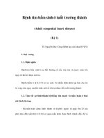
ing of the atrial wall. The morphology of the various types of ASDs is shown
in Fig. 8.1.
Secundum ASDs—defects of the oval fossa—are by far the most common. A
superior sinus venosus ASD occurs when there is a defi ciency of infolding of the
atrial wall in the environs of the superior vena cava (SVC). It is overridden by
the mouth of the SVC, which in turn has a biatrial connection. Most frequently,
the pulmonary veins from part of the right lung are also involved, connect-
ing anomalously to the SVC near to its junction with the atria. Inferior sinus
Fig. 8.1 Various types of ASD as seen from the right side of the heart. RA, right atrium; RV, right
ventricle.
Superior sinus
venous defect
Superior caval
vein
Oval fossa
defect
‘Primum’ defect
Tricuspid valve
Floor of oval
fossa
Inferior sinus
venosus defect
Coronary sinus
defect
Inferior caval
vein
RA
RV
Adult Congenital Heart Disease: A Practical Guide
Michael A. Gatzoulis, Lorna Swan, Judith Therrien, George A. Pantely
Copyright © 2005 by Blackwell Publishing Ltd
68 Chapter 8
venosus ASDs overriding the inferior vena cava (IVC) are much less common.
The rarest type of ASD is a defi ciency of the party wall between the coronary
sinus and the left atrium, producing an interatrial communication through
the mouth of the coronary sinus, a so-called coronary sinus ASD. Primum ASDs
or partial atrioventricular septal defects (AVSD) will be discussed in Chapter 10.
Large intra-atrial communications may represent a confl uence of one type of
ASD with another.
The size of the ASD and the relative compliance of the right ventricle and the
pulmonary vascular bed (in relationship to the left ventricle) determine the
degree of intra-atrial shunting (left to right under normal circumstances for
the vast majority of isolated ASDs).
Associated lesions
When ASD is the primary diagnosis, associated malformations occur in about
30% of cases.
• Partial anomalous pulmonary venous connection (almost universal with
superior sinus venosus ASDs, less common with secundum ASDs and rare
with primum ASDs).
• Pulmonary valve stenosis.
• Mitral stenosis or mitral valve prolapse.
• Ventricular septal defect.
• Patent ductus arteriosus.
• Coarctation of the aorta.
Incidence and etiology
• One of the most common congenital heart disease (CHD) defects as an iso-
lated lesion, occurring in about 6–10% of all cardiac malformations.
• ASD and a bicuspid aortic valve are the two most common CHD defects
presenting in adulthood.
• More common in females (2:1).
• There is a well-recognized association of ASD with Down syndrome (se-
cundum or primum), with Holt Oram syndrome (secundum, see glossary)
and occasionally as a familial occurrence (secundum, associated with delayed
atrioventricular conduction).
• Secundum defects are the most common (60%), with primum defects ac-
counting for 20% and superior sinus venosus defects 15%. The other types are
rare.
Presentation and course in childhood
• Most children with an ASD present with a murmur and are asymptomatic.
• Occasionally, infants may present with breathlessness, recurrent chest in-
fections and even heart failure.
• In the current era, many children are referred to a pediatric cardiologist
for spurious reasons and found to have an atrial septal defect on echocardio-
graphic testing.
Atrial Septal Defects and Anomalous Pulmonary Venous Drainage 69
• Children with sizeable ASDs and right heart dilatation should undergo elec-
tive closure of their defect for prognostic reasons during the fi rst decade of life,
irrespective of symptoms.
Course in adulthood
• Most adults present with symptoms usually in the third or fourth decade of
life. These are usually breathlessness on exertion and/or palpitations due to
atrial tachyarrhythmias. This often correlates with an increase in left-to-right
shunting seen with increasing age.
• Occasionally, adults may present with cardiac enlargement on routine chest
radiograph or a heart murmur. The latter type of presentation is particularly
common among pregnant women, due to enhanced clinical signs (more ob-
vious fl ow murmur and fi xed splitting of the second heart sound) refl ecting
increased circulating plasma volume.
• Adults with ASDs have reduced survival if ASD closure takes place after the
age of 25 years. Other late complications of unrepaired ASDs are right heart
failure, recurrent pneumonia and pulmonary hypertension, atrial fl utter and
fi brillation and paradoxical embolus and stroke.
Examination
The diagnostic work-up should:
• document the ASD, its type and size;
• determine its hemodynamic effects:
– presence and degree of right atrial and ventricular dilatation,
– status of right ventricular function,
– shunt magnitude,
– pulmonary arterial pressure;
• determine the presence of associated anomalies that need to be addressed;
and
• establish whether there is a history of sustained arrhythmia that required
arrhythmia intervention at the time of ASD closure.
It should include a detailed history and clinical examination:
• right ventricular left parasternal impulse;
• wide and fi xed splitting of the second heart sound: cardinal physical sign of an
ASD, not always present;
• pulmonary ejection systolic murmur at the upper left sternal edge;
• tricuspid mid-diastolic murmur at the lower left sternal edge, which may radi-
ate towards the cardiac apex;
• accentuated pulmonary component of the second heart sound, suggesting
raised pulmonary arterial pressure;
• cyanosis; uncommon, more likely with a large defect or virtually common
atrium, an inferior sinus venosus defect, a large coronary sinus defect, with pul-
monary vascular disease, or associated pulmonary stenosis, right ventricular
dysfunction or Ebstein’s malformation.
70 Chapter 8
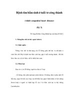
Fig. 8.2 12-lead electrocardiogram from a patient with an intra-atrial communication. Note presence of right bundle branch block (RBBB). Left axis deviation in
keeping with a primum ASD (95% of patients with a primum ASD have a superior axis, i.e. extreme right or left axis deviation). Absence of RVH suggesting no
pulmonary hypertension. Note normal P wave axis (0–60°) making a high sinus venosus ASD unlikely. 1° heart block (common with primum ASD).
Atrial Septal Defects and Anomalous Pulmonary Venous Drainage 71
Useful investigations
• Pulse oximetry: normal oxygen saturations are expected.
• EKG (see Fig. 8.2)
– Right axis deviation and incomplete right bundle branch block pattern are
common.
– Evidence of right ventricular hypertrophy, and lengthening of PR interval
may be present.
– Large P waves, suggesting atrial overload.
• Chest radiography in adults with signifi cant ASDs reveals:
– cardiac enlargement with retrosternal fi lling in the lateral fi lm
– right atrial dilatation
– prominent central pulmonary arteries and pulmonary vascular markings.
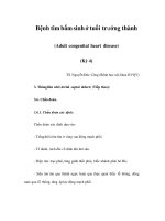
• Echocardiography (see Fig. 8.3)
– The diagnosis is usually confi rmed by cross-sectional echocardiography,
using a combination of subcostal and parasternal four-chamber sections
with colour fl ow Doppler interrogation.
– The most important fi nding is an enlarged right ventricle which might be
the only clue to an ASD in the adult with a poor window.
– The presence of tricuspid regurgitation will permit a Doppler estimate of
pulmonary artery pressure.
Fig. 8.3 Transthoracic echocardiogram from patient with a large secundum ASD. Note massive
dilatation of the right atrium (RA) and right ventricle (RV) with an echo drop-out at the level
of ASD and a squashed left ventricle (LV). Smaller ASDs can be missed with transthoracic
echocardiography. If right heart dilatation is present, transesophageal imaging may be neces-
sary. LA, left atrium.
72 Chapter 8
– A high index of suspicion is sometimes required to make the correct di-
agnosis and transesophageal studies are often needed in the adult patient to
establish the site and size of the defect and the connection of the pulmonary
veins.
– Three-dimensional and intracardiac echocardiogram may also have a role.
Management options for adults with ASD
The management of the adult with an ASD is primarily determined by the size
and type of the defect, associated lesions and the presence and degree of pulmo-
nary vascular resistance. Currently, indications for ASD closure are as follows:
• Presence of an ASD with cardiac enlargement on the chest radiography, a
dilated right ventricle on an echocardiogram and a pulmonary artery systolic
or mean pressure 50% or less than the corresponding aortic pressures. This is
irrespective of symptoms (many of these patients have symptoms such as exercise in-
tolerance without being aware of it). Patients should be considered for elective clo-
sure irrespective of age provided there are no specifi c contraindications (see
below). Younger and older patients would benefi t from ASD closure compared
to medical therapy in terms of:
– survival;
– functional class;
– exercise tolerance;
– reduction of risk of heart failure; and
– reduction of risk of pulmonary hypertension; however,
– patients older than 40 years of age and particularly those with preopera-
tive rhythm disturbance remain at risk of sustained atrial arrhythmia after
closure. For the latter group, consideration should be given to arrhythmia targeted
intervention either via transcatheter techniques, with new mapping and ablative
systems or surgical atrial ablative procedures.
• History of cryptogenic TIA or stroke in the presence of an ASD or persistent
foramen ovale and right-to-left shunting demonstrated on contrast echocar-
diogram.
Contraindications for closure include a pulmonary vascular resistance of more
than 7–8 units or a defect diameter of less than 8 mm (with no evidence of right
heart dilatation) in a patient who is symptom-free.
All secundum defects should be considered for transcatheter closure with one
of the various devices that are available. Defects up to 40 mm in diameter can
be closed with the Amplatzer® septal occluder, usually resulting in improve-
ment in symptoms at any age. Very large oval fossa defects and other types can
be closed only by surgery using cardiopulmonary bypass with the potential
for greater morbidity in the elderly with arrhythmias. Minimally invasive sur-
gery is also an alternative for selected patients.
Device closure
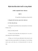
Figure 8.4 shows the sequence of device closure.
• Early and intermediate follow-up is excellent after device closure.
Atrial Septal Defects and Anomalous Pulmonary Venous Drainage 73
• The intermediate results are comparable to surgery with a high rate of shunt
closure and few major complications.
• As with the surgical group, functional capacity improves and supraven-
tricular arrhythmias are better tolerated and more responsive to medical man-
agement.
• Occasionally, residual atrial septal defects are encountered either after cath-
eter or surgical closure. Unless responsible for a signifi cant left-to-right shunt
(i.e. large residual ASDs) generally they do not require additional interven-
tion.
• Longer follow-up is needed to determine the incidence of arrhythmias and
thromboembolic complications late after device closure.
Surgical outcomes
• Secundum ASDs without pulmonary hypertension should undergo surgi-
cal closure with a very low (<1%) operative mortality.
• Early and long-term follow-up is excellent. Preoperative symptoms, if any,
should decrease or abate.
Fig. 8.4 Transcatheter closure of a secundum ASD. Catheter introduced from the right atrium
through the defect to the left atrium (top left panel). Distal part of the device released (top right
panel). Device and introducer are pulled back with the former opposing against the left atrial
aspect of the intra-atrial wall (bottom left panel). Finally, proximal part of the device is released
and deployed (bottom right panel).
74 Chapter 8
• Pre-existing atrial fl utter and fi brillation may persist unless concomitant ar-
rhythmia targeting procedures are performed. Likewise, atrial fl utter and/or
fi brillation may arise de novo after repair in the older patient, but are better
tolerated and often more responsive to antiarrhythmic therapy.
Medical management
This is primarily the management of the associated complications of right heart
failure, atrial tachyarrhythmia and occasionally pulmonary hypertension (see
management of patients with Eisenmenger physiology), when present.
Endocarditis recommendations
Endocarditis prophylaxis is only needed for primum defects and patients
with valvular regurgitation or other associated lesions. Endocarditis prophy-
laxis is also advised for patients undergoing catheter closure for a period of 6
months.
Exercise
Most adults are in New York Heart Association functional class 1 or 2 and
require no limitation on their permitted exercise.
Pregnancy and contraception
Pregnancy is well tolerated by most women with an unoperated atrial septal
defect. Cardiological review is recommended because of the small risk of para-
doxical embolus and stroke, arrhythmia and heart failure. If circumstances
allow, ASDs should be closed prior to pregnancy. However, pregnancy can
usually be allowed to continue. For a secundum defect, transcatheter device
closure can be performed during pregnancy (with transesophageal or intra-
cardiac echocardiography). The only contraindication to pregnancy in women
with ASDs, operated or not, is persisting pulmonary hypertension.
Late complications
• Premature death
• Right heart failure
• Left ventricular dysfunction
• Tricuspid and mitral valve regurgitation
• Atrial fl utter/fi brillation
• Sinus node dysfunction
• Paradoxical thromboembolism
• Endocarditis (rare)
• Systemic arterial hypertension
• Pulmonary hypertension/pulmonary vascular disease (usually a very late
complication)