Báo cáo y học: " Magnetic resonance imaging with pathological correlation in a case of mantle cell lymphoma of the parotid gland: a case report" ppsx
Bạn đang xem bản rút gọn của tài liệu. Xem và tải ngay bản đầy đủ của tài liệu tại đây (1.17 MB, 5 trang )
CAS E REP O R T Open Access
Magnetic resonance imaging with pathological
correlation in a case of mantle cell lymphoma
of the parotid gland: a case report
Mayia Pilavaki
1*
, Anastasia Athanasiadou
2
, Fotis Iordanidis
3
, Thrasivulos Karakozoglou
1
, Panagiotis Palladas
1
Abstract
Introduction: Mantle cell lymphoma is a rare non-Hodgkin’s lymphoma. It is a subtype of B-cell lymphoma with
frequent involvement of the bone marrow and the gastrointestinal tract. Isolated parotid gland involvement
seldom occurs. Here we report an unusual case of isolated infiltration of the parotid gland by mantle cell
lymphoma. The aim of our study is to correlate magnetic resonance imagi ng findings with the histological feature s
of the disease. To the best of our knowledge, no similar radiological findings of mantle cell lymph oma have been
published before.
Case presentation: A 72-year-old Caucasian woman presented with a painful left parotid enlargement. She was
diagnosed with mantle cell lymphoma involving the left submandibular gland seven years prior to presentation.
Her whole body CT scan showed the absence of pathologically enlarged lymph nodes. However, a magnetic
resonance imaging showed enlargement of her left parotid gland and an abnormal parenchyma with mixed-type
solid and cystic lesions. A biopsy of her left parotid gland and subsequent histolog ical examination confirmed a
mantle c ell lymphoma (common variant) relapse.
Conclusion: Although rare, the involvement of parotid gland with mantle cell lymphoma must be considered in
the differential diagnosis of parotid tumors.
Introduction
Mantle cell lymphoma (MCL) is a rare lymphoma that
accounts for approximately 5% to 7% of non-Hodgkin’s
lymphomas (NHL). It is us ually characterized by an aggres-
sive clinical course with a median overall survival of two to
five years [1]. The involvement of extranodal sites is not
uncommon as patients usually have an advanced-stage dis-
ease at the time of diagnosis [2]. According to Argatoff
et al., the isolated extranodal location of the disease has
been referred in 25% of cases, whereas salivary glands are
rarely affected (only 3% of reported cases) [1]. Here we pre-
sentacaseofMCLoftheparotidgland.Wehavealsocor-
related its appearance at MRI with its histological findings.
Case presentation
A 72-year-old Caucasian woman was referred to
the Department of Haematology, General Hospital
G. Papanikolaou, with a painful left parotid enlargement
but without any other physical findings. Seven years
prior to this presentation, she was diagnosed with MCL
in her left submandibular gland, which was treated with
surgical removal, radiotherapy, and a chemotherapy
regimen of cyclophosphamide, hydroxydaunorubicin
(Adriamyc in), Oncovin (vincristine) and prednisone/pre-
dnisolone (CHOP). Her white blood cell (WBC) count
of 5100/μL was represented by a normal differential of
69% neutrophils, 25% lymphocytes (with normal mor-
phology), 5% monocytes and 1% eosinophils. Her hemo-
globin, hematocrit and platelet levels were normal at
12.7 g/dL, 38.4% and 219/μL, respe ctively. Her bone
marrow smears and biopsy were both normal without
evidence of any infiltration by lymphoma cells.
An ultrasound examination of our patient r evealed
multiple cystic lesions in the parenchyma of her left par-
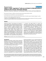
otid gland. Her MRI showed an enlargement o f her left
parotid gland and the total replacement of her normal
parenchyma with mixed-type solid and cystic lesions
* Correspondence:
1
Department of Radiology, General Hospital G Papanikolaou, Thessaloniki,
Greece
Pilavaki et al. Journal of Medical Case Reports 2010, 4:99
/>JOURNAL OF MEDICAL
CASE REPORTS
© 2010 Pilavaki et al; licensee B ioMed Central Ltd. This is an Open Access article distrib uted under t he terms o f the Cre ative Commons
Attribution License ( which permits unrestricted use, distribution, and reproduction in
any medium, provided the origin al work is properly cited.
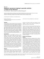
(Figure 1). The solid components were mildly enhanced
after an intravenous administration of contrast medium
(Figures 2A and 2B). A whole body computed tomogra-
phy (CT) scan showed the absence of pathologically
enlarged lymph nodes. All the other organs of her chest
and abdomen were also found to be normal.
A bi opsy of her left parotid gland and subsequent his-
tological examination showed the presence of MCL
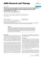
(common variant). The MCL was composed of mono-
morphous small to medium-sized lymphoid cells which
most closely resembled centrocytes with a vaguely nodu-
lar growth pattern. The prominent neoplastic nodules
were adjacent to the cystic area (Figure 3A).
Immunostaining results for CD20, CD79a, CD43,
CD5, sIgM(l+) and cyclin D1 were positive (Figures 3B,
C and 3D) and negative for CD23 and CD3.
Our patient was treated with partial parotidectomy
and chemotherapy. Her post-operative course was
uncomplicated. Eighteen months after surgery she was
asymptomatic and disease-free.
Discussion
Mantle cell lymphoma is a relati vely rare and aggressive
form of NHL. In the past, it has been referred to with
various names including intermediately differentiated
lymphocytic lymphoma, centrocytic lymphoma, and
mantle zone lymphoma [1]. MCL has a characteristic
morphological appearance consisting of small lymphoid
cells with slightly irregular nuclear cells. Its histological
growth patterns are of a nodular or diffuse type, or a
combination of these two types [2,3]. In nodular MCL,
some or many of the nodules may consist of follicles
with reactive germinal centers surrounded by broad
mantles of small lymphoid cells or the so-called mantle
zone pattern [2]. Later in the course of the disease the
mantle zonal or nodular pattern progresses to a diffuse
pattern [4].
The immunohistological features of MCL reveal a
characteristic phenotype. The cells express relatively
intense surface immunoglobulin M (IgM) and/or immu-
noglobulin D (IgD) and are usually positive for CD5,
Figure 1 Axial T2-weighted image reveals an enlargement of the left parotid gland and a total replacement of the normal
parenchyma with mixed-type solid and cystic lesions.
Pilavaki et al. Journal of Medical Case Reports 2010, 4:99
/>Page 2 of 5
Figure 2 (A) Coronal T1-weighted image shows the inhomogenic appearance of the left parotid gland. (B) Coron al T1-weighted image
after gadolinium contrast injection shows mild enhancement of the solid components, while the cystic areas remain hypodense.
Figure 3 (A) Microscopic appearance shows prominent vaguely neoplastic nodules (arrows) which are adjacent to the cystic area (red
area) (hematoxylin and eosin staining, ×25 magnification). (B) Immunohistochemical stains for CD20, (C) CD5, and (D) cyclin D1 are positive
(×100 magnification).
Pilavaki et al. Journal of Medical Case Reports 2010, 4:99
/>Page 3 of 5
FMC7 and CD43, but negative for CD10 and BCL6.
CD23 is negative or weakl y positive. Almost all cases of
MCL a re moderately to strongly positive for cyclin D1.
Cyclin D1 expression can be detected in a subset of
cases of chronic lymphocytic leukemia (CLL) and/or
small cell lymphoma (SLL) and hairy cell leukemia.
Usually, cyclin D1 is weakly positive in these neoplasms.
Cyclin D1 can also be expressed strongly in approxi-
mately one-quarter of plasma cell myeloma cases.
Conventional cytogenetic analysis demonstrates a
translocation between the immunoglobulin heavy chain
and cyclin D1 (CCND1, BCL1 and PRAD1) genes,
t(11;14)(q13;q32) in 70% t o 75% of cases. However,
almost all cases demonstrate the rearrangement of these
genes using fluorescence in situ hybridization (FISH)
probes [ 5]. This translocation can rarely occur in other
types of B-cell NHL, in lymphocytic leukemia, and in
multiple myeloma [2]. Therefore , cytogenetic findings
need to be correlated with its pathological and immuno-
logical features to confirm a diagnosis of MCL.
Clinically, MCL occurs in middl e-aged to older indivi-
duals with a median age of about 60 years, and predo-
minantly in men [2]. Most patients present with a stage
III or IV disease. According to Argatoff et al., in a clini-
copathological study of 80 cases, extranodal involvement
at presentation occurred in 76% of cases and the most
common sit es were the bone marrow (63%), peripheral
bloo d (34%), gast rointestinal tract (10%) and Waldeyer’ s
ring (10% ). In 25% of cases the extranodal location was
the disease’s primary presentation, whil e the most com-
mon sites were the Waldeyer’s ring (6%), intestine (5%),
orbit (3%) and salivary gland (3%) [1].
MRI is the method of choice when treating patients
with palpable parotid gland masses to assess the exact
extent of the tumors, the i nvasion of neighboring struc-
tures, perineural spread, and lymph nodes staging [6].
Most parotid tumors, whether benign or malignant,
rarely replace the parenchyma of the gland to tally.
Moreover, diffuse infiltration is often seen in lympho-
mas. The lymph omas have a homogenous signal pattern
with l ow intensity on T1-weighted and high intensity on
T2-weighted sequences. Although this MRI pattern is
highly suggestive of the lymphoma, there is no absolute
correlation between the imaging morphology and the
histology of the lesion [7].
In this case, our patient’s MRI showed an enlargement
of her left parotid gland and a total replacement of her
normal parenchyma with mixed-type solid and cystic
lesions. The solid components were mildly enhanced
using contrast medium. This appearance reflected the his-
tological pattern of the lesion as MCL was composed of
neoplastic nodules which were adjacent to the cystic area.
To the best of our knowledge, there are no other pub-
lished radiological findings on MCL of the parotid gland.
The differential diagnosis includes benign and malignant
parotid tumors, especially Warthin tumors and adenoid
cystic carcinomas, which may also have a solid cystic
appearance. These tumors rarely occupy the total gland
parenchyma. In particular, Warthin tumors are bilateral in
up to 10% of cases reported. They present as well-circum-
scribed, partly cystic and partly solid lesions on MRI and
are often located in the tail of the parotid gland. Adenoid
cystic carcinoma usually presents as an infiltrating mass
withahighpropensityforperineuralinvasion.OnMRI
adenoid cystic carcinoma has an irregular contour, poorly
defined margins, and a strong enhancement after the
administration of contrast medium [6].
Special caution is required in the follow-up examina-
tion of patients with primary Sjögren’s syndrome, as the
risk of developing a lymphoma is increased. In this case
the typical inhomogeneous nodular MRI picture seen in
Sjögren’ s syndrome will change into a homogeneous
pattern that can involve the parenchyma partially or
even entirely [7].
Conclusion
Isolated parotid gland involvement by MCL is very rare
but should be considered nonetheless in the differential
diagno sis when replacemen t of a patient’s normal paro-
tid parenchyma with mixed-type solid and cysti c lesions
is involved.
Consent
Written informed consent was obtained from the patient
for publicatio n of this case report and any accompany-
ing images. A copy of the written consent is available
for review by the Editor-in-Chief of this journal.
Abbreviations
CHOP: cyclophosphamide, hydroxydaunorubicin (Adriamycin), Oncovin
(vincristine) and prednisone/prednisolone; CLL: chronic lymphocytic
leukemia; CT: computed tomography; Ig: immunoglobulin; MCL: mantle cell
lymphoma; NHL: non-Hodgkin’s lymphoma; SLL: small cell lymphoma; WBC:
white blood cell.
Author details
1
Department of Radiology, General Hospital G Papanikolaou, Thessaloniki,
Greece.
2
Department of Haematology, General Hospital G Papanikolaou,
Thessaloniki, Greece.
3
Laboratory of Pathology, General Hospital G
Papanikolaou, Thessaloniki, Greece.
Authors’ contributions
MP performed the chart review and prepared the manuscript. AA evaluated
and treated our patient, and also helped prepare the manuscript. FI was the
pathologist who examined the specimens from our patient. TK and PP
participated in manuscript preparation. All authors read and approved the
final manuscript.
Competing interests
The authors declare that they have no competing interests.
Received: 4 November 2009 Accepted: 30 March 2010
Published: 30 March 2010
Pilavaki et al. Journal of Medical Case Reports 2010, 4:99
/>Page 4 of 5
References
1. Argatoff LH, Connors JM, Klasa RJ, Horsman DE, Gascoyne RD: Mantle cell
lymphoma: a clinicopathologic study of 80 cases. Blood 1997,
89:2067-2078.
2. Weisenburger DD, Armitage JO: Mantle cell lymphoma: an entity comes
of age. Blood 1996, 87:4483-4494.
3. Asaad NY, Abd El-Wahed MM, Dawoud MM: Diagnosis and prognosis of
B-cell chronic lymphocytic leukemia/small lymphocytic lymphoma
(B-CLL/SLL) and mantle cell lymphoma (MCL). J Egypt Natl Canc Inst
2005, 17:279-290.
4. Fisher RI, Dahlberg S, Nathwani BN, Banks PM, Miller TP, Grogan TM:
A clinical analysis of two indolent lymphoma entities - mantle cell
lymphoma and marginal zone lymphoma (including the mucosa-
associated lymphoid tissue and monocytoid B-cell subcategories):
a Southwest Oncology Group study. Blood 1995, 85:1075-1082.
5. Sun T, Nordberg ML, Cotelingam JD, Veillon DM, Ryder J: Fluorescence in
situ hybridization: method of choice for a definitive diagnosis of mantle
cell lymphoma. Am J Hematol 2003, 74:78-84.
6. Thoeny HC: Imaging of salivary gland tumors. Cancer Imaging 2007,
7:52-62.
7. Makula E, Pokorny G, Kiss M, Vörös E, Kovács L, Kovács A, Csernay L,
Palkó A: The place of magnetic resonance and ultrasonographic
examinations of the parotid gland in the diagnosis and follow-up of
primary Sjögren’s syndrome. Rheumatol 2000, 39:97-104.
doi:10.1186/1752-1947-4-99
Cite this article as: Pilavaki et al.: Magnetic resonance imaging with
pathological correlation in a case of mantle cell lymphoma of the
parotid gland: a case report. Journal of Medical Case Reports 2010 4:99.
Submit your next manuscript to BioMed Central
and take full advantage of:
• Convenient online submission
• Thorough peer review
• No space constraints or color figure charges
• Immediate publication on acceptance
• Inclusion in PubMed, CAS, Scopus and Google Scholar
• Research which is freely available for redistribution
Submit your manuscript at
www.biomedcentral.com/submit
Pilavaki et al. Journal of Medical Case Reports 2010, 4:99
/>Page 5 of 5