Báo cáo y học: "Acute presentation of a benign cystadenofibroma of the fallopian tube: a case report" potx
Bạn đang xem bản rút gọn của tài liệu. Xem và tải ngay bản đầy đủ của tài liệu tại đây (831.42 KB, 5 trang )
JOURNAL OF MEDICAL
CASE REPORTS
de Silva et al. Journal of Medical Case Reports 2010, 4:181
/>Open Access
CASE REPORT
© 2010 de Silva et al; licensee BioMed Central Ltd. This is an Open Access article distributed under the terms of the Creative Commons
Attribution License ( which permits unrestricted use, distribution, and reproduction in
any medium, provided the original work is properly cited.
Case report
Acute presentation of a benign cystadenofibroma
of the fallopian tube: a case report
Tania S de Silva*, Abhijeet Patil and Roy N Lawrence
Abstract
Introduction: Cystadenofibromas are rare benign tumors of the fallopian tube with only 15 reported cases worldwide.
They are usually asymptomatic and are found incidentally. This case is presented on account of its rarity and to the best
of our knowledge, is the first reported case of cystadenofibroma of the fallopian tube discovered during an
appendicectomy.
Case presentation: We report a rare case of cystadenofibroma of the fallopian tube in a 19-year-old Caucasian woman
who presented with sudden onset of right iliac fossa pain. A clinical diagnosis of appendicitis was made and she was
taken to the operating theater for an appendicectomy. Intraoperatively, the appendix appeared normal. However, the 8
cm cyst contained within the right ovary and the blood in the pelvis warranted a salpingo-oopherectomy. Our patient
made an uneventful recovery and was discharged after four days. Histology revealed a benign cystadenofibroma of the
fallopian tube. There was no evidence of recurrence in the follow-up period of 12 months.
Conclusion: Cystadenofibromas are benign tumors that may macroscopically and ultrasonographically appear
malignant. We recommend that the diagnosis of cystadenofibroma is considered prior to performing radical surgery
that may affect the fecundity of these patients. Cystadenofibromas confined to the fallopian tube can be treated
curatively with unilateral salpingo-oophorectomy, without the need for any further treatment. However, long-term
follow-up of more cases is required to draw more definitive conclusions.
Introduction
Cystadenofibromas are rare benign tumors of the fallo-
pian tube with only 15 reported cases worldwide [1].
They are usually asymptomatic and are found incidentally
[2]. Sometimes they are discovered during evaluation for
in vitro and fertilization-embryo transfer [3]. In the case
of our patient, the presentation was that of an acute abdo-
men due to hemorrhagic necrosis of the tumor. Malig-
nant potential is very rare. However, it may
macroscopically and ultrasonographically appear malig-
nant resulting in most of these younger women having
radical surgery affecting their fecundity. It is therefore
advisable to consider the possibility of cystadenofibroma
prior to selecting an aggressive surgical approach in
younger patients.
Here we report a rare case of a 19-year-old woman with
cystadenofibroma of the fallopian tube, presenting with
an acute abdomen, which was treated with a right sal-
pingo-oophorecotmy.
Case presentation
A 19-year-old nulliparous, British-Caucasian woman pre-
sented with a one-day history of worsening right iliac
fossa pain associated with nausea and vomiting. Her pre-
vious medical and gynecological history has been
uneventful. The menses of our patient (four-day duration,
28-day cycle) were regular. Abdominal examination
revealed percussion tenderness over the right iliac fossa
and a positive Rovsig's sign warranting an appendectomy.
The white cell count and C-reactive protein levels were
mildly elevated and no morphological examinations were
performed. Our patient was taken to the operating the-
ater with a view to performing an open appendectomy.
However, intra-operative findings revealed free fluid in
the pelvis with a normal-looking appendix. The right
adenexa was found to be twisted three times and was
necrotic. The ovary measured 8 cm and the fallopian tube
was found to be distended and necrotic. A right salpingo-
* Correspondence:
1
Department of General Surgery, The Great Western Hospital, Marlborough
Road, Swindon, UK
Full list of author information is available at the end of the article
de Silva et al. Journal of Medical Case Reports 2010, 4:181
/>Page 2 of 5
oophorectomy was performed and the left ovary was
checked to be normal.
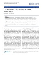
As seen in Figure 1, the histological analysis showed
congestion, hemorrhage and coagulative necrosis of the
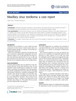
fallopian tube. The broad-based papillary lesion showed a
fibrotic stroma forming broad leaf like projections lined
by a low cuboidal ciliated epithelium as seen in Figures 2
and 3. These features are consistent with benign cystade-
nofibroma. The ovary also showed severe hemorrhage
and congestion with coagulative necrosis.
Our patient had an uneventful recovery and was dis-
charged after four days. She has since been followed up in
the out-patients clinic over a 12-month period, and was
found to do well with no evidence of recurrence of dis-
ease.
Discussion
Adenofibromas are relatively rare benign tumors with
rare malignant potential, arising from the germinal lining
and ovarian stroma. The relative amounts of the epithe-
lial and stromal constituents and the secretary activity of
the epithelial component will determine the solid, semi-
solid or liquid state of the tumor. The majority of the
reported adenofibromas are of the serous type. However,
endometrioid, clear cell and mucinous types also exist [4].
Having performed a Medline database search with the
keywords "cystadenofibroma" and "fallopian tube", we
analyzed the relevant articles and their cited references,
in order to construct Table 1. Table 1 shows a systematic
review of the cases of cystadenofibroma, specifically aris-
ing from the fallopian tube. During this search we
encountered a number of cases of the tumor arising
within the ovary, which were excluded from Table 1. The
search revealed only five cases previously reported in the
English literature. Clinico-pathological features of these
tumors, including the current case, are summarized in
Table 1.
Review of the current literature suggests that cystade-
nofibromas generally present in the fourth and fifth
decades in the life of a patient. However, they appear to
present earlier in a subset of women exposed to antenatal
diethylsilbestrol [5].
The presenting symptoms of this tumor include
abdominal pain, increased abdominal girth, dysuria, rec-
tal urgency, vaginal bleeding and feminization [6]. One
school of thought suggests that the feminization and vag-
inal bleeding symptoms are due to excessive estrogen
secretion by the tumor causing abnormal endometrial
growth [7,8]. However, other authors failed to prove
excessive endometrial growth [7,9,10].
Figure 1 A histology slide of right fallopian tube specimen taken
at time of surgery showing congestion, hemorrhage and coagu-
lative necrosis.
Figure 2 Histology slide of right fallopian tube specimen taken at
time of surgery. The broad based papillary lesion, showed a fibrotic
stroma forming broad leaf like projections lined by a low cuboidal cili-
ated epithelium.
Figure 3 A histology slide of right fallopian tube specimen taken
at time of surgery showing fibrotic stroma lined by cuboidal epi-
thelium.
de Silva et al. Journal of Medical Case Reports 2010, 4:181
/>Page 3 of 5
Table 1: Summary of current cases and their clinico-pathological features of these tumors
Author year Age Clinical symptoms Surgical intervention Outcome Pathology
[11] Silverman AY 1978 36 Finding during tubal ligation
following termination of
pregnancy
Bilateral partial
salpingectomy
3.5 cm cyst with small papillary projections
supported by central cores of fibrous tissue
and covered with ciliated cuboidal to columnar
epithelial cells
[12] Valerdiz CS et al. 1989 49 Incidental finding during salpingo
oophorectomy for leiomyomas
Salpingo oophorectomy WED after
unspecified time
cystadenofibroma
[12] Valerdiz CS et al. 1989 32 Incidental finding during early
pregnancy
salpingectomy WED after
unspecified time
2.5 cm cyst with papillations- Borderline
cystadenofibroma
[2] Gurbuz Y et al. 2003 48 Incidental finding in woman with
leiomyoma uterei
?Salpingectomy 2 serous papillary cystadennofibromas.
Immunohistochemistry suggested tumor was
an embryonic remnant of mullerian duct
[3] Sills ES et al. 2003 Infertility,
discovered during
evaluation for IVF
Laparoscopic decompression and
removal of intact cyst
WED after 3 months
follow-up
Benign serous cystadenofibroma
De Silva et al. 2009 19 Acute onset right iliac fossa pain Open salpingo
oophorectomy
WED after 12 months Benign cystadenofibroma of fallopian tube and
coagulative necrosis of ovary
IVF, in vitro fertilization; WED, Well enough for discharge from clinic.
de Silva et al. Journal of Medical Case Reports 2010, 4:181
/>Page 4 of 5
The diagnosis of cystadenofibroma is a difficult one, as
they macroscopically and ultrasonographically appear
malignant. They may grow up to 20 cm in diameter,
encapsulated and multiloculated with short broad papil-
lary projections. Laparosopy may also be used in the
diagnosis and even treatment of this condition as demon-
strated by Sills et al. In this paper, they described how the
cyst was decompressed and removed intact without inci-
dent via a 5 mm laparoscopic cannuala [3].
Czernobilsky et al. studied 34 patients with benign
serous cystadenofibromas and found the same favorable
outcome in all patients irrespective to whether they
underwent conservative cystectomy, oophorectomy or
total abdominal hysterectomy and bilateral salpingo-
oophorectomy [10].
Conclusions
Cystadenofibromas of the fallopian tube are benign
tumors with rare malignant potential. Therefore, we
advise to consider this diagnosis before employing radical
surgery in younger women, as this would impact their
fertility. However, in patients over 50 years of age, pre-
senting with any ovarian or fallopian tube tumor, there is
no need for a conservative approach. In the treatment of
younger patients of childbearing age, we found salpingo-
oophorectomy to be curative without the need for any
further treatment. However, long term follow-up of more
cases are required to make more definitive conclusions.
This case is presented on account of its rarity and we
believe this is the first reported case of cystadenofibroma
of the fallopian tube to present acutely and discovered
during an appendicectomy.
Patient's perspective
I write the following to provide assistance to the case
report written about my operation. I have no medical
knowledge or background so I only write from my own
perspective and experience.
Before the morning I was taken to hospital I had never
experienced abdominal pains, either related to my men-
strual cycle or other. I had never been submitted to hospi-
tal for any previous health concerns. It was the summer
after my first year at University, I was working as a full
time Assistant Director, working long hours, the job was
very active and predominantly outdoors (it was an out-
door production). I was 19 years old. At the time of being
submitted to hospital I was on the third day of my period,
at this age I experienced regular monthly periods lasting
seven days. I awoke very early on that morning with no
pain. I then went back to sleep but was awoken with a
severe pain in my abdomen. I also felt very hot, dizzy and
clammy. I tried to recover by taking a cool bath, drinking
water and then lying flat on the floor breathing deeply.
This did not help and the pain began to increase to an
unbearable level. An ambulance was called for, whilst
waiting for them I continued to lie flat on the cool bath-
room floor with the windows open.
When the ambulance arrived the ambulance woman
asked if I was possibly pregnant. I said no, there was no
possibility of this. They then made the presumption that
it was due to drug or alcohol abuse. Again I said it was
not. She then insisted it was food poisoning, I explained
that the pain was far more severe than food poisoning.
Finally she said that she would take me into a hospital
despite not feeling it was necessary. Despite my career in
theater I am not overly dramatic and despite the pain I
was able to converse and I suppose did not appear to be in
as much pain as I probably was. But it hurt in a way I
could never put into words. I was driven to the Accident
and Emergency unit. While in the ambulance I was giving
a mask to breathe through and told it would help the
pain; it had no affect at all. At the hospital I was put into a
cubicle. A nurse then gave me an injection in my arm, I
don't know what of. Whatever it was it relieved the pain
instantly. I could literally feel the pain dissolve as I was
given the injection - it was a heavenly experience and a
great relief. A doctor then visited me and began to apply
pressure to my abdomen, asking if I was in pain whilst he
put pressure on different areas. I explained that when he
pressed down on my abdomen, it did hurt. The pain I
experienced was mainly on the right lower side. Again the
doctor suggested I had food poisoning; I had gone to a
barbecue the night before. I was taken up to the ward and
it was then that it was suggested I possibly had appendici-
tis, I cannot remember much of this period up until it was
decided that I be operated on. I drifted in and out of sleep
and in severe pain. The morning of my operation I did
not feel in as much pain as when I first entered hospital,
but felt physically washed out and very tired. I remember
seeing the consultant who said I looked very grey and that
it was necessary to operate and remove my appendix. I
was taken down to theater and awoke later. It was then
explained to me that my appendix was removed, but also
my right ovary and fallopian tube. I was connected to a
morphine drip, which I controlled and used a lot. The
next morning I was taken to have an X-ray so that they
could find out what was wrong, this was until I explained
that I had already had an operation. Most nights I would
be sick after eating a small amount of toast and ice cream
during the day. I went home after a few days, which I
strongly pushed for because it was very uncomfortable
being in hospital on a ward with lots of elderly ladies. I
spent approximately three weeks recovering at home.
After about a week a stitch in my appendix scar became
infected, literally the wound bled severely and I was taken
to my local hospital, where they squeezed the wound
until the stitch came out. Apart from this my recovery
had no problems, it was uncomfortable to sleep, and I
de Silva et al. Journal of Medical Case Reports 2010, 4:181
/>Page 5 of 5
couldn't eat strong flavored food and felt tired. I returned
to University at the end of September, I took it easy and
felt delicate for a further four weeks until feeling fully
back to health by the end of October. I have two scars to
remind me of my experience, but both healed well. I do
now suffer from minor pain each month before my
period begins, which I never did before the operation.
Consent
Written informed consent was obtained from our patient
for the publication of this case report and any accompa-
nying images. A copy of the written consent is available
for review by the Editor-in-Chief of this journal.
Competing interests
The authors declare that they have no competing interests.
Authors' contributions
Our patient was admitted under the care of RL during this episode and was fol-
lowed up in outpatients' clinic. AP and TSdS were major contributors in writing
the manuscript. All authors read and approved the final manuscript.
Acknowledgements
Dr Lawrence John (Consultant histopathologist) for analyzing and interpreting
histological data and providing pathology images.
Author Details
Department of General Surgery, The Great Western Hospital, Marlborough
Road, Swindon, UK
References
1. WHO Classification of Tumors: Pathology and genetics of tumours of the
breast and female genital organs. 2003.
2. Gurbuz Y, Ozkara SK: Immunohistochemical profile of serous papillary
cystadenofibroma of the fallopian tube: a clue of paramesonephritic
origin. Appl Immunohistochem Mol Morphol 2003, 11(2):153-155.
3. Sills ES, Kaplan CR, Perloe M, Tucker MJ: Laparoscopic approach to an
uncommon adnexal neoplasm associated with infertility: serous
cystadenofibroma of the fallopian tube. J Am Assoc Gynecol Laparosc
2003, 10(4):545-547.
4. Wolfe SA, Seckinger DL: Various anatomical types of ovarian
adenofibroma. Am J Obstet Gynecol 1967, 99:121-125.
5. Schmidt G, Fowler WC: Ovarian cystadenofibroma in three women with
antenatal exposure to diethylstilbestrol. Gynecol Oncol 1982,
14:175-184.
6. Groutz A, Wolman I, Wolf Y, Luxman D, Sagi J, Jaffa AJ, David MP:
Cystadenofibroma of the ovary in young women. Eur J Obstet Gyanecol
Reprod Biol 1994, 54:137-139.
7. Bell DA, Scully RE: Benign and borderline clear cell adenofibromas of
the ovary. Cancer 1985, 56:2922-2931.
8. Mc Nulty JR: The ovarian serous cystadenofibroma, a report of 25 cases.
Am J Obstet Gynecol 1959, 77:1338-1347.
9. Roth LM, Langley FA, Fox H, Wheeler JE, Czernobilski B: Ovarian clear cell
adenofibromatous tumours. Cancer 1984, 53:1156-1163.
10. Czernobilsky B, Borenstein R, Lancet M: Cystadenofibroma of the ovary.
Cancer 1974, 34:1971-1981.
11. Silverman AY, Artenian B, Sabin M: Serous cystadenofibroma of the
fallopian tube: a case report. Am J Obstet Gynecol 1978, 130(5):593-595.
12. Valerdiz CS, Pardo MJ: Cystadenofibroma of fallopian tube. Appl Pathol
1989, 7(4):256-259.
doi: 10.1186/1752-1947-4-181
Cite this article as: de Silva et al., Acute presentation of a benign cystadeno-
fibroma of the fallopian tube: a case report Journal of Medical Case Reports
2010, 4:181
Received: 24 September 2009 Accepted: 17 June 2010
Published: 17 June 2010
This article is available from: 2010 de Silva et al; licensee BioMed Central Ltd. This is an Open Access article distributed under the terms of the Creative Commons Attribution License ( ), which permits unrestricted use, distribution, and reproduction in any medium, provided the original work is properly cited.Journal of Medical Case Reports 2010, 4:181