Báo cáo y học: "Bleeding from ruptured hepatic metastases as a cause of syncope in an octogenarian: a case report" pdf
Bạn đang xem bản rút gọn của tài liệu. Xem và tải ngay bản đầy đủ của tài liệu tại đây (729.48 KB, 3 trang )
JOURNAL OF MEDICAL
CASE REPORTS
Seetho et al. Journal of Medical Case Reports 2010, 4:194
/>Open Access
CASE REPORT
© 2010 Seetho et al; licensee BioMed Central Ltd. This is an Open Access article distributed under the terms of the Creative Commons
Attribution License ( which permits unrestricted use, distribution, and reproduction in
any medium, provided the original work is properly cited.
Case report
Bleeding from ruptured hepatic metastases as a
cause of syncope in an octogenarian: a case report
Ian W Seetho
1
, Simon Stinchcombe
2
and Mazen M Rizeq*
3
Abstract
Introduction: Acute hemoperitoneum as a result of hemorrhage from liver metastases is an uncommon but serious
condition. The use of appropriate imaging is important in the diagnosis and can have a profound impact on
subsequent management. This case is important because the presentation was of recurrent syncopal episodes with an
unusual underlying cause. This case highlights the need to consider this diagnosis in the differential in patients
presenting with collapse in the acute setting.
Case presentation: We present the case of an 85-year-old Caucasian man who was admitted following a collapse
episode and was found to be persistently hypotensive despite aggressive resuscitation. An acute intra-peritoneal bleed
originating from hepatic metastases from an unknown primary was identified promptly with computed tomography
imaging and was subsequently managed conservatively.
Conclusions: This case aims to convey key teaching points: (A) the need to consider intra-abdominal hemorrhage in
the differential diagnosis when assessing patients with collapse; and (B) the use of appropriate imaging such as
computed tomography can facilitate a prompt diagnosis and appropriate management steps can then be taken
accordingly.
Introduction
Spontaneous rupture of hepatic metastases leading to
hemoperitoneum may initially present as collapse in the
elderly and is a serious diagnosis. In this case report, we
present a patient who was admitted following recurrent
syncopal episodes with clinical features of persistent
hypotension. A sudden fall in his hemoglobin level sug-
gested that an acute bleed had led to his collapse. This
was an important investigation finding in determining
the cause of his syncopal episodes. The underlying diag-
nosis of acute hemorrhage from liver metastases was con-
firmed on computed tomography (CT) imaging.
Case presentation
An 85-year-old Caucasian man was admitted to hospital
following three collapse episodes with transient loss of
consciousness at home. Each episode was short-lived
lasting several minutes. Apart from mild abdominal gen-
eralized discomfort, there were no other symptoms.
There was no history of recent trauma. He had no history
of similar episodes but was known to have severe aortic
stenosis, type 2 diabetes, paroxysmal atrial fibrillation,
hypertension and a previous duodenal ulcer bleed.
At that time, he was taking aspirin, bisoprolol, omepra-
zole and ramipril. He lived with his daughter and was
independent with his activities of daily living. He had not
smoked for 35 years and his alcohol consumption was
minimal.
On examination, he was apyrexial, oxygen saturation
was 100% on air. His blood pressure was 80/40 mmHg.
He was persistently hypotensive despite aggressive fluid
resuscitation. There was an ejection systolic murmur on
cardiac auscultation. His venous pressure was not ele-
vated and there was no leg edema. The lungs were clear
on auscultation. Upper and lower limb pulses were equal
bilaterally. Examination of his abdomen revealed mild
epigastric discomfort, but there was no rebound or peri-
tonism and bowel sounds were present. Per rectal exami-
nation was normal.
Initial blood results showed a hemoglobin of 11.3 (13-
18 g/dL), white cell count of 11 (4-11 × 10
9
/L), and plate-
* Correspondence:
3
Department of Stroke Medicine and Medicine for the Elderly, King's Mill
Hospital, Sherwood Forest Hospitals NHS Foundation Trust, Mansfield Road.
Nottingham NG17 4JL, UK
Full list of author information is available at the end of the article
Seetho et al. Journal of Medical Case Reports 2010, 4:194
/>Page 2 of 3
lets of 136 (150-450 × 10
9
/L). Coagulation profile, renal
function and liver function tests were within normal lim-
its. His chest radiograph was normal and his electrocar-
diogram showed left ventricular hypertrophy. At
admission, he was taken to the coronary care unit for car-
diac monitoring because of the history of collapse with
loss of consciousness which was thought to be related to
his aortic stenosis. An urgent echocardiogram was per-
formed which showed evidence of aortic stenosis, but no
evidence of critical stenosis with good ejection fraction >
55% and good biventricular contraction.
A repeat full blood count showed that his hemoglobin
had fallen to 4.9 g/dL and he was transfused with red
cells, platelets and cryoprecipitate. The impression was
that this patient had a possible dissecting thoracic aneu-
rysm that was possibly extending into the abdomen. He
was transferred to the intensive care unit.
In view of the differential diagnosis of a possible dissec-
tion, an urgent chest and abdomen CT scan was per-
formed which showed normal appearances of the
thoracic and abdominal aorta with no evidence of aneu-
rysm or dissection. However, the scan revealed a large
amount of free intra-peritoneal fluid with areas of low
attenuation in the right lobe of the liver. The appearances
were concluded to be of metastatic disease within the
liver (Figures 1, 2, 3. No primary tumor was identified. A
diagnostic peritoneal tap was performed and frank blood
was aspirated confirming that there was hemoperito-
neum. An acute intra-abdominal bleed from the liver
metastatic disease was diagnosed.
Our patient had an esophageal gastro-duodenal endos-
copy as he had been taking aspirin and had a past history
of a duodenal ulcer. This did not show any evidence of
bleeding. A rigid sigmoidoscopy was also normal.
Whilst on the intensive care ward, our patient's blood
pressure subsequently improved and he did not require
inotropic support. He had no further hypotensive epi-
sodes and improved during his stay on the ward. Given
the advanced nature of his hepatic metastases, he did not
wish to have further investigations to identify the primary
source of the metastases and decided on conservative
supportive treatment, as advised by the oncologists. He
was referred to the Macmillan and Hospital Palliative
Care Team. Subsequently, he died three months later. A
post-mortem was not performed.
Discussion
Tumor perforation and bleeding may occur as a compli-
cation of primary hepatocellular carcinoma [1]. This
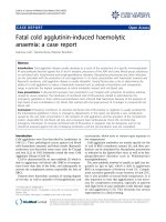
Figure 1 Coronal computed tomography view of the patient
showing intra-abdominal hemorrhage and liver metastasis (red
arrows).
Figure 2 Coronal computed tomography view of the patient
showing intra-abdominal hemorrhage (red arrows).
Figure 3 Axial computed tomography view of the patient show-
ing metastasis and intra-abdominal hemorrhage (red arrows).
Seetho et al. Journal of Medical Case Reports 2010, 4:194
/>Page 3 of 3
complication is not uncommon in primary hepatocellular
carcinoma [2]. The significance of this case is that we
describe acute rupture of hepatic metastases resulting in
acute hemoperitoneum that initially presented as a syn-
copal episode. There are only a few reported cases in the
literature of acute hemoperitoneum secondary to the
rupture of liver metastases [3-9] originating from differ-
ent sources. These sources include nasopharyngeal can-
cer [5], gastric cancer [6], lung cancer [7], renal cell
carcinoma [8] and carcinoma of the liver [9].
Intra-peritoneal hemorrhage frequently presents with
acute abdominal pain and can be life-threatening. CT is
commonly used as an imaging modality in the investiga-
tions of these patients, but ultrasound and magnetic reso-
nance imaging may also be used in the diagnosis [10]. It
should be noted that in this case, our patient presented
following recurrent collapse episodes rather than with an
acute abdomen. He did not wish for further investigations
to identify the primary source given the advanced nature
of the liver metastases. As such, the source of his liver
metastases was not identified. He survived the acute
bleeding episode with conservative management alone.
The literature reports laparotomy as a management
option for metastatic hepatic hemorrhage but this would
entail surgical risks for the patient [11,12]. In hemoperi-
toneum occurring as a result of rupture of hepatocellular
carcinoma, transcatheter arterial embolization has been
previously described as a potential therapeutic option
[1,2].
Conclusions
In conclusion, a high index of suspicion is needed in the
acute setting when considering the possibility of sponta-
neous hemoperitoneum in a patient who presents with
syncope, especially with an acute abdomen. This is par-
ticularly important if there is a known history of neoplas-
tic process. This case highlights an unusual source of
intra-abdominal bleeding which was from liver meta-
static disease. The use of appropriate CT imaging in this
case facilitated the prompt diagnosis and subsequent
management steps were then taken accordingly.
Consent
Written informed consent was obtained from our
patient's next-of-kin for publication of this case report
and any accompanying images. A copy of the written con-
sent is available for review by the Editor-in-Chief of this
journal.
Competing interests
The authors declare that they have no competing interests.
Authors' contributions
All authors contributed equally to the manuscript.
Author Details
1
Department of Medicine, City Hospital, Nottingham, Nottingham University
Hospitals NHS Trust. Hucknall Road. Nottingham NG5 1PB, UK,
2
Department of
Radiology, King's Mill Hospital, Sherwood Forest Hospitals NHS Foundation
Trust, Mansfield Road. Nottingham NG17 4JL, UK and
3
Department of Stroke
Medicine and Medicine for the Elderly, King's Mill Hospital, Sherwood Forest
Hospitals NHS Foundation Trust, Mansfield Road. Nottingham NG17 4JL, UK
References
1. Chedid AD, Klein PW, Tiburi MF, et al.: Spontaneous rupture of
hepatocellular carcinoma with haemoperitoneum: a rare condition in
Western countries. HPB 2001, 3(3):227-230.
2. Abdel Samie A, Otto G, Theilmann L: Acute haemoperitoneum due to
spontaneous tumour rupture as first manifestation of hepatocellular
carcinoma. Z Gastroenterol 2007, 45(7):615-619.
3. Schoedel KE, Dekker A: Hemoperitoneum in the setting of metastatic
cancer to the liver. A report of two cases with review of the literature.
Dig Dis Sci 1992, 37(1):153-154.
4. Fidas-Kamini A, Busuttil A: Fatal intraperitoneal haemorrhage of hepatic
origin. Postgrad Med J 1986, 62:1097-1100.
5. Dewar GA, Griffin SM, van Hasselt CA, et al.: Fatal haemoperitoneum due
to liver metastases from nasopharyngeal cancer. Aust N Z J of Surg 1991,
61(9):723-725.
6. Yoshida H, Mamada Y, Taniai N, et al.: Ruptured metastatic liver tumour
from an alpha-fetoprotein-producing gastric cancer. J Nippon Med Sch
2005, 72(4):236-241.
7. Kadowaki T, Hamada H, Yokoyama A, et al.: Hemoperitoneum secondary
to spontaneous rupture of hepatic metastasis from lung cancer. Intern
Med 2005, 44(4):290-293.
8. Wong KT, Khir AS, Noori S, et al.: Fatal haemoperitoneum due to rupture
of hepatic metastasis from renal cell carcinoma. Aust N Z J Surg 1994,
64(2):128-129.
9. Tung CF, Chang CS, Chow WK, et al.: Hemoperitoneum secondary to
spontaneous rupture of metastatic epidermoid carcinoma of liver: case
report and review of the literature. Hepatogastroenterology 2002,
49(47):1415-1417.
10. Lucey BC, Varghese JC, Anderson SW, et al.: Spontaneous
hemoperitoneum: a bloody mess. Emerg Radiol 2007, 14(2):65-75.
11. Lucha PA Jr: Spontaneous hemoperitoneum. J Am Osteopath Assoc
1996, 96(6):364-365.
12. Suber WJ Jr, Cunningham PL, Bloch RS: Massive spontaneous
hemoperitoneum of unknown etiology: a case report. Am Surg 1998,
64(12):1177-1778.
doi: 10.1186/1752-1947-4-194
Cite this article as: Seetho et al., Bleeding from ruptured hepatic metastases
as a cause of syncope in an octogenarian: a case report Journal of Medical
Case Reports 2010, 4:194
Received: 8 December 2009 Accepted: 26 June 2010
Published: 26 June 2010
This article is available from: 2010 Seetho et al; licensee BioMed Central Ltd. This is an Open Access article distributed under the terms of the Creative Commons Attribution License ( which permits unrestricted use, distribution, and reproduction in any medium, provided the original work is properly cited.Journal of Medical Case Repo rts 2010, 4:194