báo cáo khoa học: " Inversion variants in the human genome: role in disease and genome architecture" ppsx
Bạn đang xem bản rút gọn của tài liệu. Xem và tải ngay bản đầy đủ của tài liệu tại đây (1020.83 KB, 8 trang )
Inversions
Over the past 5 years there has been a major drive in
genomic research to identify submicroscopic structural
variation in the human genome, ranging from a few
hundred base pairs to approximately five megabases (Mb)
in size. Structural variation is a term describing all forms
of rearrangements, including deletions, duplications,
insertions, inversions, translocations and more complex
rearrangements. e main type of submicroscopic varia-
tion is copy number variation (CNV) [1,2], a term used to
describe gains and losses of segments of DNA. e initial
reports on CNVs as an abundant form of variation in the
human genome were published in 2004 [3,4]. Since then,
there have been multiple studies performed to charac-
terize the extent and importance of CNV in the human
genome [5-14]. e majority of these studies have been
based on microarrays, either as comparative genomic
hybridization (CGH) arrays or single nucleotide poly-
mor phism (SNP) arrays. Using array-based strategies, it
is possible to identify unbalanced changes, that is, net
gain or loss of large segments of DNA. However, other
forms of variation involving a change in orientation or
relocation of DNA, without any gain or loss, cannot
readily be detected with arrays. erefore, despite the
great success in developing human genome maps of
deletions and duplications, the mapping of inversions has
lagged behind.
It is still not clear how many common inversions exist
in the human genome, what the size distribution of
inversions variants is, and to what extent inversions are
associated with human disorders. With the recent intro-
duction of novel high-throughput sequencing techniques,
the methodology is now available to screen for inversions
in an unbiased manner. As a consequence, our under-
standing of the extent of inversion variants in the human
genome has increased dramatically in the past few years.
is review will give an overview of the current
knowledge of inversions in the human genome, the
methods used to discover and type inversions, and their
role in human disease and human genome architecture.
Cytogenetically visible inversions
It has long been possible to detect inversions of large
chromosomal regions in G-banded karyotypes. However,
this strategy is limited to identification of variants that
are several megabases in size, and even significantly
larger inversions may escape detection if the inverted
segment leads to little difference in the banding pattern.
e long history of chromosomal studies in cytogenetics
has led to the identification of several inversion variants,
Abstract
Signicant advances have been made over the past
5 years in mapping and characterizing structural
variation in the human genome. Despite this progress,
our understanding of inversion variants is still very
restricted. While unbalanced variants such as copy
number variations can be mapped using array-based
approaches, strategies for characterization of inversion
variants have been limited and underdeveloped.
Traditional cytogenetic approaches have long been
able to identify microscopic inversion events, but
discovery of submicroscopic events has remained
elusive and largely ignored. With the advent of paired-
end sequencing approaches, it is now possible to map
inversions across the human genome. Based on the
paired-end sequencing studies published to date, it is
now feasible to make a rst map of inversions across
the human genome and to use this map to explore
the characteristics and distribution of this form of
variation. The current map of inversions indicates
that many remain to be identied, especially in the
smaller size ranges. This review provides an overview
of the current knowledge about human inversions
and their contribution to human phenotypes. Further
characterization of inversions should be considered as
an important step towards a deeper understanding of
human variation and genome dynamics.
© 2010 BioMed Central Ltd
Inversion variants in the human genome: role in
disease and genome architecture
Lars Feuk*
R EV I EW
*Correspondence:
Address: Department of Genetics and Pathology, Rudbeck Laboratory, Uppsala
University, 751 85 Uppsala, Sweden
Feuk Genome Medicine 2010, 2:11
/>© 2010 BioMed Central Ltd
or heteromorphisms, that exist in the population but that
have no clinical significance [15]. Inversions are the most
common human constitutional karyotype aberration
detected in cytogenetic laboratories [16]. Pericentric
inversions are most frequent, often reported for chromo-
somes 1, 2, 3, 5, 9, 10 and 16. ese are some of the most
common cytogenetically visible rearrangements in
humans - for example, the pericentric inversion of
chromo some 9 is found in over 1% of karyotypes [17].
However, the chromosome 9 variant and many other
commonly identified hetermorphisms involve only
heterochromatic DNA.
e most frequently observed variant that includes
euchromatic sequence is the inv(2)(p11q13), which is
considered to be of no clinical significance [18]. Other
events are rarer, but still frequent enough to be seen
regularly in cytogenetic screening, especially in specific
population groups. In addition to these common
variants, numerous rare and unique inversions have been
observed in individuals with no apparent phenotype. An
illustrative example is inv(10)(q11.22q21.1), a 12 Mb
inversion with a carrier frequency of 0.11% in the
Swedish population, but with no consistent phenotype
[19]. Breakpoint and haplotype analysis indicated that
this is a rare variant in the population, originating from a
single founder event. Due to the balanced nature of
inversions, they are often of no clinical significance
unless the breakpoint disrupts a gene or falls between a
gene and its transcription regulatory elements. Excluding
the well-established cytogenetically characterized variants,
the rate of cytogenetically visible inversions reported is
significantly lower than that of translocations. However,
the exact rate of inversion formation is not known. A bias
is likely in ascertainment of inversions in comparison to
translocations, as balanced translocations lead to more
reduced fitness by increased risk for an unbalanced
transmission to the offspring than inversions do. Balanced
translocations are therefore commonly detected as part of
investigations of reproductive difficulties, while inversions
with no phenotypic effect may be transmitted through
many generations and never be detected, as there may be
no reason for cytogenetic screening.
One of the aspects that make inversions interesting as
genomic rearrangements is their role in recent primate
evolution. Comparison of the human and chimpanzee
genomes shows that there are nine cytogenetically visible
pericentric inversions [20] and many submicroscopic
inverted sequences [21]. e majority of the nine visible
inversions occurred along the chimpanzee lineage, but
inversions on chromosomes 1 and 18 are specific to the
human lineage. ese findings indicate that inversions
are a type of rearrangement that occurs quite frequently
in primate chromosomal evolution. Identification of a
large number of inversions between closely related
species, and signatures of selection associated with these,
has led to speculation that inversions have played an
important role in speciation [22].
Methods for inversion discovery and genotyping
Although inversions have long been detectable at the
resolution of cytogenetics, progress in mapping inver-
sions at the submicroscopic level is much more recent.
As inversions only lead to a change in orientation, but
not in copy number, they cannot be detected using
hybridi zation-based methods such as microarrays. Since
most strategies to map structural variation in the human
genome to date have been based on array approaches,
there is comparatively little known about the distribution
of inversions.
Although there has been a lack of methods for global
discovery of inversions, it has long been possible to test
for the presence of inversions in a targeted manner if
there is a prior hypothesis that a region may be inverted.
Testing can be done using traditional molecular
approaches such as pulse-field gel electrophoresis (PFGE)
or Southern blot. Single molecular haplotyping has also
been successfully used to screen samples for specific
inversion variants [23]. However, these strategies are
laborious and do not work for global unbiased discovery
of new inversion regions on a genome-wide scale. Despite
these limitations, a small number of studies have led to
the identification of inversion variants using ’genomic‘
strategies. One approach that led to the identification of
three polymorphic inversions was based on investigating
regions that are inverted between the human and
chimpanzee genomes. By targeting 23 such regions in
human control samples, three inversions were found to
be polymorphic in humans. In another study, Bansal et
al. [24] used the linkage disequilibrium (LD) pattern of
SNPs to map putative inversion breakpoints. By using a
statistical method to detect regions where SNPs at a
distance from each other on the reference assembly were
in higher LD than SNPs in close proximity, a number of
putative inversions were identified. Overlap with several
previously validated inversions indicated that the approach
was successful. However, the candidate variants identified
by this method require experimental validation to
distinguish real inversions from false positives. Although
the approaches outlined above have shown some success
in the discovery of novel inversion variants, recent data
indicate that only a very small fraction of frequent human
inversions were found.
A major breakthrough in the discovery of inversions
(and other forms of structural variation) came with the
intro duction of paired-end sequencing and mapping [7].
Generally, when the two ends of a cloned fragment are
sequenced, the two resulting sequences would be expected
to align to the reference genome in a + and - orientation,
Feuk Genome Medicine 2010, 2:11
/>Page 2 of 8
respectively. However, if the donor DNA carries an
inversion as compared to the reference assembly, this
would lead to the end sequences of fragments spanning
the breakpoints to align in a -/- or a +/+ orientation
(Figure 1). By searching for clusters of fragments
exhibiting this pattern of alignments to the reference
assembly, it is possible to identify putative inversion
events. e first paired-end mapping study was based on
end sequencing of fosmid clones using traditional Sanger
sequencing [7]. e study identified 56 inversion break-
points from a fosmid library representing a single human
genome (sample NA15510). e same strategy of fosmid
end sequencing was later applied to another eight
genomes, and a total of 217 inversions were identified
and validated [6]. A large number of inversions were also
reported in the first individual genome to be sequenced
(the genome of Craig Venter, called HuRef) [25]. Sanger
sequencing was employed to sequence the HuRef
genome, and an assembly was created independently
from the National Center for Biotechnology Information
(NCBI) reference assembly. An assembly comparison
analysis gave rise to 90 regions of inverted orientation
between the HuRef and NCBI assemblies. Since these
initial Sanger sequencing studies, the general strategy of
paired-end mapping has been adapted to fragment end-
sequencing with second-generation-sequencing platforms
[26,27]. Although only a small number of whole-genome
sequencing studies have so far employed this strategy to
identify inversions, this is likely to be the main approach
for identification of inversions in the near future.
Despite the success of paired-end mapping, there are
still challenges to overcome. One important feature of
the paired-end mapping approach is that it relies on the
reference assembly. It is well established that the
reference assembly represents very rare or unique alleles
at some loci in the genome. In rare instances, it is also
possible that these unique alleles represent cloning
artifacts or are a result of mis-assembly of the reference
sequence. For example, this has been suggested for an
inversion overlapping an exon of the DOCK3 gene on
chromosome 3, for which there is an inversion in the
reference assembly as compared to available mRNA
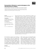
Figure 1. Overview of inversion discovery by paired-end mapping. The top part of the gure shows the alignment between the reference
assembly and an individual carrying an inversion. When paired-end mapping is performed, the donor DNA is rst sheared into several similarly
sized DNA fragments. The ends of these fragments are then sequenced (fragments are depicted in blue and red, with the boxes at the ends
showing the parts that are sequenced). The pairs of end-sequences are then mapped to the reference genome. The majority of these pairs will
map in a plus(+)/minus(-) orientation, separated by the approximate distance expected from the fragment size (labeled A and D). End-pairs labeled
B and C indicate mapping of fragment ends in a region containing an inversion compared to the reference assembly. Instead of the expected
+/- orientation of the two end-sequences, the pairs spanning the inversion breakpoints map as +/+ and -/-, respectively. Clusters of such read pairs
are indicative of an inversion. Only fragments spanning the inversion breakpoint will exhibit this pattern of alignment. Better clone coverage will
yield better resolution and more accurate mapping of the breakpoints.
Reference assembly
Inversion carrier DNA
+ + +
+
+ − − +
+
_
A B C D
A
B
+
+
C D
_
_
+
_
Paired-end
sequencing
Paired-end
mapping
Breakpoint 1 Breakpoint 2
Minimal
inversion region
Reference assembly
Feuk Genome Medicine 2010, 2:11
/>Page 3 of 8
sequences for the same gene [5]. For regions where the
reference assembly harbors a unique allele, every study
with high enough resolution and sequence coverage will
identify a homozygous inversion.
Another limitation of paired-end mapping for inversion
detection is related to the genome architecture associated
with inversions. e majority of large (>100 kb) inver-
sions described in the human genome to date are flanked
by high identity segmental duplications, that is,
sequences >1kb that exist in two or more copies of >90%
identity in the human genome [28,29]. e segmental
duplications associated with inversions cause problems
for inversion discovery using paired-end mapping. As the
method depends on alignment to the reference assembly,
highly identical sequences in the assembly will cause
problems in identifying unique placements for the
sequence reads. Many paired-end mapping pipelines
simply discard reads that cannot be uniquely mapped.
erefore, the paired-end mapping strategy often fails to
identify inversions flanked by long inverted segmental
duplications of high identity. For these regions, targeted
assays are required.
Current map of inversions in the human genome
e map of human inversions is still quite limited, and
our understanding of the number of inversions, the size
distribution and the frequency distribution is probably
biased due to biases in the approaches used for variation
identification. ere are currently 914 inversion events
reported in the Database of Genomic Variants [30], a
database resource for structural variation in the human
genome [3,31]. However, many of these overlap and
actually refer to the same locus. If only non-redundant
loci are counted, there are a total of 479 inversions in the
database. Figure 2 shows an overview of the current
inversions reported in the human genome. e inversions
are found across the size spectrum up to several
megabases. A comparison of the size distribution of
inversions and CNVs is shown in Figure 3. e size
distribution shows that most of the inversions discovered
to date are in the 10kb to 100kb interval. For CNVs, size
distribution is shifted more towards smaller size variants.
ere are many potential explanations for the differ-
ence in size distribution between inversions and CNVs
(Figure3). Biologically, large inversions are more likely to
be neutral, without obvious phenotypic consequences,
compared to large CNVs. Data from cytogenetic studies
support this. One difference between inversions and
CNVs is that the genes within an inversion can be entirely
unaffected, while genes within CNVs are always affected
by a dosage imbalance. For inversions, it is more impor-
tant where the breakpoints are located and if these
interrupt a gene or lead to disruption of the transcrip-
tional regulation of genes. If no gene or regulatory
function is interrupted by the breakpoints, inversions
that are comparatively large may be frequent in the
population. While there are very few CNVs >1Mb in size
that have reached a minor allele frequency of 1%, there
are examples of very large inversions that are frequently
observed in the population. e best-studied examples
are two inversions located on chromosomes 4 and 8,
respectively. Both these inversions have breakpoints that
Figure 2. Distribution of inversion variants in the human
genome. The blue lines in this ideogram show the human
chromosomal distribution of the 479 non-redundant inversion
variants reported in the Database of Genomic Variants.
Figure 3. Size distribution of inversions and copy number
variants. The size distribution of inversions reported in the
Database of Genomic Variants (a) shows that the majority of
inversions reported to date are in the 10 to 100kb size bin. The
size distribution of inversions diers from that reported for copy
number variants (CNVs) (b) The CNV data plotted here show the
11,700 non-redundant CNV events reported by Conrad et al. [13]. It is
currently unclear whether the dierence in size distribution between
inversions and CNVs is due to ascertainment bias, or whether there is
an actual biological dierence in size distribution. Both cytogenetic
data and evolutionary comparative genomic data indicate that large
inversions are less detrimental than large deletions and duplications.
0
50
100
150
200
250
0-1 kb 1 kb-10 kb 10 kb-100 kb 100 kb-1 Mb >1 Mb
0-1 kb 1 kb-10 kb 10 kb-100 kb 100 kb-1 Mb >1 Mb
0
1000
2000
3000
4000
5000
6000
7000
8000
(a)
(b)
Number of eventsNumber of events
Feuk Genome Medicine 2010, 2:11
/>Page 4 of 8
fall in clusters of olfactory receptors of high identity. e
inversion on chromosome 8 is approximately 3.5Mb in
size and has been reported to be present in 26% of
healthy controls, while the chromosome 4 inversion is
about 6 Mb in size and was found in 12.5% of healthy
controls [32]. ese data indicate that very large inver-
sions may exist in the human genomes without a strong
negative effect on reproductive fitness.
ere may also be a methodological explanation for the
difference in size distribution between current anno-
tations of inversions and CNVs, based on differences in
methods of discovery and limitations in technology. e
size distribution for inversions is reflective of the
resolution and limited sequence coverage of the paired-
end mapping projects published to date. For very small
inversions, deep sequence coverage would be required to
obtain several DNA fragments spanning one breakpoint.
erefore, many additional inversions will be found as
thousands of additional genomes are sequenced over the
next few years, and a large fraction of these would be
expected to increase the fraction of variants that are
<10kb in size.
Finally, it is also possible that the size distribution for
inversions differs from that of CNVs based on the
mechanisms by which the variants are created. As for
CNVs [13], it is likely that different mechanisms act across
the size spectrum and give rise to larger and smaller
inversion events, respectively. rough non-allelic homo-
lo gous recombination (NAHR) - recombina tion events
taking place between highly similar sequences - regions
located between segmental duplications or highly identical
repeat sequences may be deleted, duplicated or inverted.
Inversions can be formed by this process if the duplicated
sequences are in inverted orientation with respect to each
other. erefore, NAHR is considered the primary
mechanism by which large (tens of kilobases) inversions
are formed. However, for small inversions, the mechanisms
are not as well characterized as for smaller insertions/
deletions. Some evidence points towards replication-based
mechanisms, such as microhomology-mediated break-
induced replication (MMBIR) [33]. Other specific
mechanisms that have been suggested to be involved in
creation of inversions include fork stalling and template
switching (FoSTeS) [34] and serial replica tion slippage in
trans [35]. However, the limited number of inversions with
nucleotide resolution breakpoint information available to
date has prevented a thorough investigation of
mechanisms and sequence motifs giving rise to inversions.
As additional inversion breakpoints are identified, these
relationships should become more evident.
Inversions in human disorders
ere are many descriptions in the literature of patients
with specific phenotypes who also carry an inversion that
is cytogenetically visible. Since inversions are relatively
rare events, and it is unlikely that multiple patients with
the same inversion are found, it is often problematic to
assess whether the inversion present in the patient is
actually associated with the phenotype. e exception is
if the inversion breakpoint falls within or near a gene that
has previously been associated with the disorder through
other types of mutations. For recurrent inversions, the
association between phenotype and genotype is more
obvious, and a number of such loci have been described.
One of the best-characterized recurrent inversions giving
rise to disease causes hemophilia A, an X-linked disorder
caused by mutations in the factor VIII gene [36]. A
recurrent inversion has been found in approximately 43%
of patients [37]. Molecular characterization of the break-
points indicates that the inversion is a result of intra-
chromosomal homologous recombination, originating
almost exclusively in male germ cells. is recurrent
inversion spans approximately 400kb and is mediated by
two inverted segmental duplications, one of which is
located in intron 22 of the factor VIII gene, with two
other copies being located approximately 400 kb telo-
meric to the gene. Other examples where recurrent
inver sions have been shown to lead to a disease pheno-
type are the disruption of the idunorate 2-sulphatase
gene in mucopolysaccharidosis type II (Hunter syndrome)
[38], and disruption of the emerin gene in Emery-
Dreifuss muscular dystrophy [39].
A specific category of inversions associated with
genetic disorders is those that are not directly causative,
but rather increase the risk of further rearrangements
that cause disease. For a number of microdeletion syn-
dromes, one or both parents of probands have been
found to carry an inversion of the deleted interval. e
association was first described in Williams-Beuren
syndrome, which is most commonly caused by a 1.5Mb
microdeletion at 7q11. In a study of 12 families where the
proband carried the typical microdeletion, an inversion
was found in a parent for 33% of the patients [40]. e
inversion variant has since been shown to be relatively
frequent in the general population (approximately 5%),
and does not seem to be associated with a phenotype in
itself [41].
Another example of a disorder where an inversion has
been associated with a causative deletion is the 17q21.31
microdeletion syndrome, a genetically characterized form
of mental retardation. is region harbors a 970 kb
inversion polymorphism found at high frequency in
European populations [42]. e genetic variation pattern
within the region indicates that the inversion first
appeared before dispersal out of Africa, and that there
has been little or no recombination between the haplo-
types. Interestingly, there is some evidence that this
inversion variation is associated with higher reproductive
Feuk Genome Medicine 2010, 2:11
/>Page 5 of 8
fitness [42]. Screening patient cohorts with mental
retarda tion led to the discovery of a microdeletion
syndrome corresponding to the same region as the
common inversion polymorphism [43-45]. Studies of the
parents of microdeletion carriers showed that at least one
parent carried the inverted H2 haplotype in every case. It
was therefore initially concluded that the inversion in
itself was the cause of the increased risk for the deletion
to occur. It has been suggested that the lack of homology
across the inversion region between heterozygous
chromatids in meiosis may lead to the formation of an
‘asynaptic bubble’ that renders the region unstable and
prone to additional rearrangements [46]. However, addi-
tional characterization of the prevalent haplotypes in the
region indicates that other rearrangements present on
the inverted H2 haplotype may be the primary substrate
for the non-allelic homologous recombination giving rise
to the microdeletion [47]. Additional studies will be
needed to confirm exactly how the inversion leads to an
increased risk for deletions in the offspring.
In total, there are at least nine different microdeletion
syndromes for which the deletion region has also been
found as an inversion variant in the general population
(Table 1). For a majority of these disorders, a direct
association between the inversion carrier status and
increased risk for deletion in the offspring has been
established by comparing the inversion frequency in
parents to the frequency in the general population.
However, the exact molecular mechanisms still remain to
be elucidated and it is not confirmed whether it is the
inversion itself, or other sequence features present on the
inversion haplotype, that causes the subsequent
pathogenic rearrangement.
Conclusions and future perspectives
With the advent of deep coverage paired-end sequencing,
the number of inversions reported has increased
dramatically and the inversion breakpoints will be
pinpointed at much higher resolution. Over the next year
or two, the true extent of inversion variants in the human
genome will be revealed. Only then will it be possible to
explore the contribution of inversions to common
disease. For both inversions and other structural variants,
it has been anticipated that it would be possible to impute
these variants from high-density SNP array data.
However, recent studies indicate that this may not be the
case. Data from one study show that many large
inversions, surrounded by blocks of segmental
duplications, have arisen on more than one haplotype
background [48]. Similar data have been shown for multi-
allelic CNVs [13]. ese variants will therefore need to be
directly targeted for inclusion in association studies.
Currently, the experimental strategies for accurate high-
throughput genotyping of inversions and multi-allelic
CNVs are limited or non-existent. However, it is very
likely that smaller inversions that are not flanked by
blocks of segmental duplications will have arisen only
once and will therefore be in LD with surrounding SNPs.
is has been shown in a limited number of cases [21],
but more data are needed to confirm whether this applies
to a majority of events. Other questions that remain to be
explored in further detail include inversion formation
mechanisms, characterization of breakpoints, and
development of maps and strategies for inclusion of
inversion variants in genome-wide disease association
studies. In conclusion, we are now at the stage where we
have the tools that enable characterization of the full
extent of inversions in the human genome and their
contribution to human variation and disease.
Abbreviations
CGH, comparative genomic hybridization; CNV, copy number variation;
FoSTeS, fork stalling and template switching; kb, kilobase; LD, linkage
disequilibrium; Mb, megabase; MMBIR, microhomology-mediated break-
induced replication; NAHR, non-allelic homologous recombination; NCBI,
National Center for Biotechnology Information; PFGE, pulse-eld gel
electrophoresis; SNP, single nucleotide polymorphism.
Table 1. Rearrangements associated with inversion variants
Chromosome band Inversion size (Mb) Disorder/rearrangement Reference (syndrome : inversion)
3q29 1.9 3q29 deletion syndrome [49] : [7]
5q35.2-q35.3* 1.9 Sotos syndrome microdeletion [50] : [51]
7q11.23* 1.5 Williams-Beuren syndrome microdeletion [52] : [40]
8p23
a
4.7 Inv dup(8p) and del (8)(p23.1;p23.2) [53,54] : [32,55]
15q11-q13* 4 Angelman syndrome deletion [56] : [57]
15q13.3* 2 15q13.3 microdeletion [58] : [6,58]
15q24 1.2 15q24 microdeletion [44,59] : [6]
17q12 1.5 Renal cysts and diabetes (RCAD) microdeletion syndrome [60] : [6]
17q21.31* 0.9 17q21.31 microdeletion syndrome [43-45] : [42]
a
The inversion has been found at higher frequency in parents of probands with microdeletions than in the general population, indicating that the inversion is a risk
factor for subsequent rearrangements in the offspring.
Feuk Genome Medicine 2010, 2:11
/>Page 6 of 8
Competing interests
The author declares that he has no competing interest.
Acknowledgements
LF is supported by the Göran Gustafsson Foundation and the Future Research
Leaders Grant from the Swedish Foundation for Strategic Research.
Published: 12 February 2010
References
1. Feuk L, Carson AR, Scherer SW: Structural variation in the human genome.
Nat Rev Genet 2006, 7:85-97.
2. Sharp AJ, Cheng Z, Eichler EE: Structural variation of the human genome.
Annu Rev Genomics Hum Genet 2006, 7:407-442.
3. Iafrate AJ, Feuk L, Rivera MN, Listewnik ML, Donahoe PK, Qi Y, Scherer SW, Lee
C: Detection of large-scale variation in the human genome. Nat Genet
2004, 36:949-951.
4. Sebat J, Lakshmi B, Troge J, Alexander J, Young J, Lundin P, Maner S, Massa H,
Walker M, Chi M, Navin N, Lucito R, Healy J, Hicks J, Ye K, Reiner A, Gilliam TC,
Trask B, Patterson N, Zetterberg A, Wigler M: Large-scale copy number
polymorphism in the human genome. Science 2004, 305:525-528.
5. Khaja R, Zhang J, MacDonald JR, He Y, Joseph-George AM, Wei J, Raq MA,
Qian C, Shago M, Pantano L, Aburatani H, Jones K, Redon R, Hurles M,
Armengol L, Estivill X, Mural RJ, Lee C, Scherer SW, Feuk L: Genome assembly
comparison identifies structural variants in the human genome. Nat Genet
2006, 38:1413-1418.
6. Kidd JM, Cooper GM, Donahue WF, Hayden HS, Sampas N, Graves T, Hansen
N, Teague B, Alkan C, Antonacci F, Haugen E, Zerr T, Yamada NA, Tsang P,
Newman TL, Tuzun E, Cheng Z, Ebling HM, Tusneem N, David R, Gillett W,
Phelps KA, Weaver M, Saranga D, Brand A, Tao W, Gustafson E, McKernan K,
Chen L, Malig M, et al.: Mapping and sequencing of structural variation
from eight human genomes. Nature 2008, 453:56-64.
7. Tuzun E, Sharp AJ, Bailey JA, Kaul R, Morrison VA, Pertz LM, Haugen E, Hayden
H, Albertson D, Pinkel D, Olson MV, Eichler EE: Fine-scale structural variation
of the human genome. Nat Genet 2005, 37:727-732.
8. McCarroll SA, Hadnott TN, Perry GH, Sabeti PC, Zody MC, Barrett JC, Dallaire S,
Gabriel SB, Lee C, Daly MJ, Altshuler DM: Common deletion polymorphisms
in the human genome. Nat Genet 2006, 38:86-92.
9. McCarroll SA, Kuruvilla FG, Korn JM, Cawley S, Nemesh J, Wysoker A, Shapero
MH, de Bakker PI, Maller JB, Kirby A, Elliott AL, Parkin M, Hubbell E, Webster T,
Mei R, Veitch J, Collins PJ, Handsaker R, Lincoln S, Nizzari M, Blume J, Jones
KW, Rava R, Daly MJ, Gabriel SB, Altshuler D: Integrated detection and
population-genetic analysis of SNPs and copy number variation. Nat Genet
2008, 40:1166-1174.
10. Conrad DF, Andrews TD, Carter NP, Hurles ME, Pritchard JK: A high-resolution
survey of deletion polymorphism in the human genome. Nat Genet 2006,
38:75-81.
11. Sharp AJ, Locke DP, McGrath SD, Cheng Z, Bailey JA, Vallente RU, Pertz LM,
Clark RA, Schwartz S, Segraves R, Osero VV, Albertson DG, Pinkel D, Eichler
EE: Segmental duplications and copy-number variation in the human
genome. Am J Hum Genet 2005, 77:78-88.
12. Hinds DA, Stuve LL, Nilsen GB, Halperin E, Eskin E, Ballinger DG, Frazer KA, Cox
DR: Whole-genome patterns of common DNA variation in three human
populations. Science 2005, 307:1072-1079.
13. Conrad DF, Pinto D, Redon R, Feuk L, Gokcumen O, Zhang Y, Aerts J, Andrews
TD, Barnes C, Campbell P, Fitzgerald T, Hu M, Ihm CH, Kristiansson K,
Macarthur DG, Macdonald JR, Onyiah I, Pang AW, Robson S, Stirrups K,
Valsesia A, Walter K, Wei J, Tyler-Smith C, Carter NP, Lee C, Scherer SW, Hurles
ME: Origins and functional impact of copy number variation in the human
genome. Nature 2009 [Epub ahead of print].
14. Redon R, Ishikawa S, Fitch KR, Feuk L, Perry GH, Andrews TD, Fiegler H,
Shapero MH, Carson AR, Chen W, Cho EK, Dallaire S, Freeman JL, Gonzalez JR,
Gratacos M, Huang J, Kalaitzopoulos D, Komura D, MacDonald JR, Marshall
CR, Mei R, Montgomery L, Nishimura K, Okamura K, Shen F, Somerville MJ,
Tchinda J, Valsesia A, Woodwark C, Yang F, et al.: Global variation in copy
number in the human genome. Nature 2006, 444:444-454.
15. Thomas NS, Bryant V, Maloney V, Cockwell AE, Jacobs PA: Investigation of the
origins of human autosomal inversions. Hum Genet 2008, 123:607-616.
16. Schmidt S, Claussen U, Liehr T, Weise A: Evolution versus constitution:
differences in chromosomal inversion. Hum Genet 2005, 117:213-219.
17. Hsu LY, Benn PA, Tannenbaum HL, Perlis TE, Carlson AD: Chromosomal
polymorphisms of 1, 9, 16, and Y in 4 major ethnic groups: a large prenatal
study. Am J Med Genet 1987, 26:95-101.
18. MacDonald IM, Cox DM: Inversion of chromosome 2 (p11p13): frequency
and implications for genetic counselling. Hum Genet 1985, 69:281-283.
19. Entesarian M, Carlsson B, Mansouri MR, Stattin EL, Holmberg E, Golovleva I,
Stefansson H, Klar J, Dahl N: A chromosome 10 variant with a 12 Mb
inversion [inv(10)(q11.22q21.1)] identical by descent and frequent in the
Swedish population. Am J Med Genet A 2009, 149A:380-386.
20. Yunis JJ, Prakash O: The origin of man: a chromosomal pictorial legacy.
Science 1982, 215:1525-1530.
21. Feuk L, Macdonald JR, Tang T, Carson AR, Li M, Rao G, Khaja R, Scherer SW:
Discovery of human inversion polymorphisms by comparative analysis of
human and chimpanzee DNA sequence assemblies. PLoS Genet 2005,
1:e56.
22. Navarro A, Barton NH: Chromosomal speciation and molecular divergence-
-accelerated evolution in rearranged chromosomes. Science 2003,
300:321-324.
23. Turner DJ, Shendure J, Porreca G, Church G, Green P, Tyler-Smith C, Hurles ME:
Assaying chromosomal inversions by single-molecule haplotyping. Nat
Methods 2006, 3:439-445.
24. Bansal V, Bashir A, Bafna V: Evidence for large inversion polymorphisms in
the human genome from HapMap data. Genome Res 2007, 17:219-230.
25. Levy S, Sutton G, Ng PC, Feuk L, Halpern AL, Walenz BP, Axelrod N, Huang J,
Kirkness EF, Denisov G, Lin Y, MacDonald JR, Pang AW, Shago M, Stockwell TB,
Tsiamouri A, Bafna V, Bansal V, Kravitz SA, Busam DA, Beeson KY, McIntosh TC,
Remington KA, Abril JF, Gill J, Borman J, Rogers YH, Frazier ME, Scherer SW,
Strausberg RL, et al.: The diploid genome sequence of an individual human.
PLoS Biol 2007, 5:e254.
26. Ahn SM, Kim TH, Lee S, Kim D, Ghang H, Kim DS, Kim BC, Kim SY, Kim WY, Kim
C, Park D, Lee YS, Kim S, Reja R, Jho S, Kim CG, Cha JY, Kim KH, Lee B, Bhak J,
Kim SJ: The first Korean genome sequence and analysis: full genome
sequencing for a socio-ethnic group. Genome Res 2009, 19:1622-1629.
27. Korbel JO, Urban AE, Aourtit JP, Godwin B, Grubert F, Simons JF, Kim PM,
Palejev D, Carriero NJ, Du L, Taillon BE, Chen Z, Tanzer A, Saunders AC, Chi J,
Yang F, Carter NP, Hurles ME, Weissman SM, Harkins TT, Gerstein MB, Egholm
M, Snyder M: Paired-end mapping reveals extensive structural variation in
the human genome. Science 2007, 318:420-426.
28. Bailey JA, Gu Z, Clark RA, Reinert K, Samonte RV, Schwartz S, Adams MD,
Myers EW, Li PW, Eichler EE: Recent segmental duplications in the human
genome. Science 2002, 297:1003-1007.
29. Eichler EE: Segmental duplications: what’s missing, misassigned, and
misassembled - and should we care? Genome Res 2001, 11:653-656.
30. Database of Genomics Variants [
(accessed2 February 2010).
31. Zhang J, Feuk L, Duggan GE, Khaja R, Scherer SW: Development of
bioinformatics resources for display and analysis of copy number and
other structural variants in the human genome. Cytogenet Genome Res
2006, 115:205-214.
32. Giglio S, Calvari V, Gregato G, Gimelli G, Camanini S, Giorda R, Ragusa A,
Guerneri S, Selicorni A, Stumm M, Tonnies H, Ventura M, Zollino M, Neri G,
Barber J, Wieczorek D, Rocchi M, Zuardi O: Heterozygous submicroscopic
inversions involving olfactory receptor-gene clusters mediate the
recurrent t(4;8)(p16;p23) translocation. Am J Hum Genet 2002, 71:276-285.
33. Hastings PJ, Ira G, Lupski JR: A microhomology-mediated break-induced
replication model for the origin of human copy number variation. PLoS
Genet 2009, 5:e1000327.
34. Zhang F, Khajavi M, Connolly AM, Towne CF, Batish SD, Lupski JR: The DNA
replication FoSTeS/MMBIR mechanism can generate genomic, genic and
exonic complex rearrangements in humans. Nat Genet 2009, 41:849-853.
35. Chen JM, Chuzhanova N, Stenson PD, Ferec C, Cooper DN:
Intrachromosomal serial replication slippage in trans gives rise to diverse
genomic rearrangements involving inversions. Hum Mutat 2005,
26:362-373.
36. Lakich D, Kazazian HH Jr, Antonarakis SE, Gitschier J: Inversions disrupting
the factor VIII gene are a common cause of severe haemophilia A. Nat
Genet 1993, 5:236-241.
37. Antonarakis SE, Rossiter JP, Young M, Horst J, de Moerloose P, Sommer SS,
Ketterling RP, Kazazian HH Jr, Negrier C, Vinciguerra C, Gitschier J, Goossens M,
Girodon E, Ghanem N, Plassa F, Lavergne JM, Vidaud M, Costa JM, Laurian Y,
Lin SW, Lin SR, Shen MC, Lillicrap D, Taylor SA, Windsor S, Valleix SV, Nafa K,
Sultan Y, Delpech M, Vnencak-Jones CL, et al.: Factor VIII gene inversions in
Feuk Genome Medicine 2010, 2:11
/>Page 7 of 8
severe hemophilia A: results of an international consortium study. Blood
1995, 86:2206-2212.
38. Bondeson ML, Dahl N, Malmgren H, Kleijer WJ, Tonnesen T, Carlberg BM,
Pettersson U: Inversion of the IDS gene resulting from recombination with
IDS-related sequences is a common cause of the Hunter syndrome. Hum
Mol Genet 1995, 4:615-621.
39. Small K, Iber J, Warren ST: Emerin deletion reveals a common
X-chromosome inversion mediated by inverted repeats. Nat Genet 1997,
16:96-99.
40. Osborne LR, Li M, Pober B, Chitayat D, Bodurtha J, Mandel A, Costa T, Grebe T,
Cox S, Tsui LC, Scherer SW: A 1.5 million-base pair inversion polymorphism
in families with Williams-Beuren syndrome. Nat Genet 2001, 29:321-325.
41. Tam E, Young EJ, Morris CA, Marshall CR, Loo W, Scherer SW, Mervis CB,
Osborne LR: The common inversion of the Williams-Beuren syndrome
region at 7q11.23 does not cause clinical symptoms. Am J Med Genet A
2008, 146A:1797-1806.
42. Stefansson H, Helgason A, Thorleifsson G, Steinthorsdottir V, Masson G,
Barnard J, Baker A, Jonasdottir A, Ingason A, Gudnadottir VG, Desnica N, Hicks
A, Gylfason A, Gudbjartsson DF, Jonsdottir GM, Sainz J, Agnarsson K,
Birgisdottir B, Ghosh S, Olafsdottir A, Cazier JB, Kristjansson K, Frigge ML,
Thorgeirsson TE, Gulcher JR, Kong A, Stefansson K: A common inversion
under selection in Europeans. Nat Genet 2005, 37:129-137.
43. Koolen DA, Vissers LE, Pfundt R, de Leeuw N, Knight SJ, Regan R, Kooy RF,
Reyniers E, Romano C, Fichera M, Schinzel A, Baumer A, Anderlid BM,
Schoumans J, Knoers NV, van Kessel AG, Sistermans EA, Veltman JA, Brunner
HG, de Vries BB: A new chromosome 17q21.31 microdeletion syndrome
associated with a common inversion polymorphism. Nat Genet 2006,
38:999-1001.
44. Sharp AJ, Hansen S, Selzer RR, Cheng Z, Regan R, Hurst JA, Stewart H, Price
SM, Blair E, Hennekam RC, Fitzpatrick CA, Segraves R, Richmond TA, Guiver C,
Albertson DG, Pinkel D, Eis PS, Schwartz S, Knight SJ, Eichler EE: Discovery of
previously unidentified genomic disorders from the duplication
architecture of the human genome. Nat Genet 2006, 38:1038-1042.
45. Shaw-Smith C, Pittman AM, Willatt L, Martin H, Rickman L, Gribble S, Curley R,
Cumming S, Dunn C, Kalaitzopoulos D, Porter K, Prigmore E, Krepischi-Santos
AC, Varela MC, Koimann CP, Lees AJ, Rosenberg C, Firth HV, de Silva R, Carter
NP: Microdeletion encompassing MAPT at chromosome 17q21.3 is
associated with developmental delay and learning disability. Nat Genet
2006, 38:1032-1037.
46. Sharp AJ: Emerging themes and new challenges in defining the role of
structural variation in human disease. Hum Mutat 2009, 30:135-144.
47. Zody MC, Jiang Z, Fung HC, Antonacci F, Hillier LW, Cardone MF, Graves TA,
Kidd JM, Cheng Z, Abouelleil A, Chen L, Wallis J, Glasscock J, Wilson RK, Reily
AD, Duckworth J, Ventura M, Hardy J, Warren WC, Eichler EE: Evolutionary
toggling of the MAPT 17q21.31 inversion region. Nat Genet 2008,
40:1076-1083.
48. Antonacci F, Kidd JM, Marques-Bonet T, Ventura M, Siswara P, Jiang Z, Eichler
EE: Characterization of six human disease-associated inversion
polymorphisms. Hum Mol Genet 2009, 18:2555-2566.
49. Willatt L, Cox J, Barber J, Cabanas ED, Collins A, Donnai D, FitzPatrick DR,
Maher E, Martin H, Parnau J, Pindar L, Ramsay J, Shaw-Smith C, Sistermans EA,
Tettenborn M, Trump D, de Vries BB, Walker K, Raymond FL: 3q29
microdeletion syndrome: clinical and molecular characterization of a new
syndrome. Am J Hum Genet 2005, 77:154-160.
50. Kurotaki N, Imaizumi K, Harada N, Masuno M, Kondoh T, Nagai T, Ohashi H,
Naritomi K, Tsukahara M, Makita Y, Sugimoto T, Sonoda T, Hasegawa T, Chinen
Y, Tomita Ha HA, Kinoshita A, Mizuguchi T, Yoshiura Ki K, Ohta T, Kishino T,
Fukushima Y, Niikawa N, Matsumoto N: Haploinsufficiency of NSD1 causes
Sotos syndrome. Nat Genet 2002, 30:365-366.
51. Visser R, Shimokawa O, Harada N, Kinoshita A, Ohta T, Niikawa N, Matsumoto
N: Identification of a 3.0-kb major recombination hotspot in patients with
sotos syndrome who carry a common 1.9-Mb microdeletion. Am J Hum
Genet 2005, 76:52-67.
52. Ewart AK, Morris CA, Atkinson D, Jin W, Sternes K, Spallone P, Stock AD,
Leppert M, Keating MT: Hemizygosity at the elastin locus in a
developmental disorder, Williams syndrome. Nat Genet 1993, 5:11-16.
53. Devriendt K, Matthijs G, Van Dael R, Gewillig M, Eyskens B, Hjalgrim H, Dolmer
B, McGaughran J, Brondum-Nielsen K, Marynen P, Fryns JP, Vermeesch JR:
Delineation of the critical deletion region for congenital heart defects, on
chromosome 8p23.1. Am J Hum Genet 1999, 64:1119-1126.
54. Floridia G, Piantanida M, Minelli A, Dellavecchia C, Bonaglia C, Rossi E, Gimelli
G, Croci G, Franchi F, Gilgenkrantz S, Grammatico P, Dalpra L, Wood S,
Danesino C, Zuardi O: The same molecular mechanism at the maternal
meiosis I produces mono- and dicentric 8p duplications. Am J Hum Genet
1996, 58:785-796.
55. Giglio S, Broman KW, Matsumoto N, Calvari V, Gimelli G, Neumann T, Ohashi
H, Voullaire L, Larizza D, Giorda R, Weber JL, Ledbetter DH, Zuardi O:
Olfactory receptor-gene clusters, genomic-inversion polymorphisms, and
common chromosome rearrangements. Am J Hum Genet 2001, 68:874-883.
56. Knoll JH, Nicholls RD, Magenis RE, Graham JM Jr, Lalande M, Latt SA:
Angelman and Prader-Willi syndromes share a common chromosome 15
deletion but differ in parental origin of the deletion. Am J Med Genet 1989,
32:285-290.
57. Gimelli G, Pujana MA, Patricelli MG, Russo S, Giardino D, Larizza L, Cheung J,
Armengol L, Schinzel A, Estivill X, Zuardi O: Genomic inversions of human
chromosome 15q11-q13 in mothers of Angelman syndrome patients with
class II (BP2/3) deletions. Hum Mol Genet 2003, 12:849-858.
58. Sharp AJ, Meord HC, Li K, Baker C, Skinner C, Stevenson RE, Schroer RJ,
Novara F, De Gregori M, Ciccone R, Broomer A, Casuga I, Wang Y, Xiao C,
Barbacioru C, Gimelli G, Bernardina BD, Torniero C, Giorda R, Regan R, Murday
V, Mansour S, Fichera M, Castiglia L, Failla P, Ventura M, Jiang Z, Cooper GM,
Knight SJ, Romano C, et al.: A recurrent 15q13.3 microdeletion syndrome
associated with mental retardation and seizures. Nat Genet 2008,
40:322-328.
59. Sharp AJ, Selzer RR, Veltman JA, Gimelli S, Gimelli G, Striano P, Coppola A,
Regan R, Price SM, Knoers NV, Eis PS, Brunner HG, Hennekam RC, Knight SJ, de
Vries BB, Zuardi O, Eichler EE: Characterization of a recurrent 15q24
microdeletion syndrome. Hum Mol Genet 2007, 16:567-572.
60. Meord HC, Clauin S, Sharp AJ, Moller RS, Ullmann R, Kapur R, Pinkel D,
Cooper GM, Ventura M, Ropers HH, Tommerup N, Eichler EE, Bellanne-
Chantelot C: Recurrent reciprocal genomic rearrangements of 17q12 are
associated with renal disease, diabetes, and epilepsy. Am J Hum Genet
2007, 81:1057-1069.
Feuk Genome Medicine 2010, 2:11
/>doi:10.1186/gm132
Cite this article as: Feuk L: Inversion variants in the human genome: role in
disease and genome architecture. Genome Medicine 2010, 2:11
Page 8 of 8